
CASE REPORT/CLINICAL TECHNIQUES Qiongyi Kang, MD,* Zhengwei Huang, PhD,* and Wentao Qian, MD† SIGNIFICANCE Nicolau syndrome is a rare but serious complication of injections, causing pain and skin discoloration. This case report describes a teenager who developed Nicolau syndrome after receiving a calcium hydroxide injection during root canal treatment. The calcium hydroxide accidentally leaked beyond the intended area, blocking blood flow and causing permanent facial scarring. This case emphasizes the importance of careful injection techniques by dentists to avoid this risk, especially when dealing with bleeding during root canal procedures. From the *Department of Endodontics, Shanghai Ninth People’s Hospital, Shanghai Jiao Tong University School of Medicine, College of Stomatology, Shanghai Jiao Tong University, National Center for Stomatology, National Clinical Research Center for Oral Diseases; Shanghai Key Laboratory of Stomatology, Shanghai Research Institute of Stomatology, Shanghai, People’s Republic of China; and †Department of Oral Surgery, Shanghai Ninth People’s Hospital, Shanghai Jiao Tong University School of Medicine, College of Stomatology, Shanghai Jiao Tong University, National Center for Stomatology, National Clinical Research Center for Oral Diseases, Shanghai Key Laboratory of Stomatology, Shanghai Research Institute of Stomatology, Shanghai, People’s Republic of China 680 Nicolau Syndrome with Severe Facial Ischemic Necrosis after Endodontic Treatment: A Case Report ABSTRACT Nicolau syndrome (NS) is a rare complication resulting from intramuscular injections. It is characterized by severe pain at the injection site and the development of purplish discoloration. Only a limited number of case reports have been published documenting the adverse effects associated with the injection of calcium hydroxide (CH) beyond the apex during endodontic treatment. Here, we present the case of a 16-year-old female with NS after the injection of CH during the root canal treatment. The radiography examination revealed distal occlusion of the right maxillary and facial arteries. This caused a substantial area of skin necrosis to develop on the patient’s face, resulting in permanent scarring. NS is associated with the displacement of CH beyond the apex. To minimize the risk of NS, dentists should exercise caution by avoiding forced injection of CH duringtreatment,particularly whentheroot canal is actively bleeding. (J Endod 2024;50:680–686.) KEY WORDS Calcium hydroxide; facial necrosis; nicolau syndrome Nicolau syndrome (NS) was initially documented in 1924 as a rare cutaneous adverse reaction occurring specifically at the site of an intramuscular or intra-articular injection of a specific medication. Clinically, NS is distinguished by intense pain immediately following the injection (eg, subcutaneous, intravenous, etc.), accompanied by the development of an erythematous reticular patch. Over time, this patch progresses into a necrotic ulcer, resulting in scarring at the injection site1,2. Due to its bactericidal effects on various bacteria within the root canal, calcium hydroxide (CH) can penetrate the dentinal tubules to inactivate endotoxin, thus exerting a significant antimicrobial effect. As a result, CH has become an intracanal medicament of choice for treating apical periodontitis in permanent teeth. However, if CH is mistakenly injected into soft tissues or blood vessels, it can cause severe damage and long-term consequences. To date, the adverse reactions resulting from the displacement of CH beyond the canal terminus during root canal treatment have been documented in a relatively small number of case reports and case series. This article aims to report a case of NS that occurred after the injection of CH beyond the apex of tooth #30 during endodontic treatment.Additionally, it seeks to review previous literature to analyze and summarize the treatment approaches for this condition. CASE REPORT A 16-year-old female visited the Department of Oral Surgery, Shanghai Ninth People’sHospital, complaining of swelling and pain on the right side of the her face for 1 week. The patient went to the local hospital for root canal treatment due to pain in a lower right posterior tooth. According to the dental history, a pulp polyp was visualized on the occlusal surface of tooth #30. The treatment was conducted using local anesthesia due to pulp inflammation. During the pulp extirpation procedure, the dentist encountered bleeding and subsequently applied CH. The patient experienced a sudden onset of headache, accompanied by nausea and vomiting. This was followed by a brief episode of unconsciousness lasting approximately 10 seconds. Upon regaining consciousness, the patient reported no improvement in her headache symptoms and developed cyanosis in the face and lips. The patient was sent to the emergency department (Fig. 1A), where a magnetic resonance imaging was performed. The emergency physician suspected cavernous sinus syndrome and prescribed anti-inflammatory treatment. The next day, the Kang et al. JOE Volume 50, Number 5, May 2024 FIGURE 1–Progressionof facial necrosis after endodontic treatment. (A ) Patient immediately after treatment with facial and lip cyanosis. (B ) Worsening facial cyanosis upon arrival at clinic. (C ) Erosion and necrosis of right hard palate, buccal mucosa, and corner of mouth. (D ) Extensive facial necrosis 2 weeks after treatment. (E ) Extensive mucosal necrosis and ulceration on buccal side. (F ) Increased area of mucosal necrosis in the palate. patient was transferred to a higher-level hospital and underwent a lumbar puncture examination, Address requests for reprints to Dr Wentao Qian, Department of Oral Surgery, Shanghai Ninth People’s Hospital, Shanghai Jiao Tong University School of Medicine, College of Stomatology, Shanghai Jiao Tong University, National Center for Stomatology, National Clinical Research Center for Oral Diseases, Shanghai Key Laboratory of Stomatology, Shanghai Research Institute of Stomatology, Shanghai, People’s Republic of China. E-mail address: 589346567@139.com 0099-2399/$- see front matter Copyright © 2024 American Association of Endodontists. https://doi.org/10.1016/j.joen.2024.02.010 JOE Volume 50, Number 5, May 2024 ruling out the possibility of intracranial infection. The patient continued to receive anti inflammatory andneurotrophic treatment for her condition. After receiving treatment for 1 week, the patient’s facial cyanosis showed no signs of improvement and instead became more severe (Fig. 1B). Extensive ecchymosis ontheright side of the face, skin necrosis on the right nostril and nasal tip, swelling and cyanosis of the upper lip, and right cheek intraorally were noted. Additionally, erosion and necrosis of the mucosa were observed on the right sides of the hard palate, buccal mucosa, and oral commissure (Fig. 1C), causing significant pain on opening. As a result, the patient resisted oral examination. The patient lacked sensation in the cyanotic areas of the face. Tooth#30 had no mobility but erosion was present in the buccal gingival region. A panorama graph revealed a clearly visible high-density radiographic image along the pathway of the right mandibular canal (Fig. 2A), extending from the mental foramen to the region of the mandibular foramen. A computed tomography scan, revealed high-density images of the external carotid artery branches, including the inferior alveolar artery, facial artery, and infraorbital artery (Fig. 2B and C). Based on a thorough analysis of the patient’s medical history, clinical manifestations, and imaging findings, we diagnosed the patient as NS. A Nicolau Syndrome and Endodontic Treatment 681 DISCUSSION FIGURE 2– Imaging findings of patient. (A ) Panorama radiograph showing high-density radiolucency along right inferior alveolar nerve canal. (B ) Computed tomography scan with arrow highlighting high-density areas in facial and inferior alveolar arteries. (C ) Computed tomography scan with arrow highlighting high-density area in infraorbital artery. (D ) Digital subtraction angiography examination revealing occlusion in distal segments of maxillary and facial arteries. multidisciplinary team involving experts from neurology, plastic surgery, and oral surgery, recommended avoiding surgical debridement and prescribed a conservative treatment approach. A digital subtraction angiography examination was completed to accurately pinpoint the location of the embolism. The digital subtraction angiography revealed occlusion in the distal segments of the right maxillary and facial arteries (Fig. 2D) blocking the blood supply to the cheek and nasal regions (Fig. 1D–F). The multidisciplinary team, including vascular surgeons and neurosurgeons, concluded that revascularization interventions such as thrombolysis or thrombectomy were unlikely to 682 be successful in this case due to the significant time lapse (.1 week) since symptom onset, exceeding the established therapeutic window for these procedures. Our conservative management approach involved the use of anti-inflammatory, analgesic, and neurotrophic drugs with current psychosocial support. After 10 months, the patient experienced the shedding of necrotic tissue from the face, resulting in a tissue defect in the right nasal alar (Fig. 3A–D). Extensive scarring developed on the face, and the lower eyelid became everted due to the presence of the scar. This situation has been particularly challenging for the young woman, and as a result, she has developed depression. NS was first described by German and Romanian dermatologists Freudenthal and Nicolau in 1924 after bismuth injection in a patient with syphilis2. The first clinical case report documenting an adverse reaction to CH was published in 20003. Since then, several more cases have been reported4-7. We reviewed the literature on the association between endodontic treatment and the development of NS. NS, occurring subsequent to root canal treatment, commonly manifests in the molar area and is not constrained by gender, age, or race (Table 1). Our findings indicate a similarity among these cases: dentists encountered bleeding within the root canal during treatment and proceeded to directly inject CH without fully arresting the bleeding. Consequently, the CH entered facial vascular compromising the blood supply. Patients typically experience immediate facial pain and numbness following the injection, with whitening of the maxillofacial skin on the same day that eventually progressed to facial tissue necrosis. The pathogenesis of NS is unknown, though intra and periarterial injection of the drug is a possible cause8. The primary mechanisms implicated in skin necrosis following injection include the following9: Inflammatory vasculitis: Vascular or perivascular injectioncantriggeraninflammatory response leading to direct destruction of the arterial wall, compromising blood flow and causing skin ischemia and necrosis. Embolic occlusion: Inadvertent intra arterial injection can introduce emboli (dislodged material or medication precipitates) that occlude downstream small arteries, hindering perfusion and causing ischemic necrosis. Thrombotic occlusion: Periarterial injection with arterial wall perforation can initiate thrombus formation, occluding the vessel and leading to skin ischemia and necrosis. Vasospasm: Intra-arterial, periarterial, or perinervous injection can induce reflex sympathetic vasospasm, constricting nearby arteries and causing ischemic necrosis. It is important to note that these mechanisms often act in concert, with varying degrees of contribution depending on the specific circumstances of the injection mishap. Ultimately, all pathways converge on peripheral arterial occlusion and tissue ischemia, leading to skin necrosis. CH has gained popularity as an intracanal medicament since its introduction to dentistry by Hermann in 1920. Healing is observed in clinical situations with this medicamentwhich isduetoits antimicrobial property, induction of hard-tissue formation, and ability to promote periodontal Kang et al. JOE Volume 50, Number 5, May 2024 FIGURE 3– Follow-up after the complication occurred. (A ) One-month postevent: The right maxillofacial region exhibits an extensive area of necrotic eschar. (B ) Two months postevent: The right maxillofacial region exhibits a small area of necrotic eschar. (C ) Three months postevent: The right maxillofacial with significant recovery. (D ) Ten months postevent: Spontaneous escharolysis (shedding) of necrotic tissue has occurred, revealing a tissue defect involving the right alar base. Figure (B ) (arrow) shows a high-density blockage in the right facial artery on CT scan, indicating a blockage within the vessel. Similarly, Figure (C ) (arrow) reveals a blockage in the infraorbital vessels. These findings suggest occlusive material within both blood vessels. repair10. Due to the strongalkalinity of CH, it has a corrosive effect on the endothelial cells of blood vessels, causing vasoconstriction and thrombus formation, which in turn leads to the severe pain experienced by the patient. CH can sometimes result in the formation of an embolus in the blood. This embolization process occurs in a retrograde manner, starting from the apical blood supply, progressing to arterial branches, and finally reachingtheperipheral vesselssupplying the skin area. Consequently, it may lead to ischemic necrosis of the facial skin or mucosa. According to the literature3-7, the infraorbital artery, posterior superior alveolar artery, and anterior temporal artery are commonly reported embolized vessels in thesecases.Inthiscase,embolismoccurredat the distal branches of the maxillary facial arteries, resulting in extensive necrosis. Interestingly, despite the involvement of the mandible in our case and previous cases, no necrosis was observed in the region supplied by the inferior JOE Volume 50,
- Nicolau syndrome (NS) - overview: definition, history (Freudenthal/Nicolau 1924), clinical presentation (pain, erythema, livedo pattern progressing to necrosis)
- Pathogenesis/mechanisms of NS: inflammatory vasculitis, embolic occlusion, thrombotic occlusion, vasospasm - how injected material causes arterial occlusion and skin necrosis
- Calcium hydroxide (CH) in endodontics: why it's used as intracanal medicament, its properties (antimicrobial, alkalinity, hard-tissue induction), and how/why it becomes dangerous when forced beyond the apex during a bleeding root canal
- This specific case's clinical course: presentation, cavernous sinus syndrome workup, imaging findings (panoramic radiograph, CT, DSA showing maxillary/facial artery occlusion), and progression to facial necrosis
- Diagnosis and imaging workup for vascular occlusion/NS: role of CT, digital subtraction angiography (DSA), differentiating from other causes of facial cyanosis/necrosis
- Management of NS/CH extrusion complications: why thrombolysis/thrombectomy weren't viable (time window), rationale for conservative management (anti-inflammatory, analgesic, neurotrophic drugs), psychosocial support
- Prevention in dental practice: injection technique recommendations, avoiding forced CH injection especially with active canal bleeding, broader lessons for endodontic safety
Which topic should I go deeper on?
- Nicolau syndrome (NS) - overview: definition, history (Freudenthal/Nicolau 1924), clinical presentation (pain, erythema, livedo pattern progressing to necrosis)
- Pathogenesis/mechanisms of NS: inflammatory vasculitis, embolic occlusion, thrombotic occlusion, vasospasm - how injected material causes arterial occlusion and skin necrosis
- Calcium hydroxide (CH) in endodontics: why it's used as intracanal medicament, its properties (antimicrobial, alkalinity, hard-tissue induction), and how/why it becomes dangerous when forced beyond the apex during a bleeding root canal
- This specific case's clinical course: presentation, cavernous sinus syndrome workup, imaging findings (panoramic radiograph, CT, DSA showing maxillary/facial artery occlusion), and progression to facial necrosis
- Diagnosis and imaging workup for vascular occlusion/NS: role of CT, digital subtraction angiography (DSA), differentiating from other causes of facial cyanosis/necrosis
- Management of NS/CH extrusion complications: why thrombolysis/thrombectomy weren't viable (time window), rationale for conservative management (anti-inflammatory, analgesic, neurotrophic drugs), psychosocial support
- Prevention in dental practice: injection technique recommendations, avoiding forced CH injection especially with active canal bleeding, broader lessons for endodontic safety
Nicolau Syndrome with Severe Facial Ischemic Necrosis after Endodontic Treatment: A Case ReportQiongyi Kang, MD,* Zhengwei Huang, PhD,* and Wentao Qian, MD
What would you like me to do with this case report?
- Nicolau syndrome (NS) - overview: definition, history (Freudenthal/Nicolau 1924), clinical presentation (pain, erythema, livedo pattern progressing to necrosis)
- Pathogenesis/mechanisms of NS: inflammatory vasculitis, embolic occlusion, thrombotic occlusion, vasospasm - how injected material causes arterial occlusion and skin necrosis
- Calcium hydroxide (CH) in endodontics: why it's used as intracanal medicament, its properties (antimicrobial, alkalinity, hard-tissue induction), and how/why it becomes dangerous when forced beyond the apex during a bleeding root canal
- This specific case's clinical course: presentation, cavernous sinus syndrome workup, imaging findings (panoramic radiograph, CT, DSA showing maxillary/facial artery occlusion), and progression to facial necrosis
- Diagnosis and imaging workup for vascular occlusion/NS: role of CT, digital subtraction angiography (DSA), differentiating from other causes of facial cyanosis/necrosis
- Management of NS/CH extrusion complications: why thrombolysis/thrombectomy weren't viable (time window), rationale for conservative management (anti-inflammatory, analgesic, neurotrophic drugs), psychosocial support
- Prevention in dental practice: injection technique recommendations, avoiding forced CH injection especially with active canal bleeding, broader lessons for endodontic safety
how to present as journal club
Suggested Structure (12-15 slides, ~15-20 min + discussion)
- Recognize Nicolau syndrome (NS) as a complication of intra-arterial/periarterial drug extrusion
- Understand why calcium hydroxide (CH) extrusion during root canal treatment can cause this
- Learn the imaging workup for suspected vascular occlusion in the maxillofacial region
- Discuss prevention strategies for endodontic practice
- What is NS: rare cutaneous/soft-tissue reaction after intramuscular/intravascular injection, first described by Freudenthal and Nicolau in 1924 (bismuth injection for syphilis)
- Why CH is used in endodontics: antimicrobial action against canal bacteria, endotoxin inactivation, induces hard-tissue formation
- Why this matters: extrusion of CH beyond the apex is a known but rare complication, especially reported near molar teeth
- 16F, root canal on tooth #30 for pulp inflammation
- Bleeding encountered during pulp extirpation -> CH applied
- Immediate: headache, nausea/vomiting, ~10 sec LOC, facial/lip cyanosis on regaining consciousness
- Day 1: ED visit, MRI, suspected cavernous sinus syndrome, anti-inflammatories
- Day 2: transferred to tertiary center, lumbar puncture (ruled out intracranial infection)
- Day 7: no improvement, worsening cyanosis, skin necrosis (nostril, nasal tip), mucosal erosion of hard palate/buccal mucosa/oral commissure, sensory loss in cyanotic areas
- Panoramic radiograph: high-density radiopacity tracking the right inferior alveolar canal (mental foramen to mandibular foramen) - CH visible as radiopaque material within the vessel/canal
- CT: high-density material within inferior alveolar artery, facial artery, infraorbital artery branches
- DSA (digital subtraction angiography): occlusion confirmed in distal maxillary and facial arteries
- Teaching point: CH's radiopacity on imaging is actually diagnostic here - it lets you trace exactly where the material embolized
- Inflammatory vasculitis (direct arterial wall injury)
- Embolic occlusion (CH precipitate blocking distal vessels)
- Thrombotic occlusion (perivascular injection + wall perforation -> thrombus)
- Vasospasm (reflex sympathetic response)
- Multidisciplinary team (neurology, plastic surgery, oral surgery, vascular surgery, neurosurgery)
- Revascularization (thrombolysis/thrombectomy) ruled out - therapeutic window (>1 week) had passed
- Conservative management only: anti-inflammatory, analgesic, neurotrophic drugs, psychosocial support
- Outcome at 10 months: eschar shedding, permanent tissue defect (right nasal alar), scarring, lower eyelid ectropion, patient developed depression
- Detailed imaging documentation (panoramic, CT, DSA) rare in case reports
- Long follow-up (10 months) showing full natural history
- Clear mechanistic discussion tying CH chemistry to vascular pathology
- Single case, no control/comparison - level of evidence is lowest tier (case report)
- Causality inferred, not proven (no biopsy/histology of occluded vessels; CH identification is radiographic/circumstantial, not confirmed by chemical analysis of embolic material)
- Delayed diagnosis (initially misattributed to cavernous sinus syndrome) - worth discussing why this happened and how NS could have been suspected earlier
- No discussion of dose/volume/pressure of CH injected, or type of delivery device used, which limits reproducibility of the "lesson"
- Avoid forceful CH injection when the canal is actively bleeding (bleeding suggests a patent communication to vasculature)
- Consider NS in differential when a patient develops immediate facial pain/cyanosis after a dental injection
- Early recognition matters - the therapeutic window for revascularization is narrow (this case presented too late)
- CH's radiopacity can help trace extravasation on imaging if suspected
- Should a maximum injection pressure/technique be standardized for CH placement?
- How would you have altered management if the patient presented within 24 hours instead of a week?
- What's the differential for acute facial cyanosis after a dental procedure, and how do you rule it in/out quickly?
- Is there a role for empiric anticoagulation/thrombolysis while awaiting DSA in suspected cases?
Presentation tips
- Lead with Figure 1's progression images early - it hooks the audience emotionally and visually before you explain mechanism.
- Spend the most discussion time on mechanism (slide 7) and critical appraisal (slide 9) - that's what differentiates a journal club from a case summary.
- End with the discussion questions, not a "conclusion" slide - journal club works best when it opens debate rather than closes it.
Find PubMed literature on other complications from endodontic material extrusion0 in this
"sodium hypochlorite accident" endodontic
calcium hydroxide extrusion paresthesia
"root canal" "sealer extrusion" nerve injury
endodontic material extrusion maxillary sinus
what is 30 in this
- Teeth 1-16: upper arch (right to left)
- Teeth 17-32: lower arch (left to right)
- Tooth #30 = lower right first molar
1st give me contents for jc
Journal Club - Table of Contents
-
Introduction & Objectives
- Why this paper was chosen
- Learning objectives for the session
-
Background
- What is Nicolau syndrome (definition, 1924 origin)
- Role of calcium hydroxide (CH) in endodontics
- Why extrusion beyond the apex is a recognized risk
-
Case Presentation
- Patient history and initial procedure
- Timeline of symptom progression (Fig. 1 A-F)
- Clinical findings on presentation
-
Diagnostic Workup
- Panoramic radiograph findings
- CT findings
- Digital subtraction angiography (DSA) findings
- How imaging confirmed the diagnosis
-
Pathophysiology / Mechanism
- Four proposed mechanisms of injection-related necrosis (inflammatory vasculitis, embolic occlusion, thrombotic occlusion, vasospasm)
- CH-specific mechanism (alkalinity, endothelial damage, retrograde embolization)
-
Management and Outcome
- Multidisciplinary approach
- Why revascularization was not attempted
- Conservative management strategy
- 10-month outcome and psychosocial impact
-
Critical Appraisal of the Case Report
- Strengths (imaging detail, follow-up duration)
- Limitations (single case, no histologic confirmation, delayed diagnosis)
-
Broader Literature Review: Other Complications of Endodontic Material Extrusion
- 8a. Sodium hypochlorite accidents (extravasation causing facial ecchymosis/necrosis) - [PMID: 39418527, 18928844, 24553278, 12004727]
- 8b. Calcium hydroxide extrusion causing inferior alveolar nerve paresthesia/injury - [PMID: 40655580, 34560749, 31724802, 26877992, 38303869]
- 8c. Root canal sealer extrusion and altered sensation - systematic review - [PMID: 27133502]; outcomes cohort - [PMID: 40339896]
- 8d. Extrusion of obturation materials into the maxillary sinus - [PMID: 17368059, 3471515]
- 8e. Comparative theme: mechanism differences (chemical/caustic injury vs. mechanical nerve compression vs. foreign body reaction) across these complication types
-
Synthesis / Compare-and-Contrast
- How this case's vascular/ischemic presentation differs from the more commonly reported neurosensory (paresthesia) complications
- Common root cause across all reports: forced injection/irrigation without controlling apical patency or bleeding
-
Clinical Takeaways for Practice
- Injection technique precautions
- Early recognition signs across complication types
- When to refer/escalate
-
Discussion Questions
-
References
Create a presentation (PPTX) on Nicolau syndrome and endodontic complications for your journal club
ls -la /tmp/workspace/ 2>/dev/null || mkdir -p /tmp/workspace && ls -la /tmp/workspace/
~/nicolau-jc/build_pptx.py
from pptx import Presentation
from pptx.util import Inches, Pt, Emu
from pptx.dml.color import RGBColor
from pptx.enum.text import PP_ALIGN, MSO_ANCHOR
from pptx.enum.shapes import MSO_SHAPE

# ---------- Colors ----------
DARK = RGBColor(0x1B, 0x2A, 0x41)
ACCENT = RGBColor(0xB0, 0x2A, 0x2A)
LIGHT = RGBColor(0xF4, 0xF4, 0xF4)
GREY = RGBColor(0x55, 0x55, 0x55)
WHITE = RGBColor(0xFF, 0xFF, 0xFF)

prs = Presentation()
prs.slide_width = Inches(13.333)
prs.slide_height = Inches(7.5)
blank = prs.slide_layouts[6]

def add_slide():
    return prs.slides.add_slide(blank)

def bg_fill(slide, color=WHITE):
    bg = slide.shapes.add_shape(MSO_SHAPE.RECTANGLE, 0, 0, prs.slide_width, prs.slide_height)
    bg.fill.solid()
    bg.fill.fore_color.rgb = color
    bg.line.fill.background()
    bg.shadow.inherit = False
    slide.shapes._spTree.remove(bg._element)
    slide.shapes._spTree.insert(2, bg._element)
    return bg

def add_title_bar(slide, text, subtitle=None):
    bar = slide.shapes.add_shape(MSO_SHAPE.RECTANGLE, 0, 0, prs.slide_width, Inches(1.15))
    bar.fill.solid(); bar.fill.fore_color.rgb = DARK; bar.line.fill.background(); bar.shadow.inherit = False
    tb = slide.shapes.add_textbox(Inches(0.5), Inches(0.12), Inches(12.3), Inches(0.7))
    tf = tb.text_frame; tf.word_wrap = True
    p = tf.paragraphs[0]; p.text = text
    p.font.size = Pt(30); p.font.bold = True; p.font.color.rgb = WHITE; p.font.name = "Calibri"
    if subtitle:
        p2 = tf.add_paragraph(); p2.text = subtitle
        p2.font.size = Pt(14); p2.font.color.rgb = RGBColor(0xCF,0xD8,0xE3); p2.font.italic = True
    accent = slide.shapes.add_shape(MSO_SHAPE.RECTANGLE, 0, Inches(1.15), Inches(2.2), Inches(0.06))
    accent.fill.solid(); accent.fill.fore_color.rgb = ACCENT; accent.line.fill.background(); accent.shadow.inherit=False
    return bar

def add_bullets(slide, items, left=0.7, top=1.5, width=11.9, height=5.6, size=18, color=DARK, bold_first=False):
    tb = slide.shapes.add_textbox(Inches(left), Inches(top), Inches(width), Inches(height))
    tf = tb.text_frame; tf.word_wrap = True
    first = True
    for item in items:
        if isinstance(item, tuple):
            text, level = item
        else:
            text, level = item, 0
        p = tf.paragraphs[0] if first else tf.add_paragraph()
        first = False
        p.text = text
        p.level = level
        p.font.size = Pt(size - level*2)
        p.font.color.rgb = color if level == 0 else GREY
        p.font.bold = (level == 0)
        p.space_after = Pt(10)
    return tb

def add_footer(slide, n):
    tb = slide.shapes.add_textbox(Inches(12.5), Inches(7.15), Inches(0.7), Inches(0.3))
    p = tb.text_frame.paragraphs[0]; p.text = str(n)
    p.font.size = Pt(11); p.font.color.rgb = GREY

def pmid_note(text, pmid):
    return f"{text}  [PMID: {pmid}]"

slide_num = 0
def newslide(title, subtitle=None):
    global slide_num
    s = add_slide(); bg_fill(s); add_title_bar(s, title, subtitle)
    slide_num += 1
    add_footer(s, slide_num)
    return s

# ---------------- SLIDE 1: TITLE ----------------
s = add_slide(); bg_fill(s, DARK)
tb = s.shapes.add_textbox(Inches(0.8), Inches(2.3), Inches(11.7), Inches(2.2))
tf = tb.text_frame; tf.word_wrap = True
p = tf.paragraphs[0]; p.text = "Nicolau Syndrome and Endodontic Complications"
p.font.size = Pt(40); p.font.bold = True; p.font.color.rgb = WHITE
p2 = tf.add_paragraph(); p2.text = "Facial Ischemic Necrosis after Calcium Hydroxide Extrusion During Root Canal Treatment"
p2.font.size = Pt(20); p2.font.color.rgb = RGBColor(0xCF,0xD8,0xE3); p2.font.italic = True

tb2 = s.shapes.add_textbox(Inches(0.8), Inches(5.6), Inches(11), Inches(1.2))
tf2 = tb2.text_frame
p3 = tf2.paragraphs[0]
p3.text = "Journal Club  |  Kang Q, Huang Z, Qian W. J Endod. 2024;50:680-686."
p3.font.size = Pt(16); p3.font.color.rgb = WHITE
p4 = tf2.add_paragraph()
p4.text = "Case Report + Literature Review: Complications of Endodontic Material Extrusion"
p4.font.size = Pt(14); p4.font.color.rgb = RGBColor(0xCF,0xD8,0xE3)
accent = s.shapes.add_shape(MSO_SHAPE.RECTANGLE, Inches(0.8), Inches(2.1), Inches(2.5), Inches(0.07))
accent.fill.solid(); accent.fill.fore_color.rgb = ACCENT; accent.line.fill.background(); accent.shadow.inherit=False

# ---------------- SLIDE 2: OBJECTIVES ----------------
s = newslide("Learning Objectives")
add_bullets(s, [
    "Recognize Nicolau syndrome (NS) as a rare vascular complication of drug/material extrusion",
    "Understand how calcium hydroxide (CH) extrusion during root canal treatment can trigger facial ischemic necrosis",
    "Interpret the imaging workup used to confirm vascular occlusion (panoramic radiograph, CT, DSA)",
    "Review the broader literature on complications of endodontic material extrusion (NaOCl accidents, nerve injury, sinus extrusion)",
    "Discuss prevention strategies and critically appraise the case report's evidence",
], size=20)

# ---------------- SLIDE 3: BACKGROUND - NS ----------------
s = newslide("Background: What Is Nicolau Syndrome?")
add_bullets(s, [
    "First described 1924 by Freudenthal and Nicolau after bismuth injection for syphilis",
    "Rare cutaneous/soft-tissue reaction following intramuscular, intra-articular, subcutaneous, or intravascular injection",
    "Clinical course:",
    ("Immediate severe pain at/around the injection site", 1),
    ("Erythematous, reticular (livedo-like) patch develops", 1),
    ("Progresses to necrotic ulcer over days", 1),
    ("Heals with permanent scarring", 1),
    "Underlying event: peripheral arterial occlusion causing tissue ischemia",
], size=19)

# ---------------- SLIDE 4: BACKGROUND - CH ----------------
s = newslide("Background: Calcium Hydroxide in Endodontics")
add_bullets(s, [
    "Introduced to dentistry by Hermann (1920); standard intracanal medicament",
    "Why it's used:",
    ("Broad antimicrobial activity, inactivates bacterial endotoxin", 1),
    ("Induces hard-tissue (dentin bridge) formation", 1),
    ("Promotes periapical/periodontal healing", 1),
    "The risk: strongly alkaline (high pH) paste",
    ("Corrosive to vascular endothelium if it enters a vessel or periapical tissue", 1),
    ("Extrusion beyond the apex is a known, if uncommon, complication", 1),
    ("Risk increases when the canal is actively bleeding (patent communication to vasculature) and material is forcibly injected", 1),
], size=18)

# ---------------- SLIDE 5: CASE - TIMELINE ----------------
s = newslide("Case Presentation: Timeline", "16-year-old female, tooth #30 (lower right first molar)")
add_bullets(s, [
    "Day 0: Root canal treatment for pulp inflammation; bleeding encountered during pulp extirpation -> CH applied",
    "Immediately after: headache, nausea/vomiting, ~10 second loss of consciousness; facial and lip cyanosis on waking",
    "Day 0 (ED): MRI performed; cavernous sinus syndrome suspected; anti-inflammatory treatment started",
    "Day 1-2: Transferred to tertiary hospital; lumbar puncture ruled out intracranial infection",
    "Day 7: No improvement -- worsening cyanosis, skin necrosis (nostril, nasal tip), mucosal erosion/necrosis of hard palate, buccal mucosa, oral commissure; sensory loss in affected skin",
    "Diagnosis of Nicolau syndrome established based on history, exam, and imaging",
], size=17)

# ---------------- SLIDE 6: CASE - CLINICAL PROGRESSION ----------------
s = newslide("Clinical Progression (Figure 1, Kang et al. 2024)")
add_bullets(s, [
    "(A) Immediately after treatment: facial and lip cyanosis",
    "(B) One week later: worsening facial cyanosis on arrival at clinic",
    "(C) Erosion and necrosis of right hard palate, buccal mucosa, and oral commissure",
    "(D) Two weeks after treatment: extensive facial necrosis",
    "(E) Extensive mucosal necrosis and ulceration, buccal side",
    "(F) Increased area of mucosal necrosis in the palate",
    "Key teaching point: progression from cyanosis -> ischemia -> full-thickness necrosis unfolded over ~2 weeks despite treatment",
], size=18)

# ---------------- SLIDE 7: DIAGNOSTIC WORKUP ----------------
s = newslide("Diagnostic Workup: Imaging")
add_bullets(s, [
    "Panoramic radiograph: high-density radiopacity tracking the right inferior alveolar canal (mental foramen to mandibular foramen) -- CH itself is radiopaque and traceable",
    "CT scan: high-density material within branches of the external carotid artery -- inferior alveolar, facial, and infraorbital arteries",
    "Digital subtraction angiography (DSA): confirmed occlusion in the distal segments of the right maxillary and facial arteries",
    "Why this matters:",
    ("CH's radiopacity allowed direct visualization of the extravasation pathway", 1),
    ("DSA was the definitive study localizing the vascular occlusion and guiding the (ultimately conservative) management decision", 1),
], size=18)

# ---------------- SLIDE 8: PATHOPHYSIOLOGY ----------------
s = newslide("Pathophysiology: How Does Injected Material Cause Necrosis?")
add_bullets(s, [
    "Four proposed, often overlapping, mechanisms of injection-related skin necrosis:",
    ("Inflammatory vasculitis -- vascular/perivascular injection triggers direct arterial wall injury", 1),
    ("Embolic occlusion -- material/precipitate physically blocks distal small arteries", 1),
    ("Thrombotic occlusion -- perivascular injection + wall perforation initiates thrombus formation", 1),
    ("Vasospasm -- reflex sympathetic vasoconstriction from intra-/peri-arterial or perinervous injection", 1),
    "CH-specific mechanism in this case:",
    ("High alkalinity corrodes vascular endothelium -> vasoconstriction + thrombosis", 1),
    ("Retrograde embolization: apical vessels -> arterial branches -> peripheral skin vessels", 1),
    ("Commonly reported embolized vessels: infraorbital, posterior superior alveolar, anterior temporal arteries; this case: distal maxillary/facial artery branches", 1),
], size=16)

# ---------------- SLIDE 9: MANAGEMENT ----------------
s = newslide("Management and Outcome")
add_bullets(s, [
    "Multidisciplinary team: neurology, plastic surgery, oral surgery, vascular surgery, neurosurgery",
    "Revascularization (thrombolysis/thrombectomy) considered but rejected -- therapeutic window (>1 week since onset) had passed",
    "Conservative management chosen: anti-inflammatory, analgesic, and neurotrophic drugs + psychosocial support",
    "Outcome at 10 months:",
    ("Spontaneous shedding (escharolysis) of necrotic tissue", 1),
    ("Permanent tissue defect of right nasal alar base", 1),
    ("Extensive facial scarring; lower eyelid ectropion from scar contracture", 1),
    ("Patient developed depression -- significant psychosocial burden", 1),
], size=18)

# ---------------- SLIDE 10: CRITICAL APPRAISAL ----------------
s = newslide("Critical Appraisal of the Case Report")
tb = s.shapes.add_textbox(Inches(0.6), Inches(1.4), Inches(6.0), Inches(5.5))
tf = tb.text_frame; tf.word_wrap = True
p = tf.paragraphs[0]; p.text = "Strengths"; p.font.bold=True; p.font.size=Pt(20); p.font.color.rgb=RGBColor(0x1E,0x6E,0x3C)
for t in ["Detailed multimodal imaging (panoramic, CT, DSA) -- rare in case reports",
          "Long follow-up (10 months) capturing full natural history",
          "Clear mechanistic discussion linking CH chemistry to vascular pathology"]:
    pp = tf.add_paragraph(); pp.text = "- " + t; pp.font.size = Pt(16); pp.font.color.rgb = DARK; pp.space_after=Pt(8)

tb2 = s.shapes.add_textbox(Inches(6.8), Inches(1.4), Inches(6.0), Inches(5.5))
tf2 = tb2.text_frame; tf2.word_wrap = True
p2 = tf2.paragraphs[0]; p2.text = "Limitations"; p2.font.bold=True; p2.font.size=Pt(20); p2.font.color.rgb=ACCENT
for t in ["Single case, lowest tier of evidence (Case Report)",
          "Causality inferred, not proven -- no histology/chemical confirmation of embolic material",
          "Initial misdiagnosis (cavernous sinus syndrome) delayed recognition of NS",
          "No data on CH volume, injection pressure, or delivery device -- limits reproducibility of the practical lesson"]:
    pp = tf2.add_paragraph(); pp.text = "- " + t; pp.font.size = Pt(16); pp.font.color.rgb = DARK; pp.space_after=Pt(8)

# ---------------- SLIDE 11: LIT REVIEW INTRO ----------------
s = newslide("Broader Literature: Other Complications of Endodontic Material Extrusion")
add_bullets(s, [
    "This case is one of several documented complications when endodontic materials/irrigants escape the intended canal space",
    "Four major complication categories in the literature:",
    ("Sodium hypochlorite (NaOCl) accidents -- chemical burn / extravasation", 1),
    ("Calcium hydroxide extrusion -- nerve injury / paresthesia", 1),
    ("Root canal sealer extrusion -- altered sensation, variable prognosis", 1),
    ("Extrusion into the maxillary sinus -- sinusitis, foreign body reaction", 1),
], size=20)

# ---------------- SLIDE 12: NaOCl accidents ----------------
s = newslide("Literature: Sodium Hypochlorite Accidents")
add_bullets(s, [
    pmid_note("Fiorillo et al. (2024) -- Systematic review: clinical evidence and state of the art on NaOCl accidents", "39418527"),
    pmid_note("Kleier et al. (2008) -- Survey of ABE diplomates on NaOCl accident experience", "18928844"),
    pmid_note("Klein & Kleier (2013) -- NaOCl accident in a pediatric patient: ecchymosis, edema, facial dermatoses", "24553278"),
    pmid_note("Hales et al. (2001) -- Treatment protocol for managing a NaOCl accident during therapy", "12004727"),
    "Common features: sudden severe pain, immediate swelling/ecchymosis, tissue necrosis from chemical burn -- mechanistically distinct from NS (direct caustic injury vs. vascular occlusion) but overlapping clinical picture",
], size=17)

# ---------------- SLIDE 13: CH extrusion / nerve injury ----------------
s = newslide("Literature: Calcium Hydroxide Extrusion & Nerve Injury")
add_bullets(s, [
    pmid_note("Tripathi et al. (2025) -- Accidental CH extrusion into inferior alveolar canal", "40655580"),
    pmid_note("Sodnom-Ish et al. (2022) -- Legal liability case: IAN damage after CH extrusion", "34560749"),
    pmid_note("Montenegro Fonseca et al. (2020) -- Massive extrusion of CH/barium sulfate paste with paresthesia", "31724802"),
    pmid_note("Shin et al. (2016) -- Accidental IAN injury from CH extrusion", "26877992"),
    pmid_note("Liu et al. (2024) -- Persistent IAN paresthesia after CH + iodoform paste extrusion into mandibular canal", "38303869"),
    "Contrast with our case: most CH-extrusion reports describe neurosensory injury (paresthesia) from direct nerve compression/chemical neurotoxicity -- our case is unusual for progressing to vascular occlusion and skin necrosis rather than isolated nerve symptoms",
], size=15)

# ---------------- SLIDE 14: Sealer extrusion ----------------
s = newslide("Literature: Root Canal Sealer Extrusion")
add_bullets(s, [
    pmid_note("Rosen et al. (2016) -- Systematic review: prognosis of altered sensation after extrusion of root canal filling materials", "27133502"),
    pmid_note("Bamrungwong et al. (2025) -- Retrospective cohort: outcomes/prognostic factors after unintentional sealer extrusion", "40339896"),
    "Key findings:",
    ("Altered sensation after sealer extrusion often improves over time, but recovery is variable and material-dependent", 1),
    ("Epoxy resin-based and paraformaldehyde-containing sealers associated with worse neurotoxic potential", 1),
    ("Highlights that extrusion outcomes range from self-limited to permanent -- prognosis depends on material chemistry and proximity to neurovascular structures", 1),
], size=17)

# ---------------- SLIDE 15: Maxillary sinus extrusion ----------------
s = newslide("Literature: Extrusion into the Maxillary Sinus")
add_bullets(s, [
    pmid_note("Yamaguchi et al. (2007) -- Gross extrusion of obturation materials into the maxillary sinus complicated by aspergillosis/sinusitis", "17368059"),
    pmid_note("Bjornland et al. (1987) -- Sinusitis caused by endodontic materials displaced into the maxillary sinus", "3471515"),
    "Mechanism: proximity of maxillary posterior root apices to the sinus floor allows direct extrusion during obturation/irrigation",
    "Clinical picture is very different from NS -- chronic sinusitis, foreign body reaction, fungal superinfection rather than acute ischemic necrosis",
], size=19)

# ---------------- SLIDE 16: SYNTHESIS ----------------
s = newslide("Synthesis: Comparing Complication Types")
rows = [
    ("Complication", "Typical Mechanism", "Typical Presentation"),
    ("NaOCl accident", "Direct caustic/chemical tissue injury", "Immediate pain, swelling, ecchymosis, necrosis"),
    ("CH extrusion (nerve)", "Mechanical nerve compression / chemical neurotoxicity", "Paresthesia, altered sensation (often reversible)"),
    ("CH extrusion (vascular) -- this case", "Endothelial corrosion, embolization, vasospasm", "Cyanosis -> ischemic necrosis, scarring (often permanent)"),
    ("Sealer extrusion", "Material-dependent neurotoxicity", "Variable altered sensation, prognosis depends on sealer type"),
    ("Maxillary sinus extrusion", "Foreign body reaction +/- fungal superinfection", "Chronic sinusitis, discharge, radiographic opacity"),
]
left, top, width, height = Inches(0.5), Inches(1.4), Inches(12.3), Inches(5.4)
table_shape = s.shapes.add_table(len(rows), 3, left, top, width, height)
table = table_shape.table
table.columns[0].width = Inches(3.0)
table.columns[1].width = Inches(4.8)
table.columns[2].width = Inches(4.5)
for r, row in enumerate(rows):
    for c, val in enumerate(row):
        cell = table.cell(r, c)
        cell.text = val
        para = cell.text_frame.paragraphs[0]
        para.font.size = Pt(14) if r > 0 else Pt(15)
        para.font.bold = (r == 0)
        para.font.color.rgb = WHITE if r == 0 else DARK
        cell.fill.solid()
        cell.fill.fore_color.rgb = DARK if r == 0 else (LIGHT if r % 2 == 0 else WHITE)

# ---------------- SLIDE 17: TAKEAWAYS ----------------
s = newslide("Clinical Takeaways for Practice")
add_bullets(s, [
    "Never force CH (or any intracanal material) into a canal that is actively bleeding -- bleeding suggests a patent path to vasculature",
    "Consider Nicolau syndrome in the differential for sudden facial pain, cyanosis, or numbness immediately after a dental injection or intracanal medicament placement",
    "Recognize that the therapeutic window for revascularization (thrombolysis/thrombectomy) is narrow -- early recognition and referral are critical",
    "CH's radiopacity can be used diagnostically on plain film/CT if extravasation is suspected",
    "Document injection technique (pressure, volume, device) to support both safety review and future case reporting",
    "Multidisciplinary escalation (vascular surgery, neurology, plastics) is warranted early if vascular compromise is suspected",
], size=17)

# ---------------- SLIDE 18: DISCUSSION QUESTIONS ----------------
s = newslide("Discussion Questions")
add_bullets(s, [
    "Should a standardized maximum injection pressure/technique be adopted for CH placement in bleeding canals?",
    "How would management have differed if the patient presented within 24 hours rather than 1 week?",
    "What is your differential for acute facial cyanosis after a dental procedure, and how would you triage it?",
    "Is there a role for empiric anticoagulation or urgent vascular imaging while awaiting definitive diagnosis in suspected cases?",
    "Given the range of outcomes across extrusion complications (self-limited paresthesia vs. permanent necrosis), how should informed consent for root canal treatment address this risk?",
], size=19)

# ---------------- SLIDE 19: REFERENCES ----------------
s = newslide("References")
refs = [
    "Kang Q, Huang Z, Qian W. Nicolau Syndrome with Severe Facial Ischemic Necrosis after Endodontic Treatment: A Case Report. J Endod. 2024;50:680-686.",
    "Fiorillo L, D'Amico C, Meto A, et al. Sodium Hypochlorite Accidents in Endodontic Practice: Clinical Evidence and State of the Art. J Craniofac Surg. 2024. PMID: 39418527.",
    "Kleier DJ, Averbach RE, Mehdipour O. The sodium hypochlorite accident: experience of diplomates of the American Board of Endodontics. J Endod. 2008. PMID: 18928844.",
    "Tripathi S, Tripathi P, Misra P. Accidental Extrusion of Calcium Hydroxide into the Inferior Alveolar Canal during Endodontic Treatment: A Case Report. J Pharm Bioallied Sci. 2025. PMID: 40655580.",
    "Sodnom-Ish B, Seo MH, Yang HJ, et al. Legal Liability of a Dentist: Inferior Alveolar Nerve Damage After Calcium Hydroxide Extrusion. J Craniofac Surg. 2022. PMID: 34560749.",
    "Rosen E, Goldberger T, Taschieri S, et al. The Prognosis of Altered Sensation after Extrusion of Root Canal Filling Materials: A Systematic Review. J Endod. 2016. PMID: 27133502.",
    "Yamaguchi K, Matsunaga T, Hayashi Y, et al. Gross extrusion of endodontic obturation materials into the maxillary sinus: a case report. Oral Surg Oral Med Oral Pathol Oral Radiol Endod. 2007. PMID: 17368059.",
]
add_bullets(s, refs, size=14, height=5.6)

prs.save("/tmp/workspace/nicolau-jc/Nicolau_Syndrome_Journal_Club.pptx")
print("Saved presentation with", len(prs.slides.__iter__().__self__._sldIdLst), "slides")
cd /tmp/workspace/nicolau-jc && python build_pptx.py
ls -la /tmp/workspace/nicolau-jc/
cd /tmp/workspace/nicolau-jc && python -c " from pptx import Presentation prs = Presentation('Nicolau_Syndrome_Journal_Club.pptx') print('Slide count:', len(prs.slides)) "
Nicolau Syndrome Journal Club
Presentation · PPTX
- Title slide
- Learning objectives
- Background - Nicolau syndrome
- Background - calcium hydroxide in endodontics
- Case timeline
- Clinical progression (Figure 1 description)
- Diagnostic workup (panoramic, CT, DSA)
- Pathophysiology / mechanisms
- Management and outcome
- Critical appraisal (strengths vs. limitations, two-column)
- Literature review intro - 4 complication categories
- NaOCl accidents (with PMIDs)
- CH extrusion & nerve injury (with PMIDs)
- Sealer extrusion outcomes (with PMIDs)
- Maxillary sinus extrusion (with PMIDs)
- Synthesis comparison table (mechanism vs. presentation across all 5 complication types)
- Clinical takeaways
- Discussion questions
- References
i need picture oriented ppt
Nicolau syndrome facial skin necrosis after injection
calcium hydroxide root canal treatment intracanal medicament
This composite of three occlusal radiographs (A, B, and C) illustrates the progression and nonsurgical management of a periapical lesion in the maxillary anterior region. Image A shows a large, well-defined periapical radiolucency surrounding the apex of the maxillary right central incisor, consistent with a radicular cyst or periapical granuloma; the tooth exhibits a wide pulp canal and immature apex. Image B demonstrates the same region after 6 months of intracanal calcium hydroxide treatment, showing initial osseous regeneration and a reduction in the size of the radiolucent area. Image C shows the 20-month follow-up, where the root canal has been filled with a radiopaque medicament (calcium hydroxide), and there is significant bone healing and resolution of the periapical pathology. The patient is also seen with fixed orthodontic appliances (brackets and archwires) present throughout the treatment sequence. This comparison highlights the efficacy of long-term calcium hydroxide therapy in promoting periapical healing and apexification.
This sequence of periapical radiographs illustrates the stages of non-surgical endodontic treatment and subsequent healing of a mandibular second premolar with periapical pathology. (a) Working length determination shows a radiopaque endodontic file reaching the apical portion of the root canal. (b) Placement of calcium hydroxide intracanal medicament, appearing as a moderately radiopaque filling within the canal space. (c) Master cone selection demonstrating a radiopaque gutta-percha cone positioned at the apex. (d) Final obturation showing complete, dense radiopaque filling of the root canal system. (e, f) Follow-up radiographs at 3 and 6 months respectively. The images demonstrate progressive healing of the initial periapical radiolucency, characterized by increasing radiodensity and the reconstitution of the trabecular bone pattern and cortical lining around the apex. This timeline highlights the transition from active clinical intervention to biological bone regeneration and apical tissue repair.
An intra-operative periapical radiograph of the anterior maxilla, specifically focusing on the left central incisor (tooth 21). The image demonstrates a diagnostic image modality used in endodontics to monitor treatment progress. The root canal system of tooth 21 exhibits an irregular, tunneling appearance characterized by variable radiolucency throughout its course. This morphology is indicative of internal tunneling resorption (ITR), where resorptive channels burrow into the radicular tissue. A radio-opaque intracanal medicament, calcium hydroxide, is visible within the confines of the root canal system, appearing as a denser, more uniform material compared to the surrounding tunneling defects. The apical region of tooth 21 shows blunting and irregularity relative to the more tapered apices of adjacent teeth. The periapical bone appears healthy with an intact periodontal ligament space and lamina dura, indicating no active periapical pathosis at this stage. This visual is significant for illustrating the complex canal anatomy resulting from resorptive processes and the clinical application of inter-appointment medicaments in conservative endodontic therapy.
facial artery maxillary artery anatomy diagram
digital subtraction angiography facial artery occlusion
This set of three digital subtraction angiography (DSA) images (A, B, and C) documents the endovascular management of massive oropharyngeal hemorrhage involving the right facial artery. Panel A demonstrates a selective catheterization of the facial artery, where a liquid embolic agent, n-butyl cyanoacrylate (NBCA) mixed with Lipiodol, is being administered. The NBCA-Lipiodol mixture appears as a radiopaque, irregular cast (circled) within the arterial lumen. Panel B shows a follow-up external carotid angiography, revealing the resulting 'blind end' or abrupt truncation of the facial artery proximal to the embolization site, indicating successful initial occlusion. Panel C illustrates the final procedural stage with the placement of secondary metallic embolic coils (circled) at the same arterial segment to reinforce the occlusion and ensure definitive hemostasis. The sequence illustrates a multimodal endovascular approach (liquid embolic combined with coils) to manage high-flow arterial bleeding associated with head and neck pathology, specifically targeting branches of the external carotid artery system.
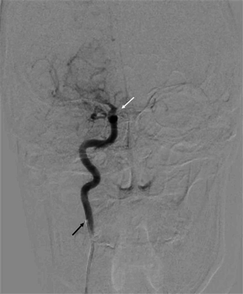
This diagnostic image is a cerebral angiogram of the right internal carotid artery (ICA) using digital subtraction angiography (DSA) in an anteroposterior (AP) view. The image demonstrates a complete vascular occlusion. The catheter tip is positioned in the proximal segment of the right ICA, indicated by a black arrow, where normal contrast opacification is initially observed. Following the course of the vessel superiorly, there is an abrupt cessation or 'cut-off' of contrast flow distal to the origin of the ophthalmic branch, marked by a white arrow. This lack of distal opacification indicates a complete occlusion of the ICA, preventing the filling of the right middle cerebral artery (MCA) and anterior cerebral artery (ACA). The faint outlines of the skull and facial bones are visible in the background due to the subtraction technique. This clinical finding is significant for diagnosing acute ischemic stroke or underlying vasculopathy in the cerebrovascular system.
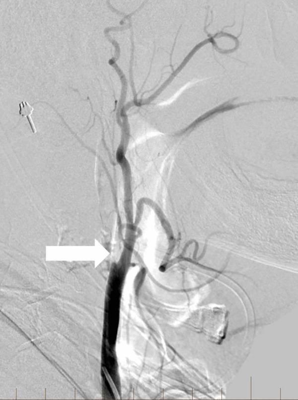
This lateral view digital subtraction angiography (DSA) image demonstrates the right extracranial carotid arterial system. The contrast-filled right common carotid artery (RCCA) is visible at the inferior aspect of the image. A white arrow points to the bifurcation, where there is an abrupt cessation of contrast flow into the internal carotid artery (ICA), indicating a complete proximal occlusion at the level of the carotid bulb. In contrast, the external carotid artery (ECA) and its distal facial and superficial temporal branches remain patent and well-opacified. The imaging is consistent with a cervical ICA occlusion, often secondary to atherosclerotic disease or dissection. This diagnostic image is used in neurointerventional radiology to evaluate the source of acute ischemic stroke symptoms, such as left-sided weakness and dysarthria, and to plan revascularization procedures like thrombectomy or stenting.
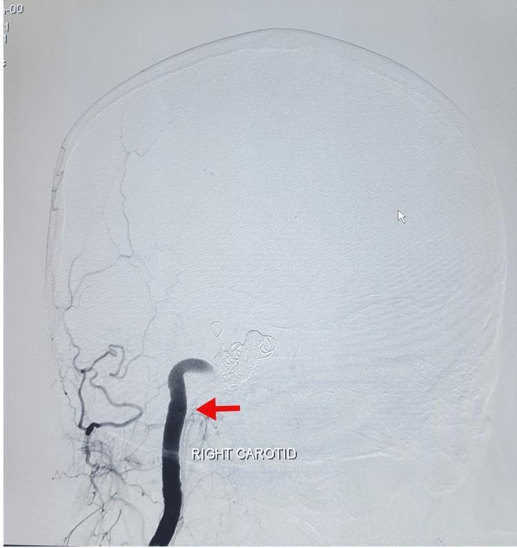
This diagnostic image is a post-operative Digital Subtraction Angiography (DSA) of the head and neck in a lateral view. The image highlights the right carotid artery, which is opacified with dark contrast material and indicated by a red arrow labeled 'RIGHT CAROTID'. At the distal aspect of the carotid artery, near the cavernous sinus region, there is a dense cluster of radio-opaque embolic coils, consistent with a parent artery occlusion for the treatment of a carotid-cavernous fistula (CCF). The surrounding cerebral vasculature appears less opacified, indicating modified hemodynamics post-embolization. The bony structures of the skull and facial bones are visible in a faint, subtracted grayscale background, providing anatomical landmarks for the vascular pathology. This image demonstrates the result of endovascular neurosurgery using metallic coils to achieve therapeutic occlusion of a high-flow vascular shunt.
sodium hypochlorite accident facial swelling ecchymosis dental
inferior alveolar nerve mandibular canal anatomy diagram
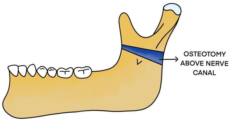
This medical illustration depicts a lateral view of the human mandible, highlighting a surgical modification for treating temporomandibular joint (TMJ) ankylosis. The diagram shows the primary anatomical features of the mandible, including the alveolar ridge with teeth, the mandibular body, and the ramus featuring the coronoid and condylar processes. A highlighted blue band across the upper portion of the mandibular ramus represents a 'Modified Osteotomy Above Nerve Canal.' This specific surgical plane is positioned superior to the inferior alveolar nerve (IAN) canal, which is indicated by a 'v' shaped marking on the ramus. The illustration demonstrates the Esmarch procedure variant where the bone cut is performed high on the ramus to separate the ankylosed condylar unit from the functional mandibular body while aiming to preserve nerve integrity. This anatomical diagram is used to explain surgical planning in maxillofacial surgery, specifically for patients with recurrent joint fusion or altered vascular anatomy.
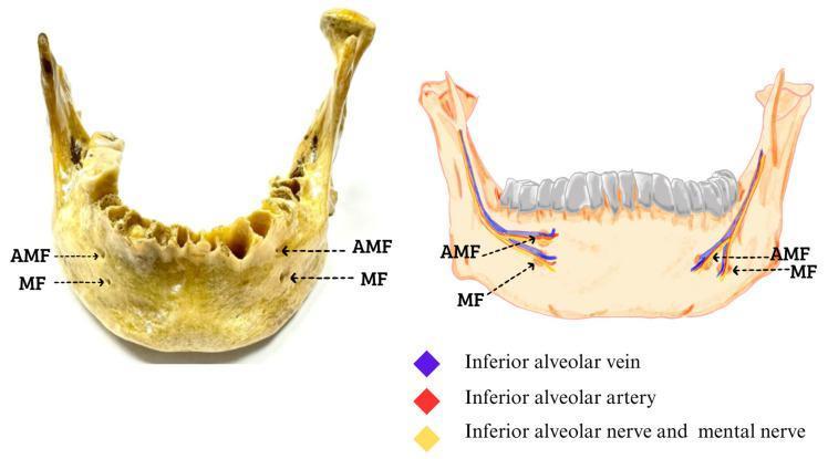
This composite educational image features a clinical photograph of a dry human mandible alongside a corresponding anatomical diagram, illustrating the bilateral presence of an accessory mental foramen (AMF). The photograph shows the anterior-lateral view of the mandible with labels pointing to the primary mental foramen (MF) and the smaller AMF located superior and slightly posterior to it on both the right and left sides. The accompanying diagram details the neurovascular bundle associated with these structures. It depicts the inferior alveolar vein (purple), inferior alveolar artery (red), and the inferior alveolar/mental nerve (yellow) traveling through the mandibular canal. The illustration highlights the branching of these neurovascular structures as they exit through both the MF and the AMF. This visual is designed for anatomy and oral surgery education, emphasizing anatomical variations of the mandibular neurovascular exit points which are critical for local anesthesia and surgical procedures in the chin and lower lip region.
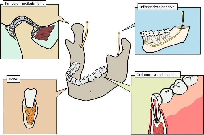
This medical anatomical diagram illustrates the composite tissue anatomy of the human mandible, intended for 3D bio-printing and surgical reconstruction planning. The central figure shows a horseshoe-shaped mandible with superior dentition and the emerging inferior alveolar nerves. Four inset boxes provide detailed structural views: 1) The 'Temporomandibular joint' panel displays the condylar articulation, articular disk, and associated ligaments and muscle attachments. 2) The 'Inferior alveolar nerve' panel shows the nerve's longitudinal pathway through the mandibular canal, providing innervation to tooth roots. 3) The 'Oral mucosa and dentition' panel depicts a cross-section of a tooth, including enamel, pulp chamber, and its anchor within the gingival tissue. 4) The 'Bone' panel illustrates the parenchymal bone frame in cross-section, highlighting the cortical shell and the internal trabecular/cancellous bone pattern. This diagram serves as an educational resource for understanding the multi-tissue complexity required in maxillofacial regenerative medicine and segmental defect reconstruction.
livedo reticularis skin lesion ischemia
gutta-percha foreign body maxillary sinus radiograph
cd /tmp/workspace/nicolau-jc && python /tmp/skills/shared/scripts/fetch_images.py \ "https://cdn.orris.care/cdss_images/pmc_clinical_VQA_6029d486bc03934b80b1754d24e802614445ed37ac930d3c675181e199f2781e.jpg" \ "https://cdn.orris.care/cdss_images/pmc_clinical_VQA_62d19f57bb7af97d17a07728a6ebd1591188dfcbe9a19b3447385791a40195db.jpg" \ "https://cdn.orris.care/cdss_images/pmc_clinical_VQA_03f60a6d0e0e5b4aab0724def545eb1a29c93e75c5911637e8395cde47bde704.jpg" \ "https://cdn.orris.care/cdss_images/pmc_clinical_VQA_5b7f0d38ae3d988312408cbdc02a5af045694aa244fcc7cfc56808fa4cbb7b03.jpg" \ "https://cdn.orris.care/cdss_images/pmc_clinical_VQA_6db00f6e56a9de24473dbcb30b77e02edbdefadbd217fa8235347401d39ad647.jpg" \ "https://cdn.orris.care/cdss_images/pmc_clinical_VQA_0488af23ebbcd15c3242b7fdee9593f356457b9767b12aa6f07a93d9e2f16017.jpg" \ > images.json python -c " import json d = json.load(open('images.json')) for x in d: print(x['url'][-20:], 'OK' if x['base64'] else 'FAIL', x.get('error')) "
~/nicolau-jc/save_images.py
import json, base64, os

d = json.load(open('/tmp/workspace/nicolau-jc/images.json'))
os.makedirs('/tmp/workspace/nicolau-jc/img', exist_ok=True)
names = ['ischemic_necrosis', 'ch_radiograph', 'dsa_facial_artery', 'ian_bundle', 'maxillary_sinus', 'mandible_composite']
for name, item in zip(names, d):
    if item['base64']:
        header, b64 = item['base64'].split(',', 1) if ',' in item['base64'] else ('', item['base64'])
        raw = base64.b64decode(b64)
        path = f"/tmp/workspace/nicolau-jc/img/{name}.jpg"
        with open(path, 'wb') as f:
            f.write(raw)
        print(name, len(raw), 'bytes saved')
    else:
        print(name, 'FAILED', item.get('error'))
cd /tmp/workspace/nicolau-jc && python save_images.py && ls -la img/
~/nicolau-jc/build_pptx_visual.py
from pptx import Presentation
from pptx.util import Inches, Pt, Emu
from pptx.dml.color import RGBColor
from pptx.enum.text import PP_ALIGN, MSO_ANCHOR
from pptx.enum.shapes import MSO_SHAPE
from PIL import Image
import os

IMGDIR = "/tmp/workspace/nicolau-jc/img"

DARK = RGBColor(0x1B, 0x2A, 0x41)
ACCENT = RGBColor(0xB0, 0x2A, 0x2A)
LIGHT = RGBColor(0xF4, 0xF4, 0xF4)
GREY = RGBColor(0x55, 0x55, 0x55)
WHITE = RGBColor(0xFF, 0xFF, 0xFF)
BOXCOLORS = [RGBColor(0x1B,0x2A,0x41), RGBColor(0xB0,0x2A,0x2A), RGBColor(0x1E,0x6E,0x3C), RGBColor(0x8A,0x5A,0x00)]

prs = Presentation()
prs.slide_width = Inches(13.333)
prs.slide_height = Inches(7.5)
blank = prs.slide_layouts[6]

def add_slide():
    return prs.slides.add_slide(blank)

def bg_fill(slide, color=WHITE):
    bg = slide.shapes.add_shape(MSO_SHAPE.RECTANGLE, 0, 0, prs.slide_width, prs.slide_height)
    bg.fill.solid(); bg.fill.fore_color.rgb = color; bg.line.fill.background(); bg.shadow.inherit = False
    slide.shapes._spTree.remove(bg._element)
    slide.shapes._spTree.insert(2, bg._element)
    return bg

def add_title_bar(slide, text, subtitle=None):
    bar = slide.shapes.add_shape(MSO_SHAPE.RECTANGLE, 0, 0, prs.slide_width, Inches(1.15))
    bar.fill.solid(); bar.fill.fore_color.rgb = DARK; bar.line.fill.background(); bar.shadow.inherit = False
    tb = slide.shapes.add_textbox(Inches(0.5), Inches(0.12), Inches(12.3), Inches(0.9))
    tf = tb.text_frame; tf.word_wrap = True
    p = tf.paragraphs[0]; p.text = text
    p.font.size = Pt(28); p.font.bold = True; p.font.color.rgb = WHITE; p.font.name = "Calibri"
    if subtitle:
        p2 = tf.add_paragraph(); p2.text = subtitle
        p2.font.size = Pt(13); p2.font.color.rgb = RGBColor(0xCF,0xD8,0xE3); p2.font.italic = True
    accent = slide.shapes.add_shape(MSO_SHAPE.RECTANGLE, 0, Inches(1.15), Inches(2.2), Inches(0.06))
    accent.fill.solid(); accent.fill.fore_color.rgb = ACCENT; accent.line.fill.background(); accent.shadow.inherit=False

def add_bullets(slide, items, left=0.6, top=1.5, width=6.3, height=5.6, size=17, color=DARK):
    tb = slide.shapes.add_textbox(Inches(left), Inches(top), Inches(width), Inches(height))
    tf = tb.text_frame; tf.word_wrap = True
    first = True
    for item in items:
        text, level = (item if isinstance(item, tuple) else (item, 0))
        p = tf.paragraphs[0] if first else tf.add_paragraph()
        first = False
        p.text = ("• " if level == 0 else "‣ ") + text
        p.font.size = Pt(size - level*2)
        p.font.color.rgb = color if level == 0 else GREY
        p.font.bold = (level == 0)
        p.space_after = Pt(10)
    return tb

def add_picture_fit(slide, path, left, top, max_w, max_h):
    im = Image.open(path)
    iw, ih = im.size
    ratio = iw/ih
    box_ratio = max_w/max_h
    if ratio > box_ratio:
        w = max_w; h = w/ratio
    else:
        h = max_h; w = h*ratio
    l = left + (max_w - w)/2
    t = top + (max_h - h)/2
    pic = slide.shapes.add_picture(path, Inches(l), Inches(t), width=Inches(w), height=Inches(h))
    # thin border
    pic.line.color.rgb = RGBColor(0xCC,0xCC,0xCC); pic.line.width = Pt(0.75)
    return pic

def add_caption(slide, text, left, top, width, size=11):
    tb = slide.shapes.add_textbox(Inches(left), Inches(top), Inches(width), Inches(0.6))
    tf = tb.text_frame; tf.word_wrap = True
    p = tf.paragraphs[0]; p.text = text
    p.font.size = Pt(size); p.font.italic = True; p.font.color.rgb = GREY
    p.alignment = PP_ALIGN.LEFT

def add_footer(slide, n):
    tb = slide.shapes.add_textbox(Inches(12.5), Inches(7.15), Inches(0.7), Inches(0.3))
    p = tb.text_frame.paragraphs[0]; p.text = str(n)
    p.font.size = Pt(11); p.font.color.rgb = GREY

slide_num = 0
def newslide(title, subtitle=None):
    global slide_num
    s = add_slide(); bg_fill(s); add_title_bar(s, title, subtitle)
    slide_num += 1
    add_footer(s, slide_num)
    return s

def image_text_slide(title, bullets, image_name, caption, subtitle=None, bullet_size=17):
    s = newslide(title, subtitle)
    add_bullets(s, bullets, left=0.6, top=1.55, width=6.3, height=5.5, size=bullet_size)
    img_path = os.path.join(IMGDIR, image_name)
    add_picture_fit(s, img_path, 7.15, 1.55, 5.6, 4.85)
    add_caption(s, caption, 7.15, 6.45, 5.6)
    return s

def pmid_note(text, pmid):
    return f"{text}  [PMID: {pmid}]"

# ================= SLIDE 1: TITLE =================
s = add_slide(); bg_fill(s, DARK)
tb = s.shapes.add_textbox(Inches(0.8), Inches(2.0), Inches(11.7), Inches(2.2))
tf = tb.text_frame; tf.word_wrap = True
p = tf.paragraphs[0]; p.text = "Nicolau Syndrome and Endodontic Complications"
p.font.size = Pt(40); p.font.bold = True; p.font.color.rgb = WHITE
p2 = tf.add_paragraph(); p2.text = "Facial Ischemic Necrosis after Calcium Hydroxide Extrusion During Root Canal Treatment"
p2.font.size = Pt(19); p2.font.color.rgb = RGBColor(0xCF,0xD8,0xE3); p2.font.italic = True
accent = s.shapes.add_shape(MSO_SHAPE.RECTANGLE, Inches(0.8), Inches(1.85), Inches(2.5), Inches(0.07))
accent.fill.solid(); accent.fill.fore_color.rgb = ACCENT; accent.line.fill.background(); accent.shadow.inherit=False
add_picture_fit(s, os.path.join(IMGDIR, "ischemic_necrosis.jpg"), 2.4, 4.1, 8.5, 2.6)
tb2 = s.shapes.add_textbox(Inches(0.8), Inches(6.85), Inches(11), Inches(0.5))
p3 = tb2.text_frame.paragraphs[0]
p3.text = "Journal Club  |  Kang Q, Huang Z, Qian W. J Endod. 2024;50:680-686."
p3.font.size = Pt(14); p3.font.color.rgb = WHITE

# ================= SLIDE 2: OBJECTIVES =================
s = newslide("Learning Objectives")
add_bullets(s, [
    "Recognize Nicolau syndrome (NS) as a rare vascular complication of drug/material extrusion",
    "Understand how calcium hydroxide (CH) extrusion during root canal treatment can trigger facial ischemic necrosis",
    "Interpret imaging used to confirm vascular occlusion (radiograph, CT, DSA)",
    "Review the broader literature on complications of endodontic material extrusion",
    "Discuss prevention strategies and critically appraise the case report",
], left=0.9, width=11.3, size=20)

# ================= SLIDE 3: NS BACKGROUND (image) =================
image_text_slide(
    "Background: What Is Nicolau Syndrome?",
    [
        "First described 1924 (Freudenthal & Nicolau) after bismuth injection",
        "Rare reaction after IM / intra-articular / subcutaneous / intravascular injection",
        "Clinical course:",
        ("Immediate severe pain at injection site", 1),
        ("Livedo reticularis / mottled discoloration", 1),
        ("Progresses to ecchymosis, then necrotic ulcer", 1),
        ("Heals with permanent scarring", 1),
    ],
    "ischemic_necrosis.jpg",
    "Representative example of injection-related ischemic skin necrosis (vascular occlusion after facial injection), illustrating the livedo -> ecchymosis -> necrosis progression seen in Nicolau syndrome."
)

# ================= SLIDE 4: CH BACKGROUND (image) =================
image_text_slide(
    "Background: Calcium Hydroxide in Endodontics",
    [
        "Introduced by Hermann (1920); standard intracanal medicament",
        "Why it's used:",
        ("Broad antimicrobial activity; inactivates bacterial endotoxin", 1),
        ("Induces hard-tissue (dentin bridge) formation", 1),
        ("Promotes periapical healing", 1),
        "The risk: strongly alkaline paste",
        ("Corrosive to vascular endothelium if it enters a vessel", 1),
        ("Risk rises when canal is bleeding + material is force-injected", 1),
    ],
    "ch_radiograph.jpg",
    "Sequential radiographs showing calcium hydroxide placement within the root canal (radiopaque fill) during normal, uncomplicated endodontic treatment."
)

# ================= SLIDE 5: CASE TIMELINE (diagram) =================
s = newslide("Case Presentation: Timeline", "16-year-old female, tooth #30 (lower right first molar)")
events = [
    ("Day 0", "RCT performed; bleeding encountered; CH applied. Headache, LOC ~10s, facial/lip cyanosis on waking"),
    ("Day 0 (ED)", "MRI performed; cavernous sinus syndrome suspected; anti-inflammatories started"),
    ("Day 1-2", "Transferred to tertiary hospital; lumbar puncture rules out intracranial infection"),
    ("Day 7", "No improvement; worsening cyanosis, skin/mucosal necrosis; NS diagnosed"),
    ("10 mo", "Eschar sheds; permanent scarring, alar defect, ectropion; depression develops"),
]
n = len(events)
margin = 0.7
avail = 13.333 - 2*margin
gap = avail/n
liney = 3.4
line = s.shapes.add_shape(MSO_SHAPE.RECTANGLE, Inches(margin), Inches(liney), Inches(avail), Inches(0.05))
line.fill.solid(); line.fill.fore_color.rgb = ACCENT; line.line.fill.background(); line.shadow.inherit=False
for i, (label, desc) in enumerate(events):
    cx = margin + gap*i + gap/2
    dot = s.shapes.add_shape(MSO_SHAPE.OVAL, Inches(cx-0.18), Inches(liney-0.15), Inches(0.36), Inches(0.36))
    dot.fill.solid(); dot.fill.fore_color.rgb = DARK; dot.line.color.rgb = WHITE; dot.line.width=Pt(1.5); dot.shadow.inherit=False
    lbl = s.shapes.add_textbox(Inches(cx-gap/2+0.1), Inches(liney-0.95), Inches(gap-0.2), Inches(0.7))
    p = lbl.text_frame.paragraphs[0]; p.text = label; p.font.bold=True; p.font.size=Pt(15); p.font.color.rgb=ACCENT; p.alignment=PP_ALIGN.CENTER
    lbl.text_frame.word_wrap = True
    dsc = s.shapes.add_textbox(Inches(cx-gap/2+0.1), Inches(liney+0.35), Inches(gap-0.2), Inches(2.6))
    tf = dsc.text_frame; tf.word_wrap = True
    p2 = tf.paragraphs[0]; p2.text = desc; p2.font.size=Pt(12.5); p2.font.color.rgb=DARK; p2.alignment=PP_ALIGN.CENTER

# ================= SLIDE 6: DIAGNOSTIC IMAGING (image) =================
image_text_slide(
    "Diagnostic Workup: Imaging",
    [
        "Panoramic radiograph: radiopacity tracking the right inferior alveolar canal",
        "CT: high-density material within inferior alveolar, facial, and infraorbital arteries",
        "DSA: confirmed occlusion of distal right maxillary and facial arteries",
        "CH's own radiopacity let clinicians trace the extravasation pathway directly on imaging",
    ],
    "dsa_facial_artery.jpg",
    "Digital subtraction angiography (DSA) demonstrating catheterization and occlusion of the facial artery -- illustrating the imaging modality used to localize the vascular occlusion in this case."
)

# ================= SLIDE 7: PATHOPHYSIOLOGY (image) =================
image_text_slide(
    "Pathophysiology: Retrograde Embolization Route",
    [
        "Proposed mechanisms of injection-related skin necrosis:",
        ("Inflammatory vasculitis - direct arterial wall injury", 1),
        ("Embolic occlusion - material blocks distal arteries", 1),
        ("Thrombotic occlusion - wall perforation -> thrombus", 1),
        ("Vasospasm - reflex sympathetic vasoconstriction", 1),
        "CH is highly alkaline -> corrodes endothelium -> vasoconstriction + thrombosis",
        "Retrograde path: apical vessels -> arterial branches -> peripheral skin vessels",
    ],
    "ian_bundle.jpg",
    "Neurovascular bundle of the mandibular canal (inferior alveolar artery, vein, and nerve) -- the anatomic corridor through which extruded material can track retrograde toward facial vasculature.",
    bullet_size=15.5
)

# ================= SLIDE 8: MANAGEMENT & OUTCOME =================
s = newslide("Management and Outcome")
add_bullets(s, [
    "Multidisciplinary team: neurology, plastic surgery, oral surgery, vascular surgery, neurosurgery",
    "Revascularization (thrombolysis/thrombectomy) rejected -- window (>1 week) had passed",
    "Conservative management: anti-inflammatory, analgesic, neurotrophic drugs + psychosocial support",
    "Outcome at 10 months:",
    ("Spontaneous eschar shedding", 1),
    ("Permanent nasal alar tissue defect", 1),
    ("Extensive scarring; lower eyelid ectropion", 1),
    ("Patient developed depression", 1),
], left=0.9, width=11.3, size=18)

# ================= SLIDE 9: CRITICAL APPRAISAL =================
s = newslide("Critical Appraisal of the Case Report")
box1 = s.shapes.add_shape(MSO_SHAPE.RECTANGLE, Inches(0.6), Inches(1.4), Inches(5.9), Inches(5.5))
box1.fill.solid(); box1.fill.fore_color.rgb = RGBColor(0xEA,0xF4,0xEC); box1.line.color.rgb = RGBColor(0x1E,0x6E,0x3C); box1.shadow.inherit=False
box2 = s.shapes.add_shape(MSO_SHAPE.RECTANGLE, Inches(6.8), Inches(1.4), Inches(5.9), Inches(5.5))
box2.fill.solid(); box2.fill.fore_color.rgb = RGBColor(0xF7,0xE9,0xE9); box2.line.color.rgb = ACCENT; box2.shadow.inherit=False
tb = s.shapes.add_textbox(Inches(0.85), Inches(1.55), Inches(5.4), Inches(5.2))
tf = tb.text_frame; tf.word_wrap = True
p = tf.paragraphs[0]; p.text = "STRENGTHS"; p.font.bold=True; p.font.size=Pt(19); p.font.color.rgb=RGBColor(0x1E,0x6E,0x3C)
for t in ["Detailed multimodal imaging (radiograph, CT, DSA) -- rare in case reports",
          "Long follow-up (10 months) capturing full natural history",
          "Clear mechanistic discussion linking CH chemistry to vascular pathology"]:
    pp = tf.add_paragraph(); pp.text = "• " + t; pp.font.size = Pt(15.5); pp.font.color.rgb = DARK; pp.space_after=Pt(10)
tb2 = s.shapes.add_textbox(Inches(7.05), Inches(1.55), Inches(5.4), Inches(5.2))
tf2 = tb2.text_frame; tf2.word_wrap = True
p2 = tf2.paragraphs[0]; p2.text = "LIMITATIONS"; p2.font.bold=True; p2.font.size=Pt(19); p2.font.color.rgb=ACCENT
for t in ["Single case -- lowest tier of evidence",
          "Causality inferred, not proven (no histology/chemical confirmation)",
          "Initial misdiagnosis (cavernous sinus syndrome) delayed recognition",
          "No data on CH volume/pressure/device -- limits reproducibility"]:
    pp = tf2.add_paragraph(); pp.text = "• " + t; pp.font.size = Pt(15.5); pp.font.color.rgb = DARK; pp.space_after=Pt(10)

# ================= SLIDE 10: LIT REVIEW INTRO (4-box) =================
s = newslide("Broader Literature: Complications of Endodontic Material Extrusion")
cats = [
    ("NaOCl Accidents", "Chemical burn / extravasation"),
    ("CH Extrusion", "Nerve injury / paresthesia"),
    ("Sealer Extrusion", "Altered sensation, variable prognosis"),
    ("Sinus Extrusion", "Sinusitis / foreign body reaction"),
]
bw, bh, gap2 = 2.9, 2.3, 0.35
startx = (13.333 - (4*bw + 3*gap2))/2
for i,(t1,t2) in enumerate(cats):
    x = startx + i*(bw+gap2)
    box = s.shapes.add_shape(MSO_SHAPE.ROUNDED_RECTANGLE, Inches(x), Inches(2.6), Inches(bw), Inches(bh))
    box.fill.solid(); box.fill.fore_color.rgb = BOXCOLORS[i % len(BOXCOLORS)]; box.line.fill.background(); box.shadow.inherit=False
    tf = box.text_frame; tf.word_wrap = True; tf.vertical_anchor = MSO_ANCHOR.MIDDLE
    p = tf.paragraphs[0]; p.text = t1; p.font.bold=True; p.font.size=Pt(18); p.font.color.rgb=WHITE; p.alignment=PP_ALIGN.CENTER
    p2 = tf.add_paragraph(); p2.text = t2; p2.font.size=Pt(13); p2.font.color.rgb=RGBColor(0xE8,0xE8,0xE8); p2.alignment=PP_ALIGN.CENTER
add_bullets(s, ["This case represents the vascular/ischemic end of a broader spectrum of extrusion complications -- most reports describe neurosensory injury or sinus disease rather than tissue necrosis."],
            left=1.0, top=5.3, width=11.3, height=1.6, size=17)

# ================= SLIDE 11: NaOCl accidents =================
s = newslide("Literature: Sodium Hypochlorite Accidents")
add_bullets(s, [
    pmid_note("Fiorillo et al. (2024) - Systematic review: clinical evidence and state of the art on NaOCl accidents", "39418527"),
    pmid_note("Kleier et al. (2008) - Survey of ABE diplomates on NaOCl accident experience", "18928844"),
    pmid_note("Klein & Kleier (2013) - NaOCl accident in a pediatric patient: ecchymosis, edema, facial dermatoses", "24553278"),
    pmid_note("Hales et al. (2001) - Treatment protocol for managing a NaOCl accident during therapy", "12004727"),
    "Common features: sudden severe pain, immediate swelling/ecchymosis, tissue necrosis from direct chemical burn -- mechanistically distinct from NS (caustic injury vs. vascular occlusion) but overlapping clinical picture",
], left=0.9, width=11.3, size=17)

# ================= SLIDE 12: CH extrusion / nerve injury (image) =================
image_text_slide(
    "Literature: Calcium Hydroxide Extrusion & Nerve Injury",
    [
        pmid_note("Tripathi et al. (2025) - CH extrusion into inferior alveolar canal", "40655580"),
        pmid_note("Sodnom-Ish et al. (2022) - Legal liability: IAN damage after CH extrusion", "34560749"),
        pmid_note("Montenegro Fonseca et al. (2020) - Massive CH/barium sulfate extrusion, paresthesia", "31724802"),
        pmid_note("Shin et al. (2016) - Accidental IAN injury from CH extrusion", "26877992"),
        pmid_note("Liu et al. (2024) - Persistent IAN paresthesia, CH+iodoform extrusion", "38303869"),
    ],
    "mandible_composite.jpg",
    "Mandibular anatomy showing the inferior alveolar nerve's course through the mandibular canal -- the structure most often injured in reported CH-extrusion complications (typically causing paresthesia rather than necrosis).",
    bullet_size=14.5
)

# ================= SLIDE 13: Sealer extrusion =================
s = newslide("Literature: Root Canal Sealer Extrusion")
add_bullets(s, [
    pmid_note("Rosen et al. (2016) - Systematic review: prognosis of altered sensation after extrusion of filling materials", "27133502"),
    pmid_note("Bamrungwong et al. (2025) - Retrospective cohort: outcomes/prognostic factors after unintentional sealer extrusion", "40339896"),
    "Key findings:",
    ("Altered sensation often improves over time, but recovery is variable and material-dependent", 1),
    ("Epoxy resin- and paraformaldehyde-based sealers carry greater neurotoxic potential", 1),
    ("Outcomes range from self-limited to permanent, depending on chemistry and proximity to neurovascular structures", 1),
], left=0.9, width=11.3, size=18)

# ================= SLIDE 14: Maxillary sinus extrusion (image) =================
image_text_slide(
    "Literature: Extrusion into the Maxillary Sinus",
    [
        pmid_note("Yamaguchi et al. (2007) - Gross extrusion of obturation materials into the maxillary sinus, complicated by aspergillosis/sinusitis", "17368059"),
        pmid_note("Bjornland et al. (1987) - Sinusitis caused by endodontic materials displaced into the maxillary sinus", "3471515"),
        "Mechanism: proximity of maxillary posterior root apices to the sinus floor allows direct extrusion during obturation/irrigation",
        "Presentation differs sharply from NS: chronic sinusitis and foreign-body/fungal reaction rather than acute ischemic necrosis",
    ],
    "maxillary_sinus.jpg",
    "CT anatomy showing the close relationship between maxillary molar/premolar root apices and the maxillary sinus floor -- the anatomic basis for iatrogenic material extrusion into the sinus."
)

# ================= SLIDE 15: SYNTHESIS TABLE =================
s = newslide("Synthesis: Comparing Complication Types")
rows = [
    ("Complication", "Typical Mechanism", "Typical Presentation"),
    ("NaOCl accident", "Direct caustic/chemical injury", "Immediate pain, swelling, ecchymosis, necrosis"),
    ("CH extrusion (nerve)", "Nerve compression / neurotoxicity", "Paresthesia, altered sensation (often reversible)"),
    ("CH extrusion (vascular) -- this case", "Endothelial corrosion, embolization, vasospasm", "Cyanosis -> ischemic necrosis, permanent scarring"),
    ("Sealer extrusion", "Material-dependent neurotoxicity", "Variable altered sensation; prognosis depends on sealer"),
    ("Maxillary sinus extrusion", "Foreign body +/- fungal reaction", "Chronic sinusitis, discharge, radiographic opacity"),
]
left, top, width, height = Inches(0.5), Inches(1.4), Inches(12.3), Inches(5.4)
table_shape = s.shapes.add_table(len(rows), 3, left, top, width, height)
table = table_shape.table
table.columns[0].width = Inches(3.0); table.columns[1].width = Inches(4.8); table.columns[2].width = Inches(4.5)
for r, row in enumerate(rows):
    for c, val in enumerate(row):
        cell = table.cell(r, c); cell.text = val
        para = cell.text_frame.paragraphs[0]
        para.font.size = Pt(14) if r > 0 else Pt(15)
        para.font.bold = (r == 0)
        para.font.color.rgb = WHITE if r == 0 else DARK
        cell.fill.solid()
        cell.fill.fore_color.rgb = DARK if r == 0 else (LIGHT if r % 2 == 0 else WHITE)

# ================= SLIDE 16: TAKEAWAYS =================
s = newslide("Clinical Takeaways for Practice")
add_bullets(s, [
    "Never force CH (or any intracanal material) into an actively bleeding canal",
    "Consider Nicolau syndrome for sudden facial pain/cyanosis/numbness after a dental injection",
    "Recognize the narrow window for revascularization -- early recognition and referral are critical",
    "CH's radiopacity can help trace extravasation on imaging if suspected",
    "Document injection technique (pressure, volume, device) for safety review",
    "Escalate to multidisciplinary care early if vascular compromise is suspected",
], left=0.9, width=11.3, size=18)

# ================= SLIDE 17: DISCUSSION QUESTIONS =================
s = newslide("Discussion Questions")
add_bullets(s, [
    "Should a standardized maximum injection pressure/technique be adopted for CH placement in bleeding canals?",
    "How would management differ if the patient presented within 24 hours rather than 1 week?",
    "What is your differential for acute facial cyanosis after a dental procedure, and how would you triage it?",
    "Is there a role for empiric anticoagulation or urgent vascular imaging while awaiting diagnosis?",
    "How should informed consent for root canal treatment address this range of rare but serious risks?",
], left=0.9, width=11.3, size=19)

# ================= SLIDE 18: REFERENCES =================
s = newslide("References")
refs = [
    "Kang Q, Huang Z, Qian W. Nicolau Syndrome with Severe Facial Ischemic Necrosis after Endodontic Treatment: A Case Report. J Endod. 2024;50:680-686.",
    "Fiorillo L, D'Amico C, Meto A, et al. Sodium Hypochlorite Accidents in Endodontic Practice. J Craniofac Surg. 2024. PMID: 39418527.",
    "Kleier DJ, Averbach RE, Mehdipour O. The sodium hypochlorite accident: experience of diplomates of the ABE. J Endod. 2008. PMID: 18928844.",
    "Tripathi S, Tripathi P, Misra P. Accidental Extrusion of Calcium Hydroxide into the Inferior Alveolar Canal. J Pharm Bioallied Sci. 2025. PMID: 40655580.",
    "Sodnom-Ish B, Seo MH, Yang HJ, et al. Legal Liability of a Dentist: IAN Damage After CH Extrusion. J Craniofac Surg. 2022. PMID: 34560749.",
    "Rosen E, Goldberger T, Taschieri S, et al. Prognosis of Altered Sensation after Extrusion of Root Canal Filling Materials: Systematic Review. J Endod. 2016. PMID: 27133502.",
    "Yamaguchi K, Matsunaga T, Hayashi Y, et al. Gross extrusion of endodontic obturation materials into the maxillary sinus. Oral Surg Oral Med Oral Pathol Oral Radiol Endod. 2007. PMID: 17368059.",
]
add_bullets(s, refs, left=0.7, width=11.9, height=5.6, size=14)

out = "/tmp/workspace/nicolau-jc/Nicolau_Syndrome_Journal_Club_Visual.pptx"
prs.save(out)
print("SAVED", out, "slides:", len(prs.slides._sldIdLst))
cd /tmp/workspace/nicolau-jc && python build_pptx_visual.py
ls -la /tmp/workspace/nicolau-jc/*.pptx
Nicolau Syndrome Journal Club Visual
Presentation · PPTX
- Title - with a representative ischemic-necrosis clinical image
- Learning objectives
- Nicolau syndrome background - clinical image of livedo → ecchymosis → necrosis progression after vascular occlusion injection injury (closest available real-world analog, captioned accordingly)
- Calcium hydroxide background - radiograph sequence showing CH placement in a canal
- Case timeline - custom horizontal timeline graphic (Day 0 → 10 months) instead of a bullet list
- Diagnostic imaging - DSA image showing facial artery catheterization/occlusion
- Pathophysiology - mandibular neurovascular bundle diagram (IAN artery/vein/nerve), showing the retrograde embolization corridor
- Management & outcome
- Critical appraisal (strengths vs. limitations, two-tone boxes)
- Literature intro - 4-color category diagram (NaOCl accidents / CH nerve injury / sealer extrusion / sinus extrusion)
- NaOCl accidents literature (PMIDs)
- CH extrusion & nerve injury - mandible/IAN anatomy composite image + PMIDs
- Sealer extrusion literature (PMIDs)
- Maxillary sinus extrusion - CT image showing molar root-to-sinus-floor relationship + PMIDs
- Synthesis comparison table
- Clinical takeaways
- Discussion questions
- References
Search PubMed for recent case reports on endodontic material complications (2020-2024)st give me introduction
endodontic material extrusion complications
Introduction (2020-2024 Case Reports: Endodontic Material Complications)
- Bone necrosis after sodium hypochlorite extrusion (Ortiz-Alves et al., 2022) - demonstrates that NaOCl accidents can progress beyond soft-tissue ecchymosis/necrosis to frank osseous necrosis, broadening the tissue-level impact beyond what is typically emphasized - [PMID: 36320668]
- Anaphylaxis after treatment with an endodontic material containing polyethylene glycol, with a fatal outcome (Alnæs et al., 2024) - introduces an immunologic/systemic complication distinct from the local ischemic or neurotoxic mechanisms discussed so far, underscoring that extrusion/exposure risk is not limited to vascular or neural injury - [PMID: 39278308]
- Altered sensation following extrusion of an endodontic file during intentional replantation (Kablan et al., 2024) - a mechanical (rather than chemical) cause of neurosensory injury, widening the differential for post-treatment paresthesia - [PMID: 38726763]
- Persistent postoperative pain from extrusion of an endodontic obturator plastic carrier, managed with periradicular microsurgery (Taschieri et al., 2021) - illustrates a foreign-body/mechanical complication requiring surgical intervention rather than conservative management - [PMID: 34322210]
give me introduction about jc
Introduction to Today's Journal Club
- It documents a rare but devastating complication that most general dentists and endodontists will never see firsthand, but should be able to recognize
- It has unusually rich imaging documentation (panoramic radiograph, CT, digital subtraction angiography) that makes the underlying vascular mechanism visually traceable
- It serves as an anchor case to discuss a broader category of endodontic material extrusion complications (sodium hypochlorite accidents, nerve injury from calcium hydroxide/sealer extrusion, sinus extrusion, and even systemic reactions like anaphylaxis)
- Background - Nicolau syndrome and calcium hydroxide's role in endodontics
- Case walkthrough - presentation, imaging, diagnosis
- Pathophysiology - how injected material causes vascular occlusion and necrosis
- Management and outcome - and why revascularization wasn't an option here
- Critical appraisal - strengths and limitations of this case report as evidence
- Literature context - how this case fits alongside other extrusion complications reported 1987-2025
- Clinical takeaways and open discussion
in detail about background whch should be introduction for my jc
"Nicolau syndrome"
Background / Introduction — Journal Club (Detailed)
Part 1: Nicolau Syndrome
- Immediate, severe pain at or near the injection site — often disproportionate to the procedure
- Rapid onset of an erythematous, reticular (livedo-like), mottled discoloration of the overlying skin — reflecting patchy arterial hypoperfusion
- Progression over hours to days to ecchymosis and then frank necrotic ulceration
- Healing by secondary intention, typically with permanent scarring, and sometimes with sensory loss in the affected territory (as seen in this case)
- Inflammatory vasculitis — vascular or perivascular injection directly injures the arterial wall, compromising flow
- Embolic occlusion — the injected material (or a precipitate it forms) physically lodges in and blocks small distal arteries
- Thrombotic occlusion — periarterial injection with wall perforation triggers local thrombus formation
- Vasospasm — reflex sympathetic vasoconstriction triggered by intra-arterial, periarterial, or perinervous injection
Part 2: Calcium Hydroxide in Endodontics
- Antimicrobial action — it dissociates into calcium and hydroxyl ions; the high pH (a saturated aqueous solution is approximately pH 12.5–12.8) is bactericidal against most bacteria found in infected canals and inactivates bacterial endotoxin (lipopolysaccharide) diffusing through dentinal tubules
- Hard-tissue induction — it promotes formation of a calcified (dentin) bridge over exposed pulp tissue, useful in apexification of immature teeth and pulp capping
- Tissue-dissolving effect — it helps dissolve residual necrotic pulp tissue and organic debris within the canal system
- Promotion of periapical healing — by creating a favorable local environment for osteoblastic/cementoblastic activity after infection is controlled
- It is caustic to vascular endothelium — direct contact can trigger vasoconstriction, endothelial injury, and secondary thrombus formation
- If forced beyond the apex (especially through an actively bleeding canal, which implies an open communication with vascular channels), it can enter the periapical vasculature and travel retrograde — from apical vessels into named arterial branches (inferior alveolar, facial, maxillary, infraorbital, posterior superior alveolar, or anterior temporal arteries, depending on the tooth) — eventually reaching the peripheral vessels that supply the overlying facial skin and mucosa
- The clinical result depends on which structure is affected: nerve involvement produces paresthesia (the more commonly reported CH-extrusion complication), while arterial involvement — as in this case — produces the ischemic cascade that defines Nicolau syndrome
give picture for this matter
calcium hydroxide pulp capping dentin bridge formation
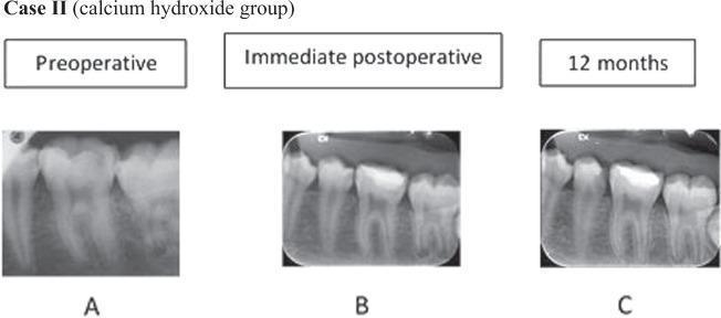
This set of diagnostic images consists of three periapical radiographs showing the longitudinal progression of a mandibular molar treatment within a dental pulp capping study (Calcium Hydroxide group). Panel A (Preoperative) reveals a deep occlusal radiolucency consistent with dental caries extending toward the pulp chamber of the first molar. Panel B (Immediate postoperative) shows the molar following indirect pulp capping and restoration with a radiopaque restorative material; a thin layer of therapeutic material (calcium hydroxide) is positioned over the pulp floor. Panel C (12 months follow-up) demonstrates the long-term clinical outcome, characterized by the formation of a distinct radiopaque bridge of reparative dentin beneath the restoration, effectively increasing the thickness of the dentin barrier between the restoration and the pulp chamber. The periapical region remains healthy with no signs of apical periodontitis or inflammatory bone resorption throughout the 12-month period. This comparison illustrates successful pulp preservation and the biological response of dentin-pulp complex regeneration.
This composite educational image illustrates the clinical applications of Acemannan (AC) in regenerative endodontics, specifically for pulp-dentin complex regeneration. Section A presents comparative periapical radiographs showing successful direct pulp capping (DPC) results in AC-treated versus calcium hydroxide-treated groups. Section B displays histopathological images comparing AC and Formocresol (FC) in deciduous tooth pulpotomy at 2, 4, and 12-week intervals, highlighting tissue inflammatory response and dentin bridge formation. Section C focuses on apexogenesis in immature permanent teeth, utilizing a temporal series of radiographs (a, b) and 3D digital reconstructions (c, d) from immediate post-op to 2 years. These visuals demonstrate progressive root lengthening, dentinal wall thickening, and narrowing of the apical foramen. Section D compares AC and Mineral Trioxide Aggregate (MTA) in pulpotomy for young permanent teeth. Longitudinal X-rays (a) and 3D root reconstructions (b) track dentine bridge formation (indicated by black arrows) and continued apical development over 12 months. The image serves as a clinical reference for biomaterial efficacy in pediatric and regenerative dentistry.
apexification immature tooth root canal treatment
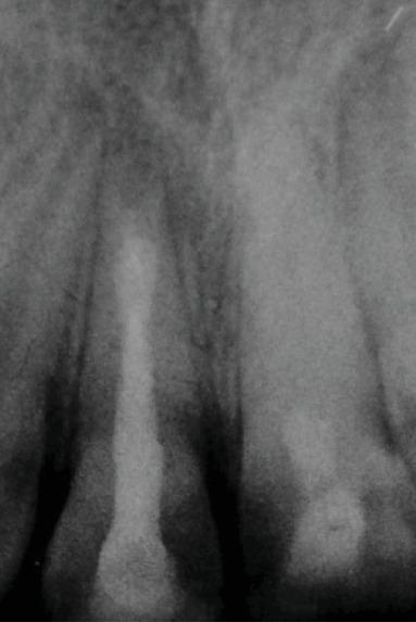
This periapical radiograph displays the maxillary central incisors (teeth 11 and 21) following endodontic treatment. Tooth 11 shows a successful apexification with a distinct apical barrier. The root canal is obturated with materials of varying radiopacity: a dense Mineral Trioxide Aggregate (MTA) plug at the apical third and a slightly less dense gutta-percha filling in the coronal two-thirds. Tooth 21 demonstrates near-complete pulp canal obliteration (calcific metamorphosis) with a visible extension of the root length, characteristic of successful pulp revascularization in an immature tooth. The surrounding periapical bone appears healthy with no signs of radiolucent inflammatory lesions. This image is an educational example of managing immature permanent teeth with open apices using different endodontic protocols: apexification for tooth 11 and regenerative endodontics/revascularization for tooth 21.
Educational comparison of endodontic root canal procedures on an immature permanent tooth model. Image A displays a diagnostic radiograph of a human tooth root specimen prepared to simulate an immature tooth with an open apex. The specimen shows a standardized 10mm cylindrical structure with an enlarged, radiolucent root canal space and thin dentinal walls. Image B shows the same specimen following orthograde obturation. A highly radiopaque calcium silicate-based filling material is visible within the apical portion of the canal, demonstrating successful apexification or apical plug placement. The filling material exhibits high density compared to the surrounding dentin. This comparison is used in endodontics to illustrate treatment protocols for teeth with incomplete root development, focusing on the visual confirmation of material placement and apical seal quality using radiographic imaging.
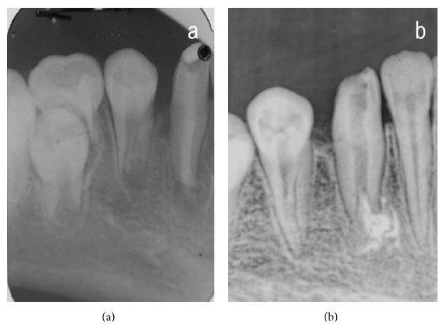
A comparison of two periapical radiographs (labeled a and b) documenting the endodontic management of an immature permanent mandibular tooth. Image (a), from the monitoring phase, reveals a tooth with significant coronal loss and an underdeveloped root characterized by a wide, divergent, or 'blunderbus' open apex. The surrounding alveolar bone is visible, and neighboring teeth show developing structures. Image (b), from the treatment phase, demonstrates the radiographic outcome after endodontic intervention. The root canal is now obturated with radiopaque filling material, likely following apexification or an apical plug procedure to address the previous open apex. The coronal portion shows signs of restoration. These images serve as an educational example of managing immature teeth with pulpal pathology, highlighting the transition from an open apex state to controlled root canal obturation.
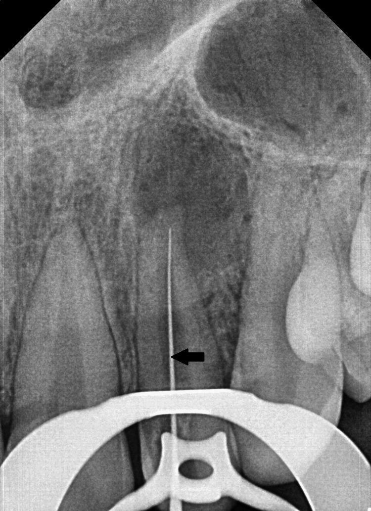
This diagnostic intraoral periapical (IOPA) radiograph demonstrates a working length determination procedure for the left maxillary lateral incisor. A radiopaque endodontic file is positioned within the root canal of the tooth, which exhibits an immature, open apex indicative of incomplete root formation or apical resorption. A significant periapical radiolucency is visible surrounding the apex, suggesting chronic periapical pathology or abscess. The periodontal ligament (PDL) space appears widened along the root surface. Anatomically, the radiolucent shadow of the nasal cavity and the floor of the nasal fossa are visible superior to the root apices. A highly radiopaque rubber dam clamp is placed over the cervical region of the tooth for isolation during the endodontic treatment. This image serves as an educational example of managing non-vital immature teeth requiring apexification and illustrates the radiographic method of calculating canal length in a pediatric endodontic context.
livedo reticularis mottled skin discoloration
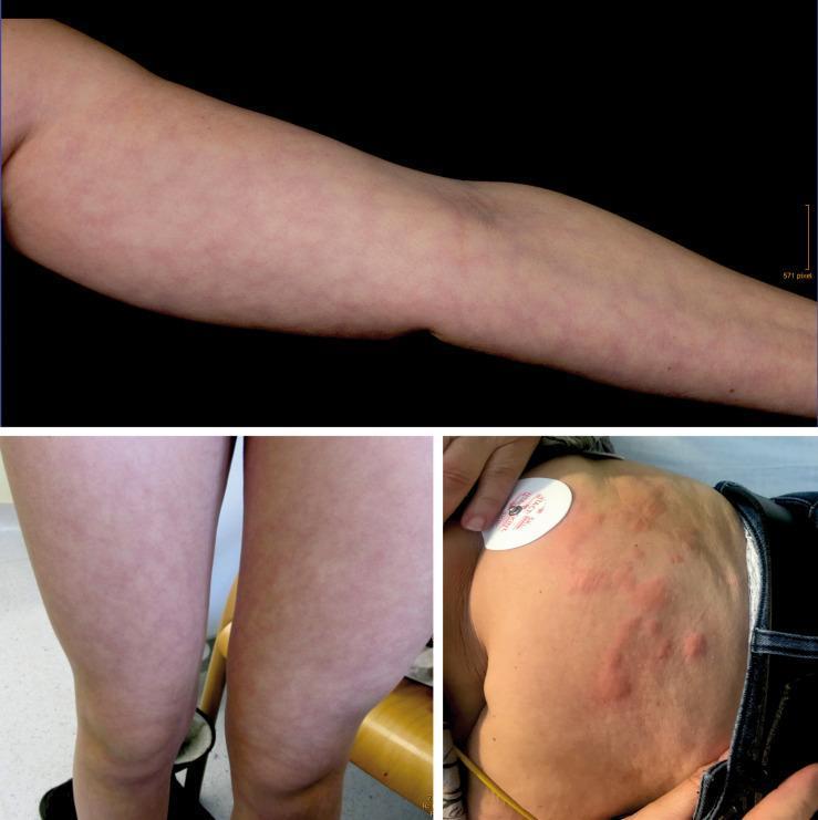
This composite of clinical photographs illustrates two distinct dermatological manifestations: livedo reticularis and urticaria. The top and bottom-left images display livedo reticularis on the arm and thighs, respectively. These images demonstrate a characteristic symmetric, mottled, and net-like (reticular) pattern of reddish-blue cyanotic discoloration. The pattern consists of closed rings or polygons, representing physiological or pathological changes in blood flow within the cutaneous vascular cones. The bottom-right image shows urticaria on the trunk, characterized by multiple raised, erythematous wheals and edematous plaques of varying sizes. Unlike the vascular mottling of livedo reticularis, these lesions are circumscribed, elevated, and represent localized dermal edema. These visuals serve as a comparative tool for differentiating vascular vasomotor changes from inflammatory skin reactions, which is clinically relevant in diagnosing conditions ranging from physiological cold response to systemic vascular disorders and allergic reactions.
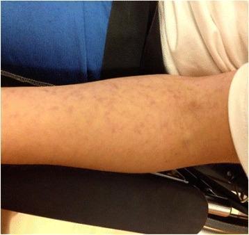
Clinical photograph of a human upper extremity demonstrating livedo reticularis. The image shows the forearm area with a characteristic reddish-blue to purplish, net-like (reticular) vascular pattern. This mottled appearance consists of irregular, interconnected rings or blotches of cyanotic discoloration surrounding central areas of normal-toned skin. The lesions appear flat and non-blanching in this view, consistent with dermal vascular congestion or vasospasm. This cutaneous finding is often associated with systemic conditions such as vasculitis, connective tissue diseases, or Susac syndrome, where it may present alongside neurological and ophthalmological symptoms. The photograph serves as an educational example of dermatological manifestations of microvascular disease, highlighting the distinctive distribution and morphology used to distinguish livedo reticularis from other forms of purpura or exanthema.
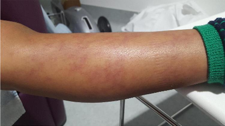
This clinical photograph displays a patient's forearm demonstrating livedo racemosa, a distinctive dermatological sign characterized by a persistent, broken, and irregular reticular (net-like) pattern. The rash consists of reddish-purple to violaceous discoloration that forms an asymmetric, branching network across the skin. Unlike livedo reticularis, which typically presents as complete circles, the patterns here are open and irregular in shape. The surrounding skin appears otherwise normal in texture, though the vascular pattern creates a mottled, non-uniform appearance. Clinically, this visual finding is a hallmark of vasculopathy and systemic inflammatory conditions, such as Deficiency of Adenosine Deaminase-2 (DADA2). It represents compromised blood flow in the medium and small vessels of the dermis and subcutis. This image is an educational example of autoinflammatory skin manifestations used to differentiate between benign physiological livedo and pathological vasculitic processes in a pediatric or adult rheumatology context.
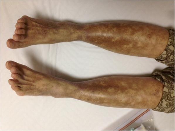
This clinical photograph displays a bilateral and symmetrical dermatological condition on the lower extremities, extending from the feet and ankles up to the proximal lower legs. The primary finding is livedo reticularis, characterized by a cyanotic, reddish-brown, reticular (net-like) pattern of skin discoloration. This mottled appearance consists of irregular rings of dusky erythema surrounding central areas of relatively pale or normal-appearing skin. The distribution is most concentrated and confluent around the distal ankles and dorsal surfaces of the feet, becoming more diffuse as it progresses toward the knees. This vascular pattern is a classic sign of impaired cutaneous blood flow, often associated with systemic conditions such as vasculitis or, as in this clinical context, type II cryoglobulinemia. There are no visible ulcerations or focal gangrene in the provided view, though the diffuse nature of the livedo suggests significant underlying small-vessel pathology.
tooth anatomy root apex pulp chamber diagram
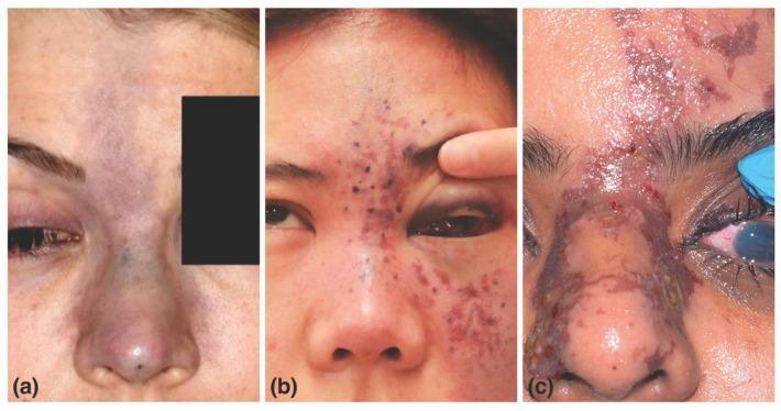
/tmp/workspace/nicolau-jc/img/ if you want them pulled directly into the PPTX. Want me to insert the three new ones (livedo reticularis, dentin bridge, apexification) into the existing slide deck's Background section now?