

How to fix this case
intertrochanteric femur fracture intramedullary nail fixation x-ray
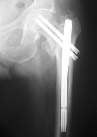
Anteroposterior (AP) X-ray of the proximal femur demonstrating internal fixation of an intertrochanteric fracture using a short cephalomedullary nail. The radiographic study shows a titanium alloy intramedullary nail positioned within the femoral shaft, secured proximally by two parallel cephalic lag screws extending into the femoral head to stabilize the fracture site. A single distal locking screw is visible at the inferior end of the nail to prevent rotation and axial displacement. The image displays acceptable closed reduction with evidence of radiographic healing, characterized by the continuity of the cortical bone and callus formation across the fracture line at the 4-month postoperative stage. Anatomical landmarks including the acetabulum, femoral head, and greater trochanter are clearly visible, illustrating the proper alignment and tip-apex distance of the orthopedic hardware for hip fracture management.
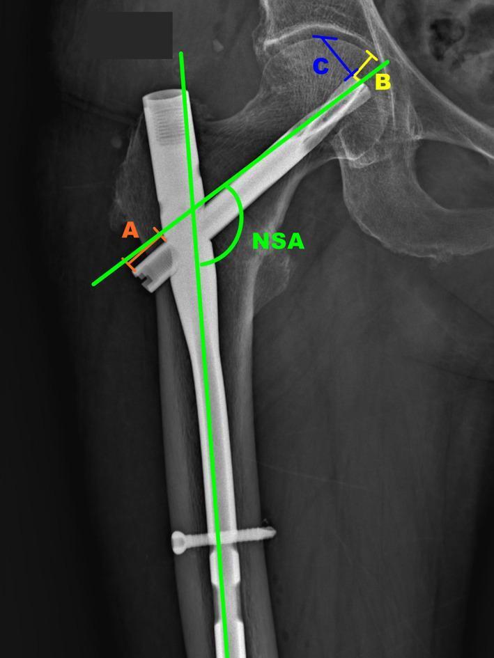
Anteroposterior (AP) X-ray of the proximal femur demonstrating an intramedullary nail fixation for an intertrochanteric fracture. The diagnostic image shows a cephalomedullary nail positioned within the femoral canal, secured by a proximal helical blade extending into the femoral head and a distal transverse locking screw. Annotated green lines indicate the Neck-Shaft Angle (NSA), measuring the alignment between the femoral shaft and neck axes. Specific geometric measurements are labeled for post-operative monitoring: Line A measures the distance from the helical blade's distal end to the nail's lateral cortex to monitor blade backout; Line B (yellow) measures distance to the medial cortex to assess femoral neck shortening or cut-through; and Line C (blue) measures distance to the superior cortex to evaluate for superior cutout. This radiograph illustrates key orthopedic parameters used to evaluate the mechanical stability and potential complications of proximal femoral internal fixation during clinical follow-up.
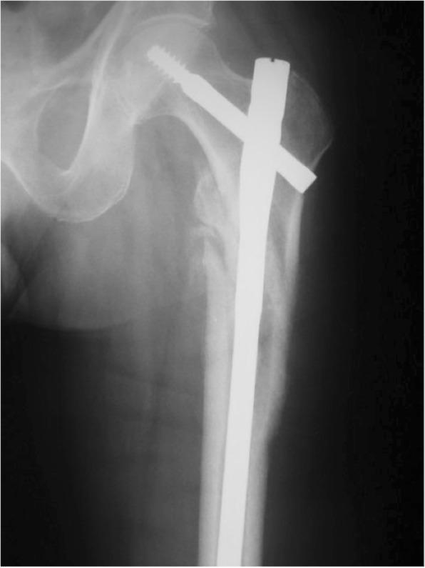
This anteroposterior (AP) x-ray radiography shows the surgical fixation of an intertrochanteric femoral fracture using a long cephalomedullary nail. The orthopedic construct consists of a radiopaque intramedullary nail inserted into the femoral shaft, with a thick cephalic lag screw and a secondary integrated stabilization pin (anti-rotation pin) extending proximally through the femoral neck and into the femoral head. The imaging demonstrates the fracture site located between the greater and lesser trochanters, showing signs of surgical reduction and stabilization. The cortical margins of the proximal femur appear irregular and disrupted consistent with a high-energy or unstable fracture pattern (OTA/AO type 31). This diagnostic image illustrates the standard of care for stabilizing extracapsular hip fractures to facilitate weight-bearing and bone union while preventing collapse or varus deformity. Relevant for orthopedic trauma education, specifically in understanding intramedullary fixation mechanics and proximal femoral anatomy.
X-Ray Findings: B. Ramudu, 60M
- Left femur: Significantly displaced fracture with proximal migration (shortening), lateral displacement, and varus angulation. The femoral head remains in the acetabulum (no hip dislocation).
- Right hip: Normal alignment, no fracture.
- Pelvic ring: Intact - symmetric SI joints, no pubic symphysis diastasis.
- Bone quality: Generalized osteopenia, which predisposed to this injury.
- Lumbar spine: Mild degenerative changes.
Classification
- Proximal segment: flexed, abducted, externally rotated (pull of iliopsoas, gluteus medius, short external rotators)
- Distal segment: shortened and medialized (unopposed adductors)
Management
Immediate / Pre-operative
- Resuscitation and pain control - IV analgesia, assess for blood loss (thigh/pelvis can hide significant hemorrhage)
- Traction splint - to reduce displacement, control pain, limit further soft tissue injury
- Pre-operative imaging: AP pelvis + lateral hip + contralateral hip (for neck-shaft angle template)
- Labs: CBC, coagulation, metabolic panel; cross-match blood
- Surgery within 24-48 hours once medically optimized (delay beyond 48h increases mortality in elderly)
Definitive Surgical Treatment: Cephalomedullary Intramedullary Nail (IMN)
Surgical Steps (ORIF with IMN)
- Position: Supine on fracture table or flat table with fluoroscopy
- Reduction: Closed reduction first; if inadequate, use pointed reduction forceps, Schanz screws, or consider mini-open. Correct the flexion-abduction deformity of the proximal segment.
- Entry portal: Tip of greater trochanter (trochanteric entry) with awl/guidewire
- Nail insertion: Appropriately sized long nail (diameter matched to IM canal)
- Cephalic fixation: Lag screw or helical blade into femoral head/neck - center-center on AP, center or slightly inferior on lateral
- Distal locking: At least 2 distal locking screws (static locking) to control rotation and length
- Verify: AP and lateral fluoroscopy confirming reduction and implant position
Post-Operative Care
| Timeline | Management |
|---|---|
| Day 1-2 | Mobilize with physiotherapy, TTWB (toe-touch weight bearing) |
| 6 weeks | Follow-up X-ray; advance weight bearing if callus forming |
| 3 months | Progressive weight bearing; repeat X-ray |
| 6-12 months | Full weight bearing; monitor for hardware failure or AVN |
- DVT prophylaxis: LMWH (e.g., enoxaparin) starting 12h post-op
- Osteoporosis treatment: Start calcium + Vitamin D; consider bisphosphonates after fracture healing (avoid in acute phase)
- Secondary fracture prevention: Falls risk assessment, physiotherapy
Complications to Watch For
| Complication | Prevention |
|---|---|
| Varus malreduction ("cut-out") | Center-center placement, TAD <25mm |
| Non-union | Anatomic reduction, stable fixation |
| Implant failure | Long nail to prevent stress riser |
| DVT/PE | Early mobilization + anticoagulation |
| AVN femoral head | Unlikely here (extracapsular fracture) |
| Infection | Prophylactic antibiotics (cefazolin) |