
Draw shoulder orthosis diagram
A clear labeled medical diagram of a shoulder orthosis (shoulder brace/support), showing the anatomical components including: abduction pillow, humeral cuff, chest strap, axillary pad, arm sling component, and adjustment straps. Show anterior and lateral views. Include labels pointing to each component. Clean white background, medical illustration style.
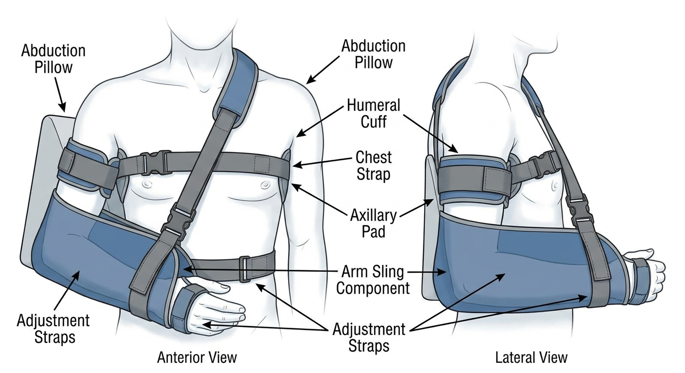
| Component | Function |
|---|---|
| Humeral cuff | Wraps around the upper arm to support and position the humerus |
| Abduction pillow | Holds the arm away from the body at a set angle (commonly 15–90°) |
| Axillary pad | Cushions the axilla and distributes pressure |
| Chest strap | Anchors the orthosis to the torso for stability |
| Arm sling | Supports the forearm and elbow weight |
| Adjustment straps | Allow customization of abduction angle and fit |
Drawable diagram
What do you mean by 'drawable diagram'?
Simple clean black line-art sketch of a shoulder orthosis (shoulder abduction brace) on a human torso, drawn in minimal outline style suitable for hand tracing or coloring. Bold black outlines only, no shading, no color, white background. Show the abduction pillow holding the arm out to the side, chest strap across the torso, humeral cuff around the upper arm, axillary pad, and forearm sling. Include simple handwritten-style labels with leader lines pointing to each part: Humeral Cuff, Abduction Pillow, Axillary Pad, Chest Strap, Forearm Sling, Adjustment Buckle. Anatomical diagram worksheet style.
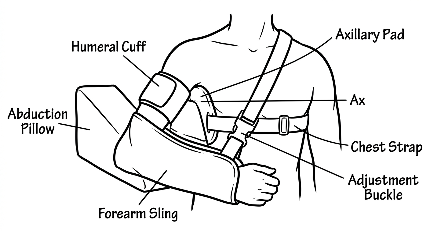
- Humeral Cuff — wraps the upper arm
- Abduction Pillow — holds the arm elevated laterally
- Axillary Pad — cushions the underarm
- Chest Strap — secures the brace to the torso
- Forearm Sling — supports the weight of the forearm
- Adjustment Buckle — sets the abduction angle
Draw finger orthosis
Simple clean black line-art sketch of finger orthoses (finger splints/braces), drawn in minimal outline style suitable for hand tracing. Bold black outlines only, no shading, no color, white background. Show multiple types of finger orthoses on a hand diagram: 1) Stack splint (mallet finger) on the index finger DIP joint, 2) Figure-of-8 splint on the middle finger PIP joint, 3) Dynamic extension splint on the ring finger, 4) Buddy strap/splint between fingers. Include a side view of a single finger showing the joints labeled: DIP, PIP, MCP. Add simple handwritten-style labels with leader lines: Stack Splint, Figure-of-8 Splint, Dynamic Splint, Buddy Strap, Dorsal surface, Volar surface, Velcro strap, Thermoplastic shell. Anatomical worksheet style, educational medical illustration.
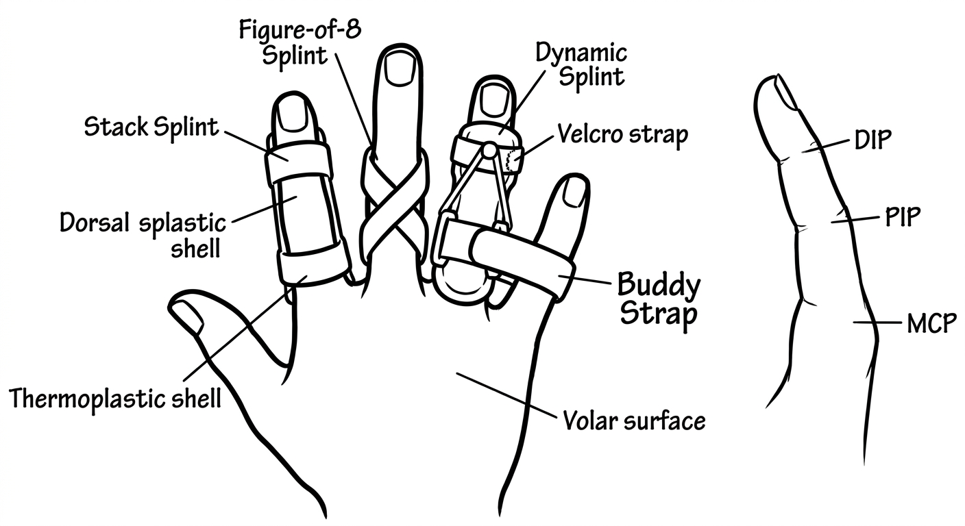
| Orthosis | Joint Targeted | Indication |
|---|---|---|
| Stack Splint | DIP | Mallet finger, extensor tendon injury |
| Figure-of-8 Splint | PIP | Swan neck deformity, PIP hyperextension |
| Dynamic Extension Splint | PIP / MCP | Flexion contracture, post-surgical mobilization |
| Buddy Strap | PIP / DIP | Collateral ligament sprain, fracture |
- MCP — Metacarpophalangeal joint
- PIP — Proximal interphalangeal joint
- DIP — Distal interphalangeal joint
Hand splint
Simple clean black line-art sketch of hand splints, drawn in minimal outline style suitable for hand tracing and printing. Bold black outlines only, no shading, no color, white background. Show three types of hand splints side by side: 1) Resting hand splint (volar view) - wrist in neutral, fingers slightly flexed, thumb abducted; 2) Cock-up wrist splint (dorsal and volar view) - wrist extended 20-30 degrees, fingers free; 3) Thumb spica splint - immobilizing thumb CMC and MCP joints. Include anatomical landmarks labeled with leader lines: Wrist (radiocarpal joint), Thumb CMC joint, MCP joints, Thermoplastic shell, Velcro straps, Palmar bar, Thumb post, Forearm trough. Also show a small inset diagram of the hand skeleton labeling: Distal phalanx, Middle phalanx, Proximal phalanx, Metacarpal, Carpals, Radius, Ulna. Educational medical worksheet style, clean anatomical illustration.
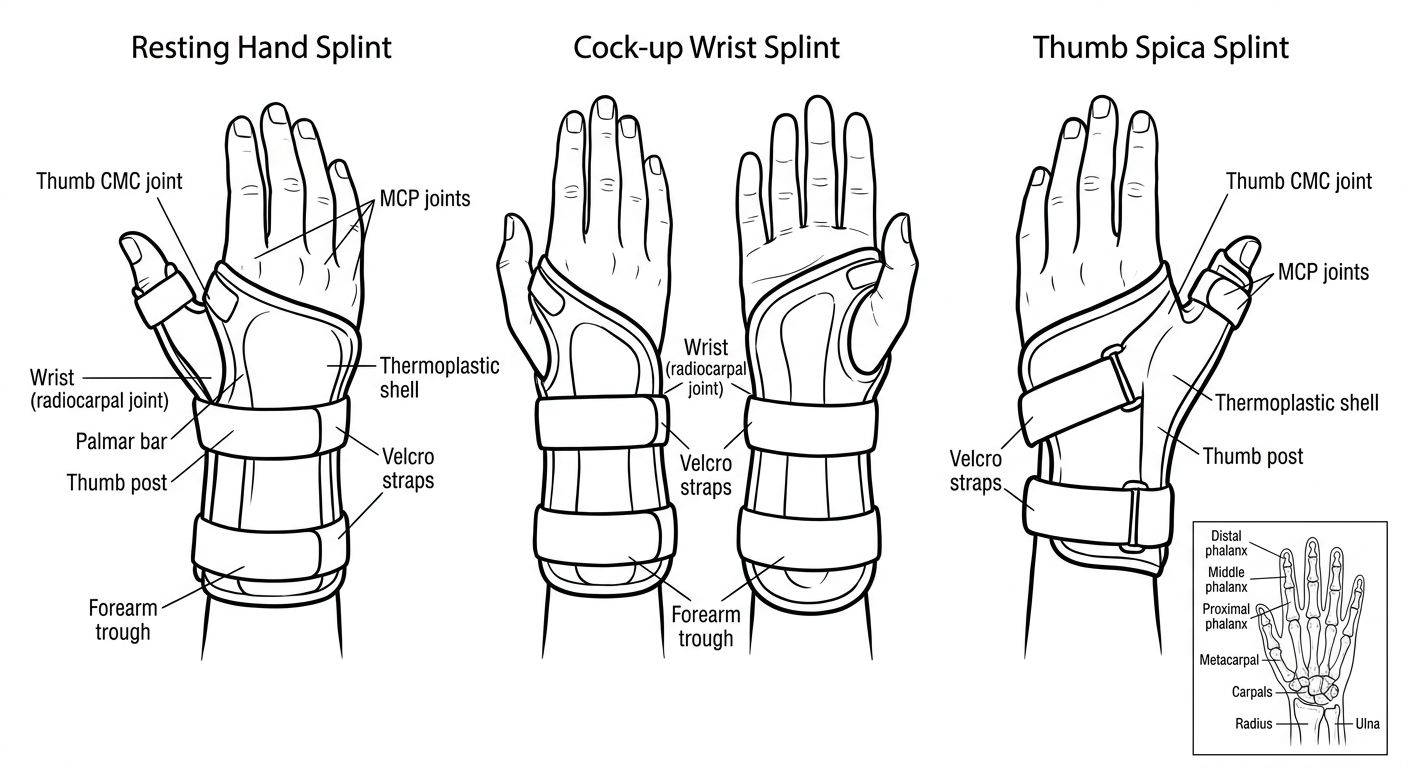
Types Shown
| Splint | Position | Indication |
|---|---|---|
| Resting Hand Splint | Wrist 10–20° extension, MCPs 45° flexion, IPs slight flexion, thumb abducted | Stroke, RA, burns, spasticity, nerve injury |
| Cock-Up Wrist Splint | Wrist 20–30° extension, fingers free | Carpal tunnel syndrome, wrist sprain, radial nerve palsy |
| Thumb Spica Splint | Thumb CMC + MCP immobilized, wrist neutral | De Quervain's tenosynovitis, scaphoid fracture, thumb UCL injury |
Key Components Labeled
- Forearm trough — proximal support base
- Palmar bar — maintains transverse arch
- Thumb post — positions and immobilizes thumb
- Velcro straps — secure fit at forearm, wrist, and hand
- Thermoplastic shell — custom-molded low-temperature material
Elbow orthosis
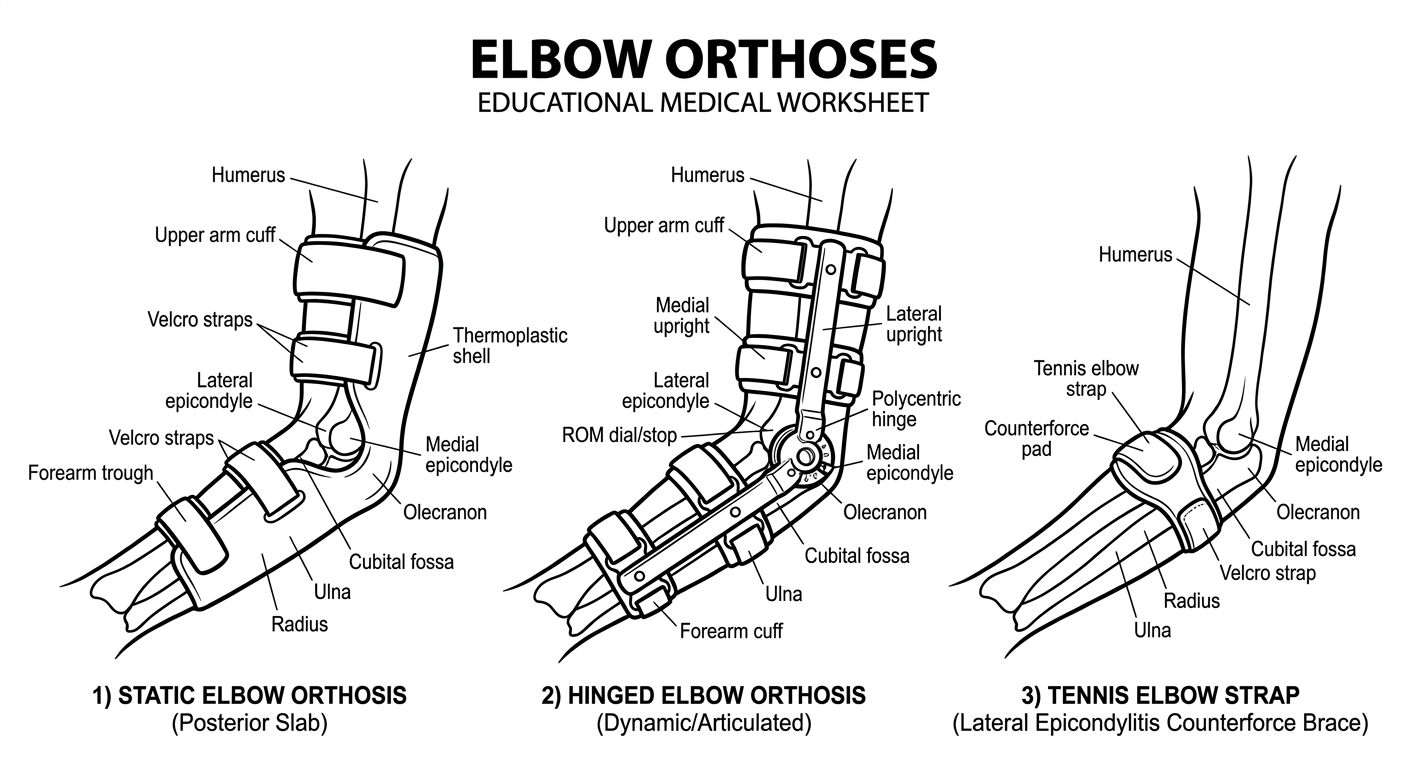
Simple clean black line-art sketch of elbow orthoses, drawn in minimal outline style suitable for hand tracing and printing. Bold black outlines only, no shading, no color, white background. Show three types of elbow orthoses on arm diagrams: 1) Static elbow orthosis - posterior slab holding elbow at 90 degrees flexion, with forearm trough and upper arm cuff; 2) Hinged elbow orthosis (dynamic/articulated) - showing medial and lateral uprights, polycentric hinge joint at elbow, upper arm and forearm cuffs, adjustable ROM stops; 3) Tennis elbow strap (lateral epicondylitis counterforce brace) - circular strap just below elbow. Include anatomical landmarks labeled with leader lines: Lateral epicondyle, Medial epicondyle, Olecranon, Humerus, Radius, Ulna, Cubital fossa. Label brace components: Upper arm cuff, Forearm cuff, Polycentric hinge, ROM dial/stop, Velcro straps, Counterforce pad, Thermoplastic shell. Educational medical worksheet style, clean anatomical illustration.
Types Shown
| Orthosis | Position / Function | Indication |
|---|---|---|
| Static Elbow Orthosis | Elbow fixed at 90° flexion, no movement | Post-fracture immobilization, ulnar nerve transposition, cubital tunnel syndrome |
| Hinged Elbow Orthosis | Adjustable ROM stops on polycentric hinge; allows controlled flex/extension | Post-surgical rehab, elbow instability, contracture management |
| Counterforce Strap | Compressive band ~5 cm distal to lateral epicondyle | Lateral epicondylitis (tennis elbow), medial epicondylitis (golfer's elbow) |
Key Components Labeled
- Upper arm cuff — proximal anchor on humerus
- Forearm cuff — distal anchor on radius/ulna
- Polycentric hinge — mimics elbow's moving axis of rotation
- ROM dial/stop — sets extension and flexion limits
- Counterforce pad — offloads stress at epicondyle
- Olecranon cutout — relieves pressure on bony prominence