
acute and chronic cholecystits
acute cholecystitis gallbladder pathology
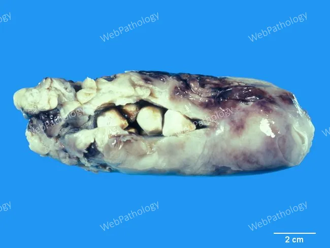
Gross pathology photograph of a gallbladder specimen displaying features of acute calculous cholecystitis. The organ is enlarged with marked wall edema and thickening; the serosal surface is dull and may be variably congested, with patchy fibrinopurulent exudate adherent to the serosa. The specimen shows omental adhesions and pericholecystic fluid, reflecting surrounding inflammatory response. The lumen contains one or more gallstones, establishing cholelithiasis as the inciting factor. Outer gallbladder surfaces exhibit hemorrhagic discoloration and, in advanced disease, may demonstrate gangrenous changes ranging from deep red to black. This image, taken as a fixed macrographic photograph against a standardized blue background with a 2 cm scale bar, captures the gross correlates of biliary inflammation due to cystic duct obstruction. The gross features correlate with histopathologic processes such as mucosal irritation, edema, and transmural inflammation; if unrecognized or prolonged, progression to perforation or gangrene is possible. Clinically, these findings align with acute cholecystitis presentations: right upper quadrant pain, fever, leukocytosis, and leukogram abnormalities. Radiologic evaluation often demonstrates gallstones and pericholecystic fluid; surgical pathology confirms the diagnosis and informs subsequent management, including cholecystectomy. This image is educational for medical students, residents, and pathology trainees, illustrating typical macroscopic gallbladder pathology.
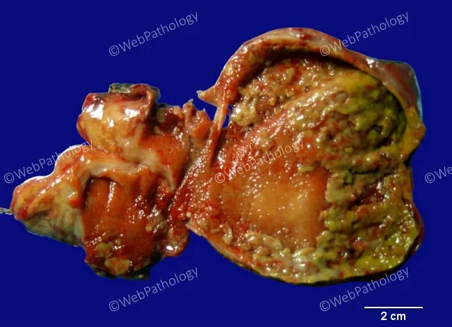
Gross pathology of a gallbladder with acute calculous cholecystitis. The specimen shows a distended, inflamed gallbladder wall with mucosal edema and erythema. The mucosa is congested and irregular, with focal necrosis and yellow-green exudate admixed with fibrinous material. Patchy greenish-yellow pseudomembranes overlying necrotic mucosa are evident, consistent with pseudomembranous cholecystitis. The serosa may be variably irritated, and adherent inflammatory adhesions can be seen along the gallbladder bed. The exterior surface demonstrates hyperemia; the wall may be thickened due to edema. A portion of the lumen is exposed, showing necrotic debris and exudate filling the lumen interface. The 2 cm scale bar provides context for dimensions; overall changes correlate with acute inflammatory insult, often precipitated by gallstone obstruction of the cystic duct. The appearance aligns with fulminant cholecystitis and may precede gangrenous transformation if infection progresses. These macroscopic findings are supported by typical histology of mucosal necrosis, neutrophilic infiltrate, edema, and fibrinous exudate on the surface. This image highlights hallmark features of pseudomembranous cholecystitis with inflammatory exudate forming pseudomembranes, mucosal ulceration, and mural necrosis within the gallbladder wall. Correlation with clinical history of gallstones, fever, RUQ pain, leukocytosis, and imaging findings reinforces diagnosis and guides emergent management for surgical intervention planning.
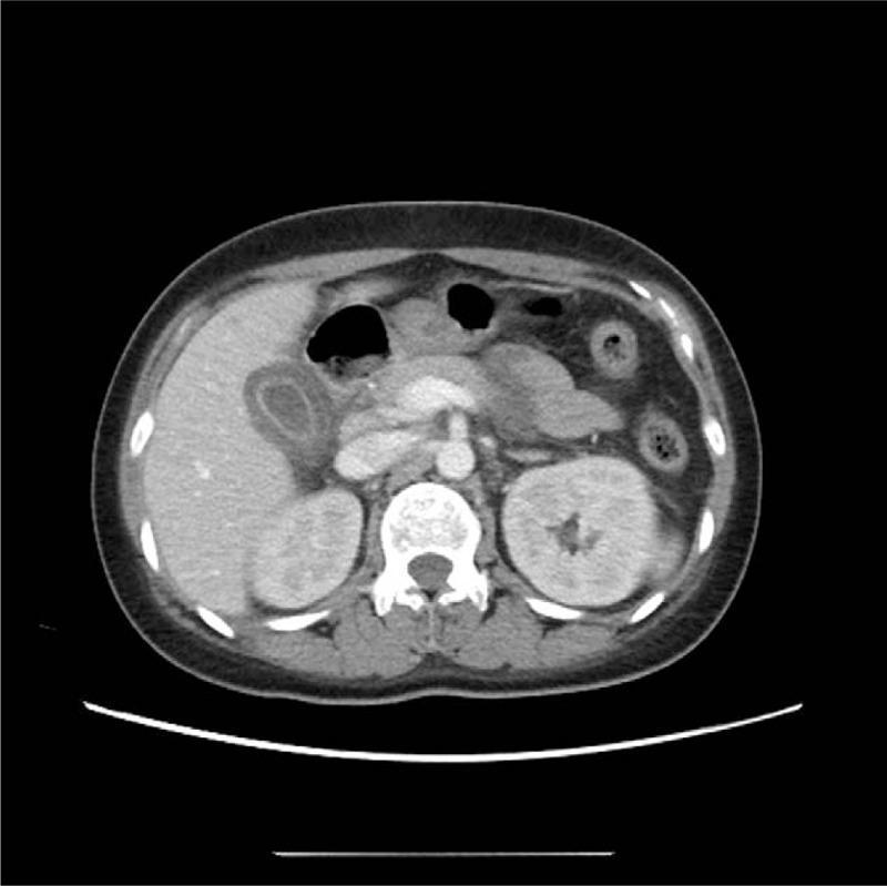
This diagnostic image is an axial slice from a contrast-enhanced abdominal computed tomography (CT) scan. The primary pathology is located in the right upper quadrant, showing significant gallbladder wall thickening and mural edema, creating a 'halo' or layered appearance. There is associated pericholecystic fluid and fat stranding, which are hallmarks of acute cholecystitis. Notably, there are no radiopaque gallstones or sludge visible within the gallbladder lumen, consistent with a diagnosis of acalculous cholecystitis. Other visible abdominal structures include the liver parenchyma, which appears homogeneous; both kidneys, which show normal cortical enhancement; the pancreas; the stomach containing air; and the abdominal aorta. The image serves as a clinical example of secondary gallbladder changes often associated with systemic inflammatory conditions or acute acalculous cholecystitis (AAC). This material is relevant for medical education regarding gastrointestinal imaging and the differential diagnosis of acute abdominal pain.
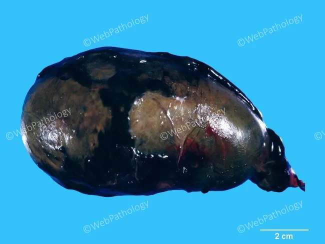
Gross pathology photograph of an excised gallbladder, captured as an external macroscopic view with a scale reference to convey size and color change. Anatomical context places this within the hepatobiliary system, the gallbladder in the right upper quadrant with wall thickening and distension typical of severe acute cholecystitis. Macroscopic features include enlargement and greenish-black discoloration, with the lumen filled by necrotic debris admixed with purulent, thick bile. The exterior may show an irregular serosal surface and subtle hemorrhagic streaks at the neck. Histologic correlation (described in accompanying notes) indicates complete mucosal and muscularis necrosis, replaced by acute inflammatory infiltrate and granulation tissue, i.e., gangrenous transformation. Pathological significance is the progression from acute inflammation to gangrene, observed in patients with diabetes mellitus, cardiovascular disease, or trauma, and is associated with a high risk of gallbladder perforation, bile collection, and sepsis. Clinically relevant implications include need for urgent cholecystectomy and broad-spectrum antibiotics; surgical pathology confirms necrosis and inflammatory sequelae. Differential considerations include emphysematous cholecystitis and acalculous or calculous cholecystitis. The image supports educational purposes in gross pathology, biliary infection, sepsis risk assessment, and prognosis determination. This depiction aids trainees in recognizing gangrenous changes and correlating gross findings with clinical staging and outcome.
"cholecystitis" AND "management"
chronic cholecystitis histology Rokitansky-Aschoff sinuses
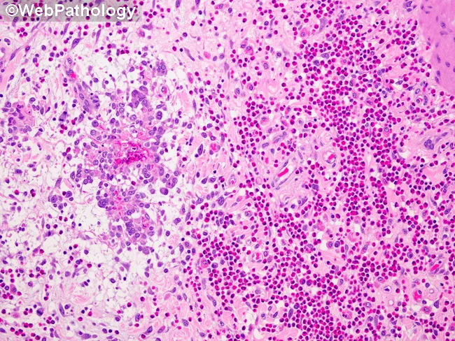
Imaging modality and technique: Light microscopy of a paraffin-embedded gallbladder mucosa section stained with Hematoxylin and Eosin (H&E). Primary subject: eosinophilic cholecystitis with destruction of Rokitansky-Aschoff sinus glands by an intense eosinophilic infiltrate. The visible lamina propria demonstrates dense clusters of eosinophils (bilobed nuclei with bright pink granules) intermixed with other inflammatory cells, accompanied by glandular distortion and gland rupture within Rokitansky-Aschoff sinuses. Mucosal damage is evident as focal erosion and edema, with disruption of normal mucosal architecture and crypt-like glands in the gallbladder mucosa. The eosinophilic inflammation drives tissue destruction, contributing to gallbladder dyskinesia and potential biliary colic. Notable features include perivascular eosinophils, tissue edema, and preservation of downstream serosa in this field, though transmural involvement may occur in advanced cases. This histology is diagnostic for eosinophilic cholecystitis, a rare inflammatory variant of cholecystitis linked to systemic eosinophilia, atopy, parasitic infection, or drug hypersensitivity. Diagnostic significance rests on recognizing eosinophil predominance and mucosal injury rather than neutrophilic invasion. Differential considerations include acute bacterial cholecystitis, chronic cholecystitis with eosinophils, parasitic inflammation, and drug-induced hypersensitivity. Clinically, correlate with peripheral eosinophil count, allergic history, and imaging findings; guide management and immunologic workup; educate pathology trainees and researchers about eosinophil-mediated biliary disease.
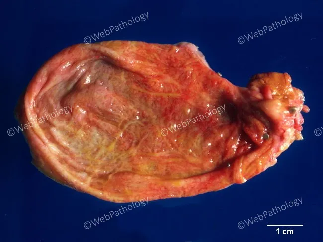
This gross pathology photograph depicts a gallbladder from a patient with chronic acalculous cholecystitis. The organ shows pronounced wall thickening with fibromuscular hypertrophy of the muscularis and expansion of intramural folds. The mucosal surface exhibits multiple Rokitansky-Aschoff sinuses, mucosal outpouchings that extend into the muscular layer, a hallmark of chronic inflammatory outflow obstruction. No radiopaque stones are evident on this exterior view, consistent with the acalculous pattern; subtle serosal vascularity and mild edema may be present. The overall texture is firm rather than edematous, reflecting chronicity. Inflammatory infiltrates are suggested by the pink-staining tissue bands and the presence of scattered inflammatory cells within the mural layers. The specimen lacks discrete calculus but displays features compatible with histologic subtypes of acalculous cholecystitis, including eosinophilic, lymphoplasmacytic, or lymphocytic predominance, as described in literature. The gross pattern aligns with clinical contexts of gallbladder outflow obstruction and chronic inflammation, providing diagnostic context for subsequent histopathology. Clinically, these findings are relevant for differential diagnoses that include chronic cholecystitis without stones, biliary dyskinesia, and inflammatory gallbladder diseases. This image is useful for education, slide correlation, and radiologic-pathologic correlation in GI pathology and hepatobiliary surgery. Correlation with histology enhances diagnostic confidence and educational value for trainees worldwide.
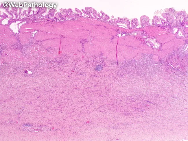
This is a low-magnification bright-field light microscopic image of gallbladder wall in chronic calculous cholecystitis, stained with Hematoxylin and Eosin (H&E). The mucosa is hyperplastic with epithelium showing mucosal metaplasia and folded mucosal ridges. The lamina propria contains a dense mononuclear inflammatory infiltrate (lymphocytes and plasma cells) that extends through the muscularis propria into the subserosa, reflecting a transmural chronic inflammatory process. The gallbladder wall is markedly thickened due to persistent inflammation and interstitial fibrosis, with loss of normal wall architecture. Rokitansky-Aschoff sinuses are evident as invaginations of mucosa through the muscularis, a hallmark of chronic cholecystitis. Pan-mural involvement with fibrotic remodeling can be seen. There may be focal areas of edema and vascular congestion without acute neutrophilic microabscesses, distinguishing this from acute cholecystitis. The image underlines the histopathologic hallmarks: chronic inflammatory infiltrate in lamina propria, fibrosis of the gallbladder wall, and Rokitansky-Aschoff sinuses; these features relate to gallstone disease and may predispose to complications such as gallbladder scarring and porcelain gallbladder in long-standing disease. Clinically, this pattern supports a history of biliary colic with chronic gallstone irritation and potentially guides surgical pathology interpretation and prognosis. This histology supports calcified and fibrotic remodeling typical of chronic inflammatory cholecystitis.
Acute and Chronic Cholecystitis
Overview
ACUTE CHOLECYSTITIS
Types
Pathogenesis (Acute Calculous)
- Mucosal phospholipases hydrolyze biliary lecithin → lysolecithin (toxic to mucosa)
- The normal protective glycoprotein mucous layer is disrupted, exposing mucosa to the detergent action of bile salts
- Prostaglandins released from the distended gallbladder wall amplify mucosal and mural inflammation
- Distention and increased intraluminal pressure compromise mucosal blood flow → ischemia
- Bacterial infection (E. coli, anaerobes in up to 40%) may be superimposed later, but is not the initiating event
Pathogenesis (Acute Acalculous)
- Major surgery / severe trauma
- Severe burns
- Sepsis with hypotension and multisystem organ failure
- Immunosuppression
- Diabetes mellitus
- AIDS (often related to Cryptosporidium infection)
Morphology
| Feature | Description |
|---|---|
| Gross | Enlarged, tense gallbladder; red, violaceous, or green-black discoloration from subserosal hemorrhage |
| Serosa | Fibrinopurulent exudate |
| Lumen | Turbid bile mixed with fibrin, pus, and hemorrhage |
| Histology (early) | Edema, congestion, mucosal erosion; neutrophils sparse unless bacterial superinfection |
- Empyema: lumen filled with virtually pure pus
- Gangrenous cholecystitis: wall becomes green-black and necrotic, small to large perforations
- Emphysematous cholecystitis: gas-forming organisms (Clostridia, coliforms) invade the wall; air in gallbladder wall/lumen; dirty acoustic shadowing on ultrasound
- Hemorrhagic cholecystitis / hemobilia: bleeding into the gallbladder
- Perforation: usually near the fundus → pericholecystic collection or intrahepatic abscess
Clinical Features
| Feature | Detail |
|---|---|
| Pain | Progressive RUQ or epigastric pain lasting >6 hours; may radiate to right shoulder or right scapular tip |
| Associated symptoms | Fever, anorexia, nausea, vomiting, tachycardia, sweating |
| Jaundice | Usually absent; if present, suggests common bile duct obstruction |
| Murphy's sign | Inspiratory arrest with RUQ palpation; positive predictive value >90% when combined with stones and wall thickening on US |
| Leukocytosis | Mild to moderate; may be absent in up to ~50% |
| LFTs | Alkaline phosphatase and aminotransferases mildly elevated or normal |
Note on acalculous: Symptoms are more insidious because they are obscured by underlying critical illness. A higher proportion have no gallbladder-specific symptoms. Incidence of gangrene and perforation is significantly higher than in calculous disease. Diabetic patients with denervated gallbladders may lack Murphy's sign.
Diagnosis
- Gallstones + positive sonographic Murphy's sign + pericholecystic fluid + wall thickening >3 mm → PPV >92%
- Absence of stones + negative sonographic Murphy's sign → NPV 95%
- Color Doppler may show mural hyperemia
- Wall striation (multilaminar hyperechoic/hypoechoic alternating layers) + sloughed membranes → suggest gangrenous cholecystitis
Management
- IV fluids, NPO, analgesia (ketorolac/opioids)
- Antibiotics (broad-spectrum covering gram-negatives and anaerobes, e.g., piperacillin-tazobactam) for moderate-severe disease or suspected bacterial superinfection
- Laparoscopic cholecystectomy is definitive treatment - early cholecystectomy (within 24-72 hours of admission) is preferred over delayed surgery
- Percutaneous cholecystostomy for patients too ill for surgery (drains the gallbladder as a bridge to definitive surgery)
- Patients with acalculous cholecystitis: early operative intervention reduces morbidity and mortality - early diagnosis is critical
CHRONIC CHOLECYSTITIS
Pathogenesis
Morphology
| Feature | Description |
|---|---|
| Gross | Gallbladder may be contracted or normal-sized; wall thickened, fibrotic |
| Mucosa | Flattening and atrophy of mucosal folds |
| Rokitansky-Aschoff sinuses | Herniation of mucosal epithelium through the thickened muscular wall - pathognomonic feature |
| Histology | Chronic mononuclear infiltrate (lymphocytes, plasma cells) in lamina propria ± transmural extension; interstitial fibrosis; loss of normal architecture |
| Porcelain gallbladder | Dystrophic calcification of the wall in long-standing disease |
Clinical Features
- Recurrent RUQ pain (biliary "colic") - constant, not truly colicky - often precipitated by fatty meals (stimulates gallbladder contraction against stones)
- Nausea, fatty food intolerance, bloating, belching
- Symptoms may be indistinguishable from acute cholecystitis during exacerbations
- Many patients have a long history of episodic pain before diagnosis
Diagnosis
- Ultrasound: thickened gallbladder wall, gallstones (often multiple), contracted or shrunken gallbladder, reduced or absent contractility
- HIDA scan: reduced ejection fraction (<35%) in acalculous chronic cholecystitis / biliary dyskinesia
- Histologic confirmation on the cholecystectomy specimen
Management
- Elective laparoscopic cholecystectomy is definitive
- Symptomatic relief with antispasmodics and dietary fat restriction pending surgery
Comparison Summary
| Feature | Acute Cholecystitis | Chronic Cholecystitis |
|---|---|---|
| Onset | Sudden | Insidious / recurrent |
| Stone association | 90% (calculous) | >90% |
| Key pathogenesis | Cystic duct obstruction → chemical inflammation | Repeated injury + bile supersaturation |
| Dominant histology | Edema, neutrophils, mucosal erosion | Mononuclear infiltrate, fibrosis, Rokitansky-Aschoff sinuses |
| Murphy's sign | Usually positive | Variable |
| Fever / leukocytosis | Typically present | Often absent between attacks |
| Urgency | Emergency (if severe/complicated) | Elective surgery |
| Definitive treatment | Laparoscopic cholecystectomy (early) | Elective laparoscopic cholecystectomy |