
Write above the articulating bone of temporomandibular joint.
The temporomandibular joint (TMJ) involves the condylar head of the mandible articulating with the mandibular fossa (glenoid fossa) of the temporal bone. The articular disc sits between them, dividing the joint into upper and lower compartments.
Searching sources...
Reading File
Finding Sources
Searching the Web
articulating bones of temporomandibular joint anatomy
Reading File
Reading File
Reading File
Loading Image
Here is a thorough account of the articulating bones (and surfaces) of the temporomandibular joint (TMJ):
Articulating Bones of the Temporomandibular Joint
The TMJ is a bilateral synovial joint formed by the articulation of two bones:
- Temporal bone (above / superiorly)
- Mandible (below / inferiorly)
1. Temporal Bone - Superior Component
The temporal bone contributes two articular surfaces:
a. Mandibular Fossa (Glenoid Fossa)
- A concave hollow on the inferior surface of the squamous part of the temporal bone.
- Receives the head (condyle) of the mandible.
- The posterior part of the fossa (behind the articular eminence) is thin and non-articular; the actual weight-bearing articular portion is the anterior slope of the fossa.
b. Articular Eminence (Articular Tubercle)
- A convex ridge just anterior to the mandibular fossa, also on the squamous temporal bone.
- This is the true articular surface - during mouth opening and protrusion, the mandibular condyle slides forward and articulates against this eminence.
- Prevents the condyle from entering the middle cranial fossa during forceful biting.
Key point: The articular surfaces of the temporal bone are covered by dense fibrous tissue (not hyaline cartilage), which is unusual for synovial joints. This reflects the joint's intramembranous (not endochondral) developmental origin.
2. Mandible - Inferior Component
Condylar Process (Head of the Mandible)
- The superior rounded projection of the ramus of the mandible.
- The articular surfaces of the condyle are its anterior and superior surfaces.
- The condyle is roughly ellipsoid, measuring approximately 15-20 mm mediolaterally and 8-10 mm anteroposteriorly.
- Its long axis is oriented slightly obliquely (roughly perpendicular to the ramus but angled medially when viewed from above).
- Below the condyle lies the neck of the condyle - a common fracture site.
- On the anterior surface of the neck is the pterygoid fovea, the attachment point for the lateral pterygoid muscle.
- In growing individuals (up to early teens), a layer of condylar cartilage (secondary hyaline cartilage) lies immediately beneath the fibrous articular surface and serves as a growth centre.
The Articular Disc - The Third "Component"
Although not a bone, the articular disc (meniscus) is anatomically inseparable from the discussion because it sits between the two bony surfaces and divides the joint into two distinct compartments:
| Compartment | Surfaces | Movement |
|---|---|---|
| Upper (superior) | Disc vs. temporal bone (fossa + eminence) | Gliding / translational (protrusion, retraction) |
| Lower (inferior) | Disc vs. mandibular condyle | Hinge / rotational (mouth opening, elevation) |
The disc is composed of dense fibrous connective tissue (avascular centrally, vascular at the periphery). Its shape is concavo-convex superiorly and concave inferiorly to accommodate the mismatched shapes of the fossa and condyle.
Summary Diagram
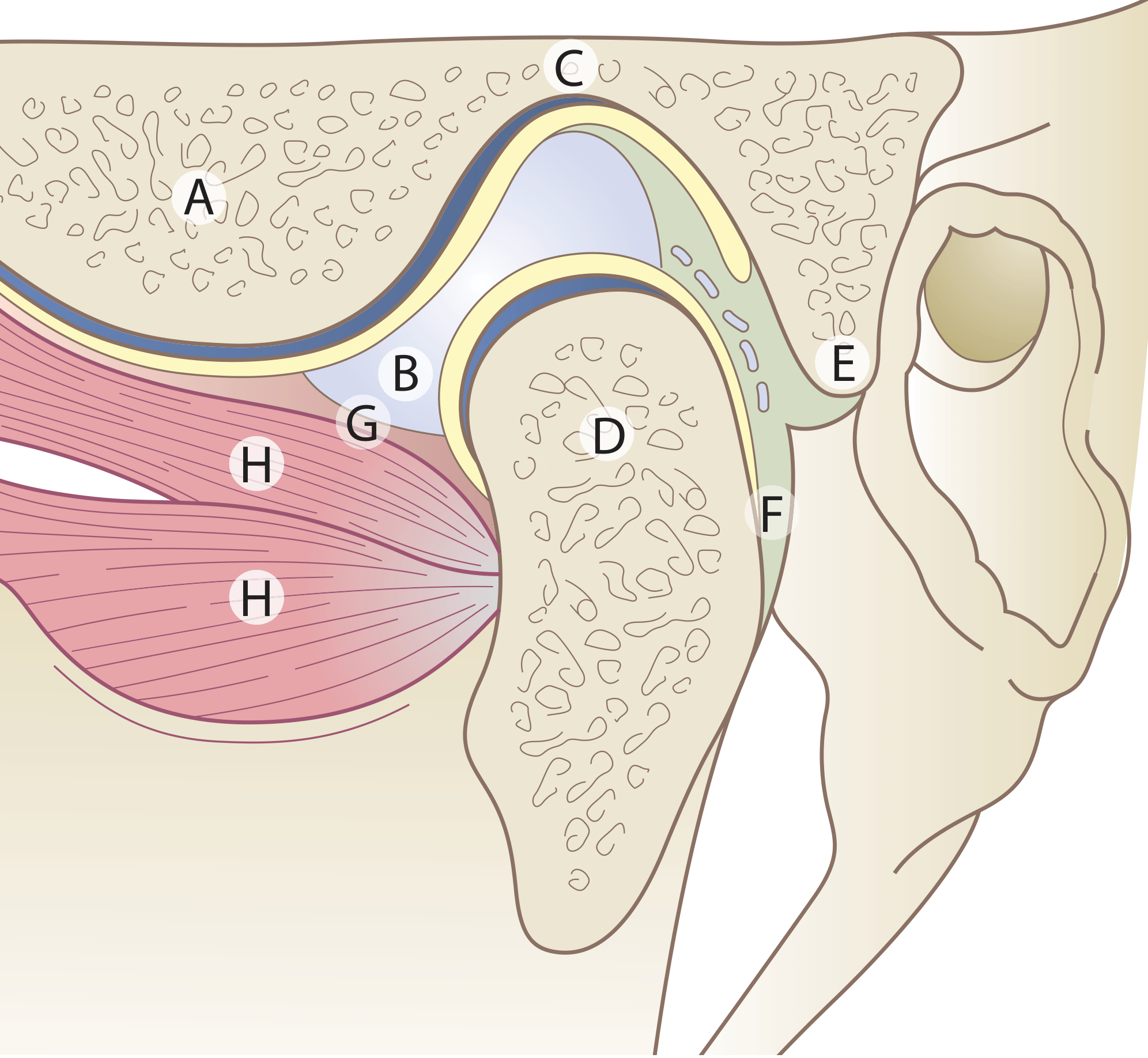
Key: A = Articular eminence, B = Articular disc, C = Mandibular fossa (temporal bone), D = Condyle (mandible), E = Upper lamina (fibro-elastic retrodiscal), F = Lower lamina, G = Joint capsule, H = Lateral pterygoid muscle
Unique Features of the TMJ Articulating Surfaces
- Not hyaline cartilage - articular surfaces are covered by fibrous tissue (reflects intramembranous ossification origin).
- Incongruent surfaces - the shapes of the fossa and condyle do not match, which is why the articular disc is needed.
- Growth centre - the condylar head contains secondary cartilage in youth, unlike most other synovial joints.
- Bilateral articulation - the two TMJs always function together; movement on one side necessarily affects the other.
Sources: Scott-Brown's Otorhinolaryngology Head & Neck Surgery; TeachMeAnatomy - TMJ; Kenhub - TMJ Anatomy
Can you give me short answer for 2 marks only
Articulating Bones of the Temporomandibular Joint
The TMJ is formed by the articulation of two bones:
-
Temporal bone (above) - contributes the mandibular (glenoid) fossa and the articular eminence (articular tubercle).
-
Mandible (below) - contributes the head of the mandibular condyle.
The articular surfaces are covered by dense fibrous tissue (not hyaline cartilage), and the joint space is divided into upper and lower compartments by an intra-articular disc.
Capsular attachment of TMJ
The TMJ capsule attaches above to the margins of the mandibular fossa and articular eminence, and below to the neck of the mandibular condyle.
Searching sources...
Finding Sources
Reading File
Capsular Attachment of the TMJ
The joint capsule is a thin, loose fibrous envelope attached as follows:
Above (Superior Attachment) - Temporal Bone
- Anteriorly - just in front of the crest of the articular eminence
- Posteriorly - to the squamotympanic and petrotympanic fissures
- Encloses the margins of the mandibular fossa all around
Below (Inferior Attachment) - Mandible
- Attached to the neck of the mandibular condyle
Key Points
- The capsule is a thin, slack cuff - it does not significantly limit mandibular movements and provides little joint support on its own.
- The inner surface is lined by synovial membrane, which is reflected over the disc margins but does not cover the articular surfaces.
- The synovial membrane secretes synovial fluid that lubricates both joint compartments.
The capsule is reinforced laterally by the temporomandibular (lateral) ligament, which is the main supporting ligament of the joint.
Source: Scott-Brown's Otorhinolaryngology Head & Neck Surgery
Give me short answer
Capsular Attachment of TMJ
Above (Temporal bone):
- Anteriorly - just in front of the crest of the articular eminence
- Posteriorly - to the squamotympanic and petrotympanic fissures
Below (Mandible):
- Attached to the neck of the mandibular condyle
The capsule is a thin, slack cuff lined internally by synovial membrane.
This is a shared conversation. Sign in to Orris to start your own chat.