
Bed sore ointment
- Silver sulfadiazine (antimicrobial)
- Hydrocolloid/hydrogel dressings
- Zinc oxide paste (for protection)
- Povidone-iodine (debridement/antisepsis - controversial)
- Collagenase-based ointments (enzymatic debridement)
"pressure ulcer" AND "topical treatment"
pressure ulcer bed sore staging classification diagram

Clinical photograph of a Grade IV sacral pressure sore on a patient with dark skin. The image demonstrates a deep, roughly oval-shaped ulcer in the sacral region. The wound bed shows extensive tissue loss with visible red granulation tissue and central areas of white-yellowish tissue, consistent with slough or exposed deep structures such as fascia or bone. The wound margins are irregular and slightly raised, with pale, macerated edges. The periwound skin exhibits hyperpigmentation and inflammatory changes. Adjacent to the ulcer, on the left side of the image, surgical markings are present, including a dashed curvilinear line and a central circular mark, indicating preoperative planning for a reconstructive skin flap (such as a superior gluteal artery perforator flap). The photograph serves as a visual reference for chronic wound assessment, staging of pressure injuries, and surgical flap design in plastic and reconstructive surgery.
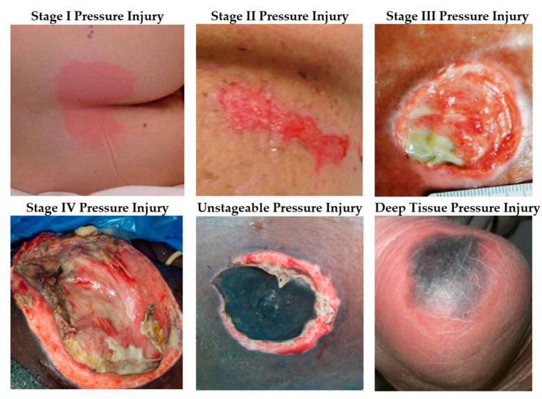
This clinical photographic comparison illustrates the diverse visual presentations of pressure injuries (decubitus ulcers) according to the National Pressure Injury Advisory Panel (NPIAP) staging system. The image is organized into a six-panel grid. Stage I: Shows intact skin with localized non-blanchable erythema. Stage II: Displays partial-thickness skin loss with a shallow, open ulcer and a red-pink wound bed. Stage III: Exhibits full-thickness skin loss where subcutaneous fat is visible within a deeper, crater-like wound. Stage IV: Demonstrates extensive full-thickness tissue loss with exposed muscle, tendon, or fascia. The Unstageable Pressure Injury panel shows a wound bed completely obscured by dark eschar or yellow slough, preventing depth assessment. The Deep Tissue Pressure Injury (DTPI) panel presents as a localized area of intact skin with deep purple or maroon discoloration, indicating underlying soft tissue damage. This educational resource is intended for dermatology and nursing students to assist in the clinical classification, wound assessment, and management of pressure-related skin lesions.
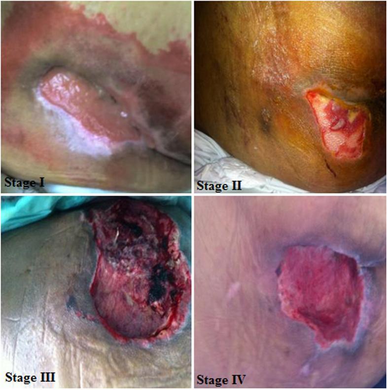
A multi-panel clinical photograph illustrating the four-stage classification of pressure ulcers (decubitus ulcers) in human skin. Panel 'Stage I' depicts intact skin with localized non-blanchable erythema and significant surrounding discoloration. Panel 'Stage II' shows a partial-thickness skin loss involving the epidermis and dermis, presenting as a shallow open ulcer with a red-pink wound bed and visible slough. Panel 'Stage III' demonstrates full-thickness tissue loss with visible subcutaneous involvement; the wound is deeper with evidence of necrotic tissue and dark eschar. Panel 'Stage IV' illustrates advanced full-thickness tissue loss with extensive destruction, revealing deep structures such as muscle or fascia, and potentially exposing bone or tendon. This educational resource is used in dermatology and nursing education to differentiate wound severity and guide staging based on tissue depth, color morphology, and integrity of anatomical layers.
Bed Sore (Pressure Ulcer) - Topical Treatments & Ointments
Staging (NPUAP Classification)
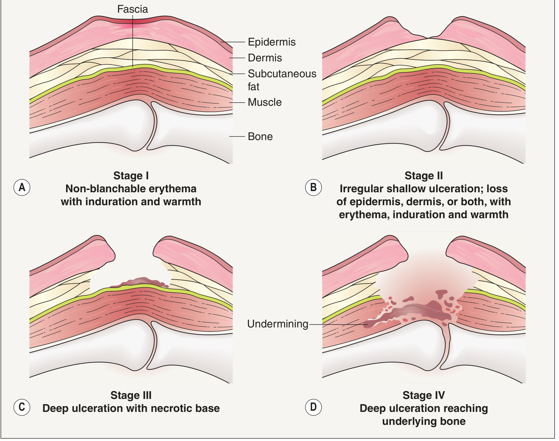
| Stage | Description |
|---|---|
| I | Non-blanchable erythema, intact skin |
| II | Partial-thickness skin loss (shallow ulcer/blister) |
| III | Full-thickness skin loss into subcutaneous fat |
| IV | Full-thickness with destruction of muscle, bone, or tendon |
Topical Agents by Category
1. Antimicrobial Ointments (for infected/colonized wounds)
| Agent | Details |
|---|---|
| Silver sulfadiazine (Silvadene) | Broad-spectrum antibacterial; commonly used. Note: can retard epithelial migration in clean healing wounds - better reserved for infected ulcers. |
| Cadexomer iodine (Iodosorb, Iodolex) | Preferred over povidone-iodine - slowly releases iodine from dextran beads; antimicrobial for several days without cytotoxic effects on keratinocytes; highly absorptive (1g absorbs up to 7 mL fluid). Use with caution in thyroid disease. |
| Silver nitrate | Broad antimicrobial spectrum; used as topical solution/cream. |
| Mafenide acetate (Sulfamylon) | Broad antibacterial; penetrates eschar. |
| Metronidazole gel | Applied topically for anaerobic-colonized ulcers with foul odor - eliminates odor within 36 hours. Very useful in palliative/nursing home settings. |
Avoid plain povidone-iodine and hydrogen peroxide directly in pressure ulcers - both are cytotoxic to keratinocytes and impair healing. Normal saline irrigation is preferred. - Dermatology 2-Volume Set 5e, Fitzpatrick's Dermatology
2. Enzymatic Debridement Agents
| Agent | Details |
|---|---|
| Collagenase (Santyl) | Proteolytic enzyme that selectively digests necrotic collagen; applied once daily to devitalized tissue; does not harm healthy tissue. First-line enzymatic debridement. |
| Papain-urea preparations | Enzymatic debriding agents; less commonly used today. |
3. Barrier & Protective Ointments (mainly Stage I-II, periwound skin)
| Agent | Details |
|---|---|
| Zinc oxide paste | Protects periwound skin from moisture, urine, feces; forms a physical barrier. Not applied inside the wound bed. |
| Petroleum jelly (Vaseline) | Simple occlusive; keeps intact/superficial wounds moist. |
| Dimethicone-based creams | Skin barrier protection; prevents maceration from incontinence. |
4. Moisture-Retentive Dressings (not ointments but equally important)
| Dressing Type | Best For | Notes |
|---|---|---|
| Hydrocolloid (DuoDERM) | Stage II, light-moderate exudate | Occlusive; promotes autolytic debridement |
| Hydrogel | Dry/necrotic wounds | Donates moisture; good for eschar softening |
| Foam dressings | Moderate-heavy exudate (Stage III) | Absorbs without desiccating |
| Alginate/gelling fiber | Stage III-IV, heavy exudate | Highly absorptive; haemostatic |
| Transparent film | Stage II only, light drainage | Not for deep or infected wounds |
| Hydrofiber (Aquacel) | Stage III-IV, full-thickness | High absorbency |
5. Newer / Adjunct Topical Agents
- Manuka honey - Natural antimicrobial (high osmolality + flavonoids); promotes debridement and macrophage activation; a Cochrane review found benefit for superficial burns, and it is used in pressure ulcers too.
- Growth factors (e.g., becaplermin/Regranex - PDGF) - Investigational/approved for diabetic ulcers; promising for pressure ulcers.
- Copper oxide dressings - Pro-angiogenic + antimicrobial; enhances vascularization.
- Methylene blue + gentian violet sponge (Hydrofera Blue) - Broad-spectrum including Candida; dyes are not released so no tissue toxicity.
Treatment Principles (Summary)
- Relieve pressure first - Reposition every 2 hours; use pressure-redistribution mattresses/cushions. No ointment works if pressure continues.
- Clean with normal saline - Avoid cytotoxic antiseptics (hydrogen peroxide, povidone-iodine) in the wound bed.
- Debride necrotic tissue - Sharp (surgical), mechanical, enzymatic (collagenase), or autolytic (hydrocolloid/hydrogel).
- Control infection - Use antimicrobial ointments only when there are signs of infection (increased exudate, odor, erythema, pain). Do not use prophylactically.
- Maintain moist environment - Use appropriate dressings; keep periwound skin dry with barrier paste (zinc oxide).
- Address systemic factors - Nutrition (protein, Vitamin C, zinc), anemia, diabetes, circulatory status.
- Stage IV ulcers often need surgical flap repair.