
Functions of rpe
- Visual cycle - regenerates 11-cis retinal from all-trans retinal after phototransduction
- Phagocytosis - daily removal of shed photoreceptor outer segment discs
- Blood-retinal barrier - tight junctions form the outer BRB
- Nutrient transport - delivers glucose, fatty acids, and other nutrients from choroid to photoreceptors
- Ion/fluid transport - maintains subretinal space homeostasis by pumping ions and water
- Light absorption - melanin granules absorb stray light, reducing optical scatter
- Growth factor secretion - produces VEGF (toward choroid) and PEDF (toward retina)
retinal pigment epithelium RPE functions diagram
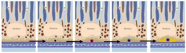
This pathophysiology diagram illustrates the sequential formation of sub-retinal pigment epithelium (RPE) deposits, a key process in the pathogenesis of Age-Related Macular Degeneration (AMD). The five-panel progression depicts the retinal layers from top to bottom: photoreceptor outer segments receiving light, the RPE cell layer (containing a central nucleus and brown melanocytes), and Bruch's membrane above the choriocapillaris. The metabolic mechanism begins with the appearance of micrometer-sized, black cholesterol-containing extracellular lipid droplets beneath the RPE. The sequence then shows the precipitation of magenta-colored hydroxyapatite (HAP) onto these droplets, followed by the recruitment and binding of blue-colored proteins to the HAP surface. The final panel demonstrates a self-driven oligomerization process resulting in a macroscopic yellow sub-RPE deposit (drusen). This educational model highlights the role of lipid accumulation and mineralization in Bruch's membrane aging and the development of early-stage AMD lesions.
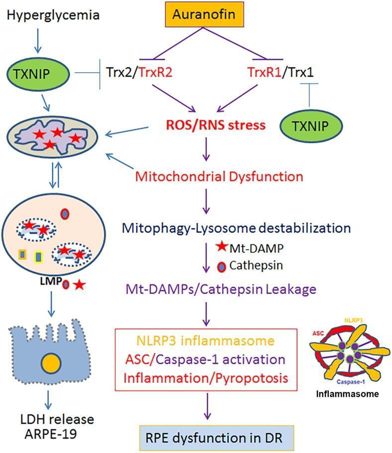
A pathophysiology diagram illustrating the signaling pathway through which auranofin and hyperglycemia-induced TXNIP contribute to Retinal Pigment Epithelium (RPE) dysfunction in Diabetic Retinopathy (DR). The flowchart depicts the inhibition of Thioredoxin/Thioredoxin Reductase systems (Trx2/TrxR2 and TrxR1/Trx1) by auranofin and TXNIP. This inhibition triggers Reactive Oxygen Species/Reactive Nitrogen Species (ROS/RNS) stress, leading to mitochondrial dysfunction. Downstream events include mitophagy-lysosome destabilization characterized by the leakage of mitochondrial Damage-Associated Molecular Patterns (Mt-DAMPs) and Cathepsins. This leakage activates the NLRP3 inflammasome, ASC, and Caspase-1, resulting in pro-inflammatory pyroptosis and cellular damage, evidenced by LDH release from ARPE-19 cells. The diagram uses anatomical illustrations of mitochondria, lysosomes, and the NLRP3 inflammasome complex to show cellular organelle involvement. This schematic summarizes the mitochondria-lysosome axis dysfunction and its role in chronic retinal neurodegenerative diseases including Diabetic Retinopathy, Age-related Macular Degeneration (AMD), and Retinitis Pigmentosa (RP).
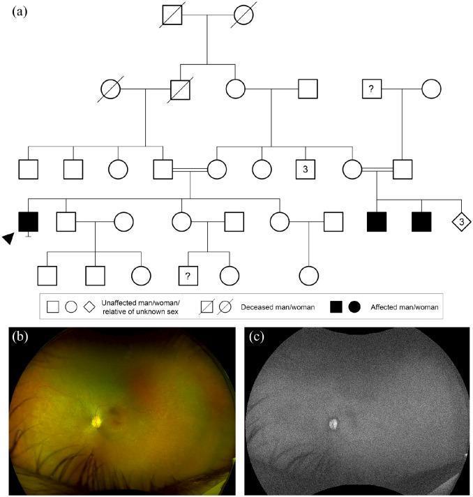
This composite figure provides a genetic and multimodal imaging analysis of retinitis pigmentosa (RP). Panel (a) is a complex consanguineous pedigree diagram illustrating an inheritance pattern associated with a homozygous RPE65 variant. Filled symbols indicate affected individuals across generations, with a double line indicating consanguinity. Panel (b) is a widefield color fundus photograph of a 42-year-old patient's right eye. It demonstrates characteristic features of retinal degeneration, including prominent retinal pigment epithelium (RPE) granularity and subtle scattered white dots, with notable pigmentation changes across the posterior pole and periphery. Panel (c) is a widefield fundus autofluorescence (FAF) image of the same eye, showing a profound and global lack of autofluorescence. This generalized hypoautofluorescence indicates severe dysfunction or loss of fluorophores in the RPE and photoreceptor layers, a hallmark finding in RPE65-related retinopathies. The combined clinical and genetic data illustrate the diagnostic correlation between inheritance patterns and progressive retinal degeneration.
Functions of the Retinal Pigment Epithelium (RPE)
1. Outer Blood-Retinal Barrier (Outer BRB)
- Prevents extracellular fluid from the choriocapillaris leaking into the subretinal space
- Actively pumps ions and water out of the subretinal space, keeping it dry and maintaining photoreceptor apposition
2. Phagocytosis of Photoreceptor Outer Segments
3. Maintenance of Optimal Retinal Milieu
- Inward transport of metabolites (amino acids, glucose) from the choriocapillaris to photoreceptors
- Outward transport of metabolic waste products back toward the choroid, via Bruch's membrane as a route
- Regulates ionic composition of the subretinal space (especially K⁺ buffering)
4. Visual Cycle (Vitamin A Metabolism)
- After light bleaches rhodopsin, all-trans retinal is released into the subretinal space
- RPE takes it up, isomerizes it back to 11-cis retinal, and returns it to photoreceptors for rhodopsin regeneration
- Also stores and transports vitamin A (retinol), as described in Goodman & Gilman's
5. Light Absorption (Optical Function)
- The dense melanin granules (melanosomes) in the apical RPE absorb stray light
- This reduces light scatter, optical noise, and photo-oxidative damage to the outer retina
- The foveal RPE contains more numerous and larger melanosomes than the periphery
6. Secretory Functions (Growth Factors)
- VEGF (vascular endothelial growth factor) - secreted basally toward the choriocapillaris, essential for choriocapillaris maintenance
- PEDF (pigment epithelium-derived factor) - secreted apically, provides neuroprotective and anti-angiogenic support to photoreceptors
- Other cytokines involved in immune privilege of the subretinal space
7. Immune Privilege
- Expressing immunosuppressive molecules
- Forming the outer BRB that excludes circulating immune cells from the subretinal space
Summary Table
| Function | Mechanism |
|---|---|
| Outer blood-retinal barrier | Tight junctions (zonula occludentes) |
| Phagocytosis of OS discs | Lysosomal degradation of shed outer segments |
| Nutrient/waste transport | Bidirectional transport across Bruch's membrane |
| Visual cycle | Retinoid isomerization (all-trans → 11-cis retinal) |
| Light absorption | Melanin granules absorb stray photons |
| Growth factor secretion | VEGF (basal), PEDF (apical) |
| Subretinal fluid regulation | Active ion/water pumping out of subretinal space |
Clinical relevance: RPE dysfunction underlies many major retinal diseases - age-related macular degeneration (AMD, drusen accumulation beneath RPE), Stargardt disease (impaired visual cycle), Best disease (vitelliform dystrophy), and geographic atrophy (RPE cell death). The outer BRB breakdown is also the mechanism of cystoid macular edema and central serous chorioretinopathy.