

This is cbc of buffalo does you suspect any haemoprotozoa ?
haemoprotozoa buffalo CBC findings anaemia thrombocytopenia leukocytosis Theileria Babesia
Theileria annulata blood smear buffalo haemoprotozoa
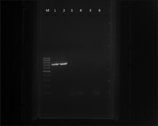
Diagnostic diagnostic image showing agarose gel electrophoresis of Polymerase Chain Reaction (PCR) products used for detecting Theileria annulata. The image consists of seven lanes. Lane M contains a 100 bp DNA ladder (marker) with multiple distinct bands of varying intensities used for fragment size estimation. Lane 1 displays a sharp, bright band at approximately 721 bp, serving as the positive control for T. annulata. Lane 2 shows a similar bright band at the same vertical migration level, indicating a positive clinical sample. Lanes 3, 4, and 5 contain negative clinical samples, showing no visible amplification bands. Lane 6 represents the negative control and is devoid of bands, confirming the absence of reagent contamination. This visual demonstrates molecular diagnostic techniques in infectious disease, specifically identifying the presence of parasitic DNA in biological samples through targeted primer amplification and size-based separation.
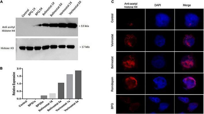
Educational figure illustrating the acetylation profiles of Theileria annulata-infected host cells treated with various histone deacetylase inhibitors (HDACi). Panel A presents a Western blot comparing protein expression across seven groups: Control, BPQ (1X, 5X), Belinostat (1X, 5X), and Vorinostat (1X, 5X). The Anti-acetyl Histone H4 bands (~13 kDa) show a dose-dependent increase in intensity following HDACi treatment, while the Histone H3 bands (~17 kDa) serve as a consistent loading control. Panel B is a bar graph quantifying the relative expression of acetylated H4, confirming the significant hyperacetylation induced by Vorinostat and Belinostat compared to control groups. Panel C displays immunofluorescence (IFA) microscopy of cells treated with Vorinostat, Belinostat, and Romidepsin. Three channels are shown: Anti-acetyl histone H4 (red), DAPI nuclear stain (blue), and a merged image. The IFA reveals enhanced red fluorescence in treated cells compared to the Control and BPQ groups, indicating concentrated histone acetylation within and around the parasite-infected host cell nucleus. This content demonstrates the molecular mechanism of HDAC inhibition in parasitology and cellular biology.
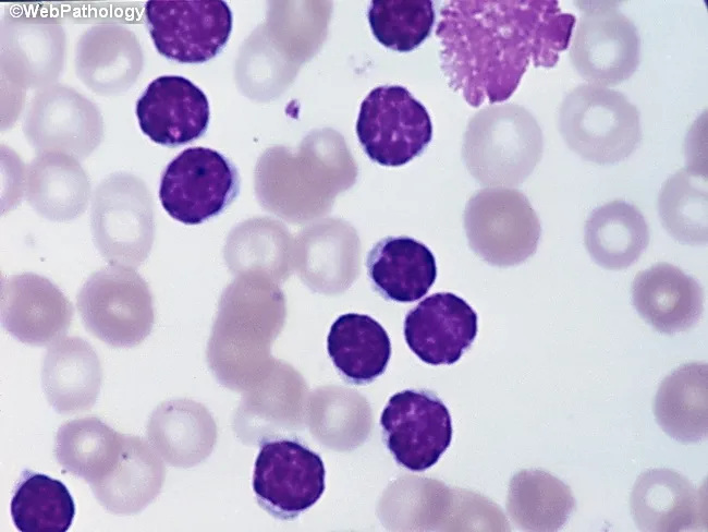
Peripheral blood smear from a patient with suspected chronic lymphocytic leukemia (CLL) shows a predominance of small, mature-appearing lymphocytes with scant cytoplasm and clumped nuclear chromatin, accompanied by fragile cells that readily disrupt during smear preparation, producing numerous smudge or basket cells visible on the slide. The image depicts a single smudge cell in the upper right, with numerous small darkly staining lymphocytes occupying the field. The smear was prepared with Wright-Giemsa stain and examined under light microscopy at high magnification (approximately 1000x total magnification with oil immersion). Clinically, CLL is defined by absolute mature lymphocytosis of ≥5.0 × 10^9/L sustained for ≥3 months, often asymptomatic and detected incidentally on complete blood count. Morphology alone is insufficient for diagnosis; immunophenotyping showing co-expression of CD5, CD19, CD23 with weak surface immunoglobulin and light chain restriction supports CLL diagnosis. Smudge cells result from fragile leukemic cells and reflect underlying cytoskeletal fragility, though they are not specific. The image illustrates typical features: monomorphic lymphocytes, scant cytoplasm, condensed chromatin, and absent prominent nucleoli. This finding has diagnostic significance, guiding further workup, including flow cytometry, cytogenetics, and molecular studies, to distinguish CLL from other chronic lymphoproliferative disorders such as small lymphocytic lymphoma (SLL), mantle cell lymphoma, prolymphocytic leukemia, and reactive lymphocytosis.
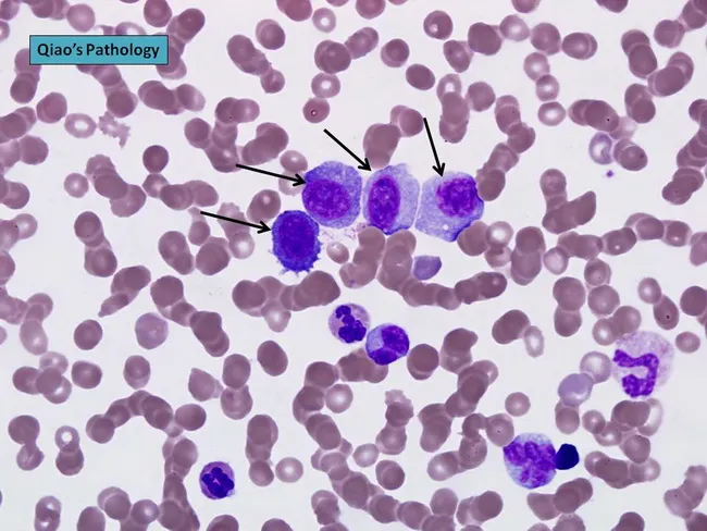
Peripheral blood smear prepared from a patient with suspected plasma cell leukemia (PCL) was examined by bright‑field light microscopy after Wright‑Giemsa staining. The smear shows several circulating plasma‑cell–like elements, including four conspicuous proplasmacytes with dispersed nuclear chromatin and prominent nucleoli, as indicated by arrows in the original figure. In addition, background erythrocytes exhibit rouleaux formation, a common feature in paraproteinemic states. The cells display eccentrically placed nuclei with pronounced basophilic cytoplasm and a coarse chromatin pattern; occasional cells show a perinuclear clearing (Golgi rim) suggestive of plasmacytic differentiation. Immunoglobulin light‑chain restriction is not determined on this smear alone but flow cytometry or immunohistochemistry would typically confirm clonal plasma cells. Relative proportions indicate that neoplastic plasma cells constitute a substantial fraction of leukocytes, consistent with plasma cell leukemia rather than reactive plasmacytosis. PCL is an aggressive myelomatous process often presenting with cytopenias, organomegaly, and sometimes lymphadenopathy; prognosis is poor, and treatment responsiveness is limited. This image illustrates characteristic morphologic features used for differential diagnosis against multiple myeloma, Waldenström macroglobulinemia, and other causes of plasmacytosis. Clinically relevant keywords include plasma cells, rouleaux, Wright‑Giemsa stain, 100X oil, peripheral blood, leukemia, myeloma, and hematologic malignancy.
CBC Analysis - Buffalo (Nihon Kohden, 27 Jul 2026)
Important note: The analyzer is set to "Dog" species - the reference ranges shown are canine. For accurate interpretation, buffalo reference ranges should be used. The flags (H/L) on this report are relative to canine normals, not buffalo normals. Keep this in mind.
Extracted CBC Values
| Parameter | Value | Flag | Buffalo Normal Range |
|---|---|---|---|
| WBC | 39.6 | H | 5-12 × 10³/µL |
| RBC | 2.31 | L | 5-8 × 10⁶/µL |
| HGB | 5.9 | L | 9-14 g/dL |
| HCT | 16.1 | L | 31-45% |
| MCV | 69.7 | Normal | 42-55 fL |
| MCH | 25.5 | H (instrument) | varies |
| MCHC | 36.6 | H | 30-37 g/dL |
| PLT | 94 | L | 200-800 × 10³/µL |
| LY% | 9.5 | L | 45-75% |
| GR% | 85.2 | H | 20-45% |
| MPV | 6.2 | L | - |
| RBC histogram flag | (7)(AT) | - | Abnormal cell flag |
Key Abnormalities
- Severe anaemia - HGB 5.9 g/dL, HCT 16.1%, RBC 2.31 × 10⁶/µL - all dramatically low. This is haemolytic-pattern anaemia.
- Marked leukocytosis - WBC 39.6 × 10³/µL with predominant granulocytosis (85.2%) - severe neutrophilia suggesting systemic inflammation/infection.
- Thrombocytopenia - PLT 94 × 10³/µL - below normal, consistent with platelet consumption or destruction.
- RBC histogram flag (7)(AT) - The "AT" flag stands for Atypical cells - this is the machine's automated alert that abnormal/atypical particles were detected in the RBC channel, which in haemoprotozoan infections often corresponds to parasitized erythrocytes being counted differently.
- Lymphopenia (9.5%) - common in acute overwhelming infection.
- Elevated MCHC (36.6) - may reflect haemoconcentration or falsely elevated due to haemolysis releasing free Hb.
Suspected Haemoprotozoa - YES, Strongly Suspected
1. Theileria annulata (Tropical Theileriosis) - Top Suspect
- Most common and deadly haemoprotozoan in Indian buffaloes
- Transmitted by Hyalomma ticks
- Causes severe haemolytic anaemia, marked leukocytosis (lymphocyte lysis and inflammatory response), thrombocytopenia, high fever, lymph node enlargement
- The profound anaemia (HCT 16%) with neutrophilia pattern fits perfectly
- Piroplasms appear as small rings/dots inside RBCs; Koch's blue bodies (schizonts) appear in lymphocytes
2. Babesia bigemina / Babesia bovis - Possible
- Transmitted by Rhipicephalus (Boophilus) ticks
- Causes intravascular haemolysis, anaemia, haemoglobinuria ("red-water fever")
- Anaemia tends to be very severe with regenerative response
- Large pear-shaped paired merozoites visible on smear
3. Trypanosoma evansi (Surra) - Consider
- Transmitted by biting flies (Tabanus)
- Causes progressive anaemia, leukocytosis, thrombocytopenia, wasting
- Trypanosomes visible in wet blood preparations or smears
4. Anaplasma marginale - Differential
- Rickettsia-like organism (not strictly a protozoan but a haemotropic pathogen)
- Causes haemolytic anaemia in cattle/buffalo; marginal inclusions in RBCs
What You Must Do Next
| Step | Action |
|---|---|
| 1. Peripheral blood smear | Giemsa/Wright stain - look for piroplasms inside RBCs, Koch's blue bodies in lymphocytes, trypanosomes in plasma |
| 2. Buffy coat smear | Increases sensitivity for trypanosomes |
| 3. Thin smear × 3 times | Multiple smears improve detection rate |
| 4. PCR | Gold standard - can differentiate species precisely |
| 5. Temperature | Record rectal temperature - fever >104°F supports active parasitaemia |
| 6. Lymph node aspirate | If Theileria suspected, FNA of prescapular lymph node for schizont detection |
Clinical Urgency
- Theileria: Buparvaquone (Butalex) 2.5 mg/kg IM - single dose
- Babesia: Diminazene aceturate (Berenil) 3.5 mg/kg IM or Imidocarb dipropionate 1.2-3 mg/kg
- Trypanosoma: Diminazene aceturate 3.5-7 mg/kg or Isometamidium chloride