
aterial supply of the mouth, including the lingual artery, facial artery, and maxillary artery and its supplies.
Reading File
Finding Sources
Finding Sources
Finding Sources
Finding Sources
Reading File
Reading File
Reading File
Reading File
Reading File
Reading File
Loading Image
Loading Image
Loading Image
Loading Image
Reading File
I now have comprehensive information from Gray's Anatomy for Students and Scott-Brown's Otorhinolaryngology. Here is a detailed, well-structured answer:
Arterial Supply of the Mouth
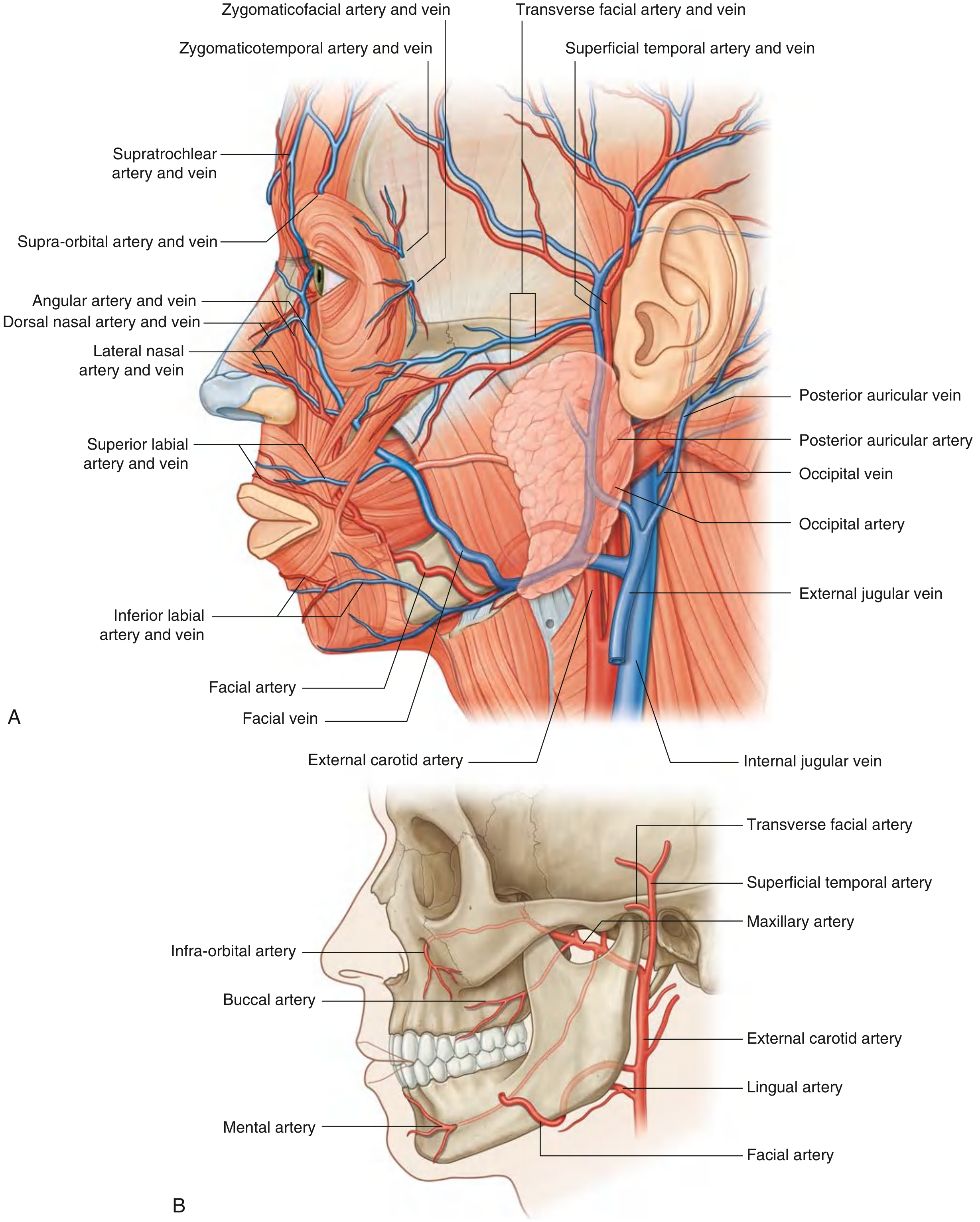
The mouth and its associated structures receive blood from three main branches of the external carotid artery: the lingual artery, the facial artery, and the maxillary artery. All three are pictured below in lateral view.
1. Lingual Artery
Origin: External carotid artery in the neck, adjacent to the tip of the greater horn of the hyoid bone.
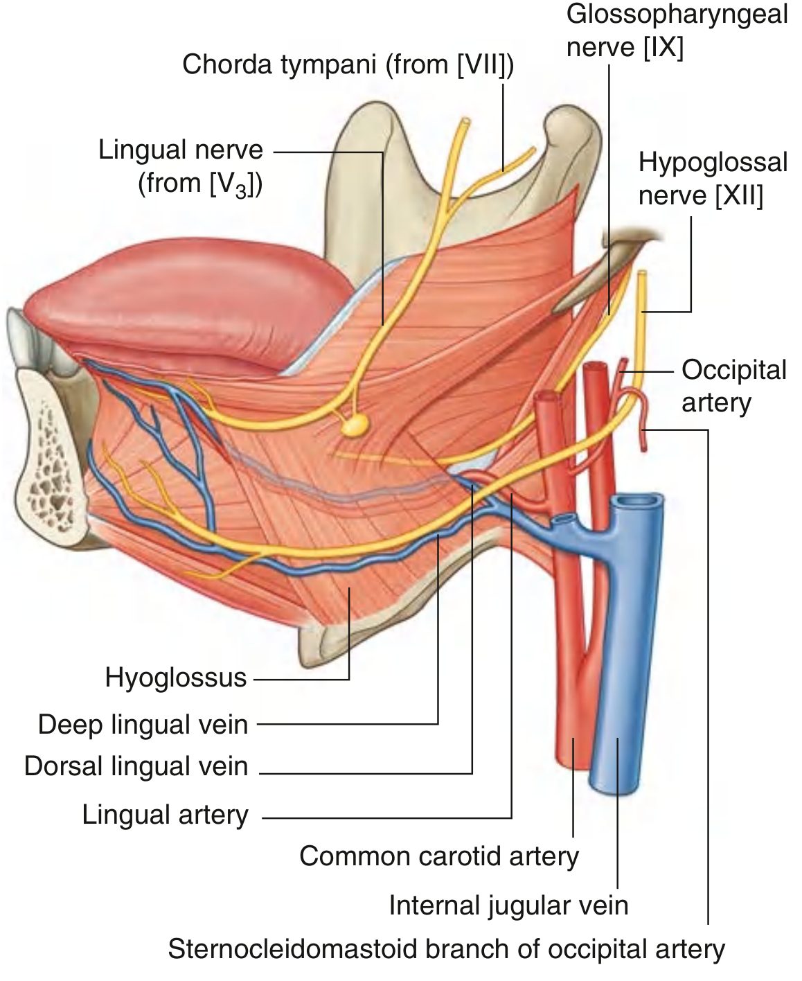
Course: It forms an upward bend, then loops downward and forward to pass deep to the hyoglossus muscle, entering the floor of the oral cavity through the oropharyngeal triangle (bounded by the mylohyoid, superior constrictor, and middle constrictor). It then travels forward in the plane between the hyoglossus and genioglossus to the apex of the tongue.
Branches:
| Branch | Supply |
|---|---|
| Dorsal lingual arteries (2-3) | Arise medial to hyoglossus; supply the mucous membrane of the dorsum of tongue, palatoglossal arch, soft palate, tonsil, and epiglottis |
| Sublingual artery | Arises at the anterior margin of hyoglossus; passes between genioglossus and mylohyoid to supply the sublingual gland; also enters the lingual foramen of the mandible |
| Deep lingual artery | Terminal branch; runs on the inferior surface of the tongue near the lingual frenum; near the tip, anastomoses with its fellow from the opposite side |
Additional supply to the tongue root: The tonsillar and ascending palatine branches of the facial artery and ascending pharyngeal artery also supply tissue at the root of the tongue. In the region of the valleculae, epiglottic branches of the superior laryngeal artery anastomose with the inferior dorsal lingual branches.
Summary of structures supplied: Tongue, sublingual gland, gingiva, and oral mucosa of the floor of the mouth. - Gray's Anatomy for Students; Scott-Brown's Otorhinolaryngology
2. Facial Artery
Origin: Anterior surface of the external carotid artery in the neck.
Course: Passes up through the deep structures of the neck, then behind the submandibular gland, hooks around the inferior border of the mandible just anterior to masseter (where its pulse is palpable), and enters the face. It runs in a tortuous upward and medial course - deep to platysma, risorius, and zygomaticus major and minor, superficial to buccinator and levator anguli oris - and terminates as the angular artery at the medial corner of the eye.
Branches supplying the mouth:
| Branch | Supply |
|---|---|
| Inferior labial artery | Supplies the lower lip |
| Superior labial artery | Supplies the upper lip; also gives a branch to the nasal septum |
| Lateral nasal branch | Supplies the lateral surface and dorsum of the nose |
Anastomoses: Near the midline, the superior and inferior labial branches from each side anastomose with their counterparts. This provides an important connection between the facial arteries and external carotid arteries of opposite sides.
Note: The transverse facial artery (a branch of the superficial temporal artery) also contributes to the face - it arises within the parotid gland and crosses the face on the superficial surface of masseter, between the zygomatic arch and parotid duct. - Gray's Anatomy for Students, pp. 1057-1058
3. Maxillary Artery
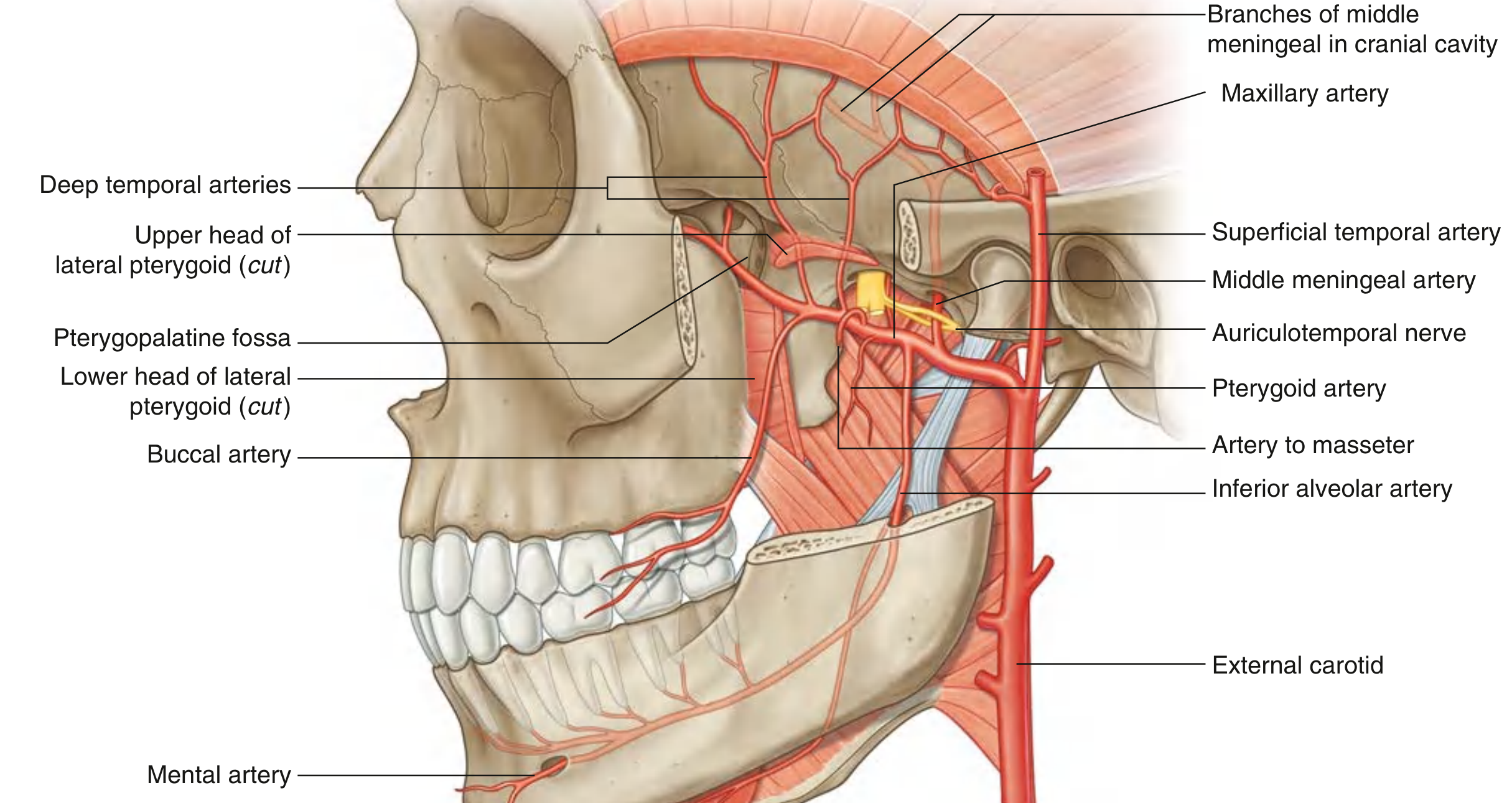
The maxillary artery is the largest branch of the external carotid artery. It is the major source of blood supply to the nasal cavity, lateral wall and roof of the oral cavity, all teeth, and the dura mater. It is classically divided into three parts based on its relationship to the lateral pterygoid muscle.
Origin & Course: Originates within the parotid gland, passes forward between the neck of the mandible and sphenomandibular ligament into the infratemporal fossa, then enters the pterygopalatine fossa through the pterygomaxillary fissure.
Part 1 - Mandibular (between neck of mandible and sphenomandibular ligament)
| Branch | Supply |
|---|---|
| Middle meningeal artery | Ascends through foramen spinosum; largest meningeal vessel; supplies dura mater, cranial vault bone and marrow |
| Inferior alveolar artery | Enters the mandibular canal; gives the mental branch (exits mental foramen to supply chin and lower lip); supplies mandibular teeth, gingiva, and lower jaw |
| Deep auricular artery | External acoustic meatus |
| Anterior tympanic artery | Tympanic membrane |
| Accessory meningeal artery | Dura and infratemporal fossa structures |
Part 2 - Pterygoid (related to lateral pterygoid muscle)
| Branch | Supply |
|---|---|
| Deep temporal arteries | Temporalis muscle |
| Masseteric artery | Masseter muscle |
| Buccal artery | Buccinator muscle and cheek mucosa |
| Pterygoid branches | Pterygoid muscles |
Part 3 - Pterygopalatine (in the pterygopalatine fossa)
| Branch | Supply |
|---|---|
| Posterior superior alveolar artery | Exits via alveolar foramina; supplies molar and premolar teeth, adjacent gingiva, and maxillary sinus |
| Infra-orbital artery | Passes through inferior orbital fissure and infra-orbital canal; supplies lower eyelid, cheek, and upper lip; gives anterior superior alveolar arteries (incisor and canine teeth, maxillary sinus) |
| Greater palatine artery | Descends through palatine canal; gives lesser palatine branch (soft palate); continues through greater palatine foramen to supply the hard palate; loops through incisive canal to supply anterior nasal septum |
| Lesser palatine artery | Soft palate |
| Pharyngeal branch | Via palatovaginal canal; supplies posterior roof of nasal cavity, sphenoidal sinus, and pharyngotympanic tube |
| Sphenopalatine artery | Terminal branch; exits through sphenopalatine foramen; gives posterior lateral nasal arteries (lateral nasal wall, paranasal sinuses) and posterior septal branches (nasal septum) |
| Artery of pterygoid canal | Travels through pterygoid canal; supplies mucosa of nasopharynx |
Summary: What Each Artery Supplies to the Mouth
| Structure | Arterial Supply |
|---|---|
| Tongue (body) | Lingual artery (deep lingual + dorsal lingual branches) |
| Tongue (root/base) | Lingual a., tonsillar branch of facial a., ascending pharyngeal a. |
| Floor of mouth | Sublingual branch of lingual artery |
| Sublingual gland | Sublingual branch of lingual artery |
| Upper lip | Superior labial branch of facial a.; infra-orbital a. (maxillary a.) |
| Lower lip | Inferior labial branch of facial a.; mental branch of inferior alveolar a. |
| Cheek (buccal mucosa) | Buccal artery (maxillary a.) |
| Hard palate | Greater palatine artery (maxillary a.) |
| Soft palate | Lesser palatine artery (maxillary a.); ascending palatine branch of facial a. |
| Upper teeth | Posterior superior alveolar a. + Anterior superior alveolar a. (maxillary a.) |
| Lower teeth | Inferior alveolar artery (maxillary a.) |
| Gingiva | Superior/inferior alveolar branches (maxillary a.); facial a. |
Sources: Gray's Anatomy for Students, pp. 1057-58, 1130-1138; Scott-Brown's Otorhinolaryngology Head & Neck Surgery

Gemini_Generated_Image_xwljejxwljejxwlj.png
please provide with diagram from authenticated source for lingual artery which includes all the supplies.
Finding Sources
Reading File
Reading File
Reading File
Loading Image
Loading Image
Here are two authenticated textbook diagrams of the lingual artery with full annotation:
Lingual Artery - Authenticated Textbook Diagrams
Diagram 1 - Gray's Anatomy for Students (Fig. 8.263)
Arteries, Veins, and Nerves of the Tongue
This medial sagittal view shows the lingual artery coursing between the hyoglossus and genioglossus muscles, with the companion deep and dorsal lingual veins (blue) alongside it. The hyoglossus muscle is clearly visible as the key anatomical landmark deep to which the lingual artery travels. The hypoglossal nerve [XII] runs with the deep lingual vein on the lateral surface of hyoglossus.
Diagram 2 - Scott-Brown's Otorhinolaryngology Head & Neck Surgery (Figure 23.4)
Blood vessels of the tongue - the four segments of the lingual artery
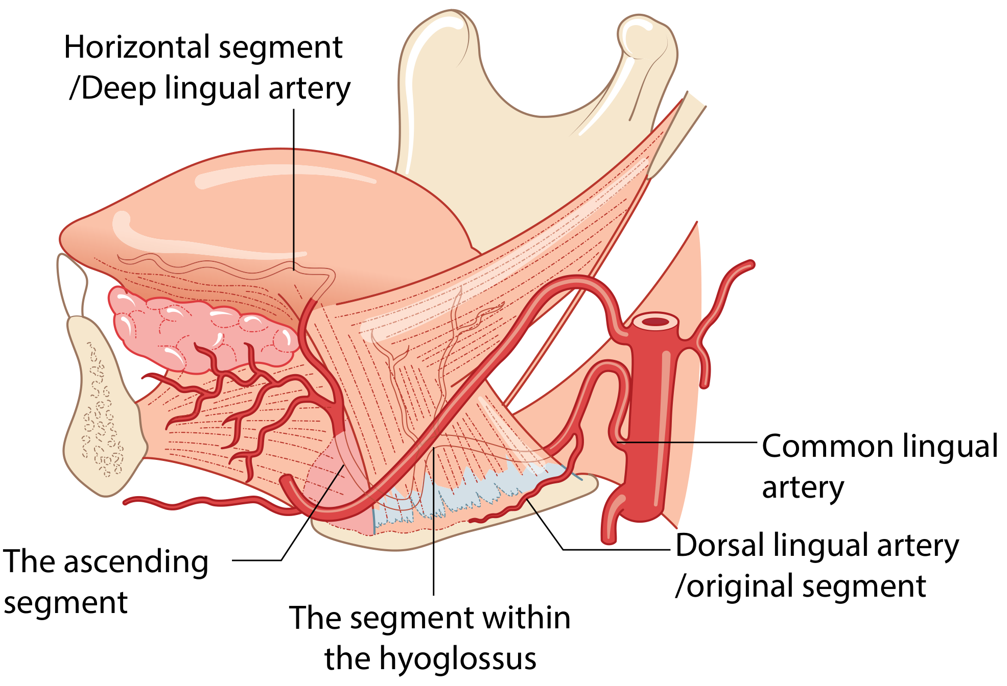
This diagram from Scott-Brown's is the most detailed illustration of the lingual artery's segments and branches, showing:
| Segment | Description | Branch arising |
|---|---|---|
| Original segment (Dorsal lingual / original) | Root of the lingual artery; lies 3-4 mm supero-lateral to the greater horn of the hyoid | - |
| Segment within hyoglossus | Passes obliquely from posterosuperior to anteroinferior under hyoglossus, medial to hypoglossal nerve | Dorsal lingual artery - arises ~0.7 cm from the anterior border of hyoglossus; supplies root of tongue, glossoepiglottic folds, palatoglossal arch, epiglottis |
| Ascending segment | Ascends in a tortuous manner along the anterior border of hyoglossus | Sublingual artery - arises ~0.8 cm above hyoid; supplies ventral tongue, anterior/lateral floor of mouth, vestibular and lingual gum, sublingual gland |
| Horizontal segment / Deep lingual artery | Terminal part; runs forward between longitudinal muscle of tongue and genioglossus, extending to ~1 cm from the tip | Supplies inferior surface of tongue; anastomoses with fellow of opposite side near the tip |
What Each Branch Supplies - Complete Summary
| Branch | Structures Supplied |
|---|---|
| Dorsal lingual arteries (2-3) | Mucous membrane of dorsum of tongue, palatoglossal arch, soft palate, tonsil, epiglottis |
| Sublingual artery | Sublingual salivary gland, floor of mouth mucosa, ventral tongue, gingiva; also enters lingual foramen of mandible |
| Deep lingual artery (terminal) | Inferior surface and apex of tongue; anastomoses across midline with contralateral deep lingual artery |
| Lingual artery overall | Tongue (body), sublingual gland, gingiva, and oral mucosa of the floor of the mouth |
Additional supply to tongue root (not from lingual artery directly): tonsillar and ascending palatine branches of the facial artery, branches of the ascending pharyngeal artery, and epiglottic branches of the superior laryngeal artery (which anastomose with the inferior dorsal lingual branches at the valleculae).
Sources: Gray's Anatomy for Students, Fig. 8.263; Scott-Brown's Otorhinolaryngology Head & Neck Surgery, Fig. 23.4


mouth.jpg
+2 more
Using these notes, find out diagram from authenticated source for arterial supply of oral cavity.
Finding Sources
Finding Sources
Finding Sources
Finding Sources
Finding Sources
Finding Sources
Finding Sources
Reading File
Reading File
Reading File
Finding Sources
Finding Sources
Finding Sources
Loading Image
Loading Image
Loading Image
Loading Image
Here are all four authenticated textbook diagrams matched to your notes, organised by artery:
Arterial Supply of the Oral Cavity - Authenticated Textbook Diagrams
Diagram 1 - Overall View: All Three Arteries Together
Gray's Anatomy for Students, Fig. 8.70 - Vasculature of the Face and Mouth
This is the best single overview diagram matching your notes. Panel (A) shows the facial artery winding up the face with its superior labial, inferior labial, lateral nasal, and angular branches. Panel (B) shows the skull in lateral view with all three arteries labelled together:
- Lingual artery - arising from external carotid
- Facial artery - arising just above the lingual artery
- Maxillary artery - terminal branch of external carotid, with infra-orbital, buccal, and mental (branch of inferior alveolar) branches visible
Diagram 2 - Maxillary Artery Part 3 (Pterygopalatine Fossa Branches)
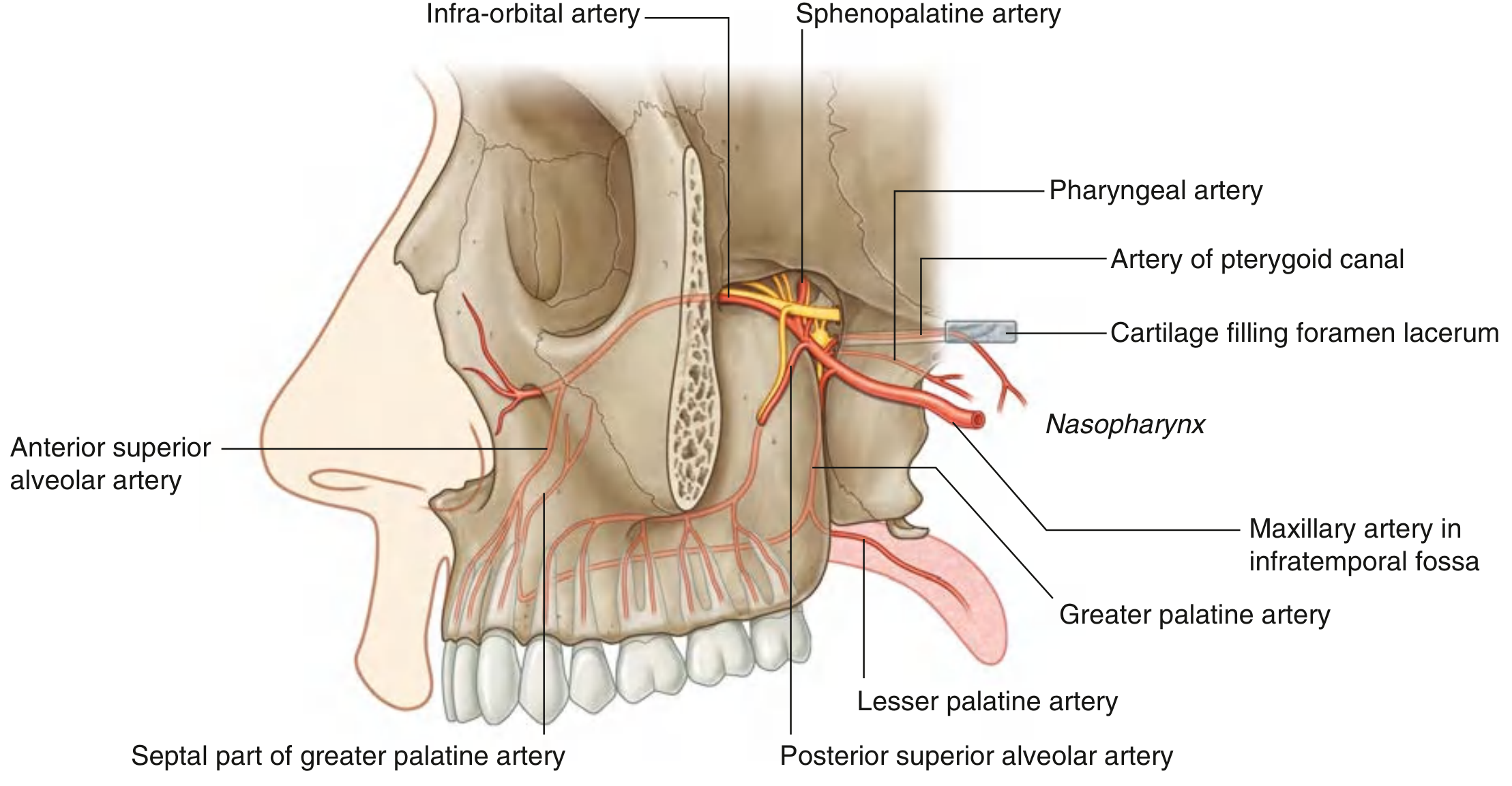
Gray's Anatomy for Students, Fig. 8.161 - Maxillary Artery in the Pterygopalatine Fossa
Directly matches your notes on Maxillary Artery Part 3 (Pterygopalatine). Shows clearly:
- Posterior superior alveolar artery - premolar and molar teeth, maxillary sinus
- Infra-orbital artery + anterior superior alveolar artery - upper canine and incisor teeth
- Descending palatine → greater palatine (hard palate) + lesser palatine (soft palate)
- Sphenopalatine artery - terminal branch, nasal cavity
- Pharyngeal artery and artery of pterygoid canal
Diagram 3 - Infratemporal Fossa: Maxillary Artery Parts 1 & 2
Scott-Brown's Otorhinolaryngology, Figure 41.5 - The Buccinator Muscle and Infratemporal Fossa (cadaveric dissection)
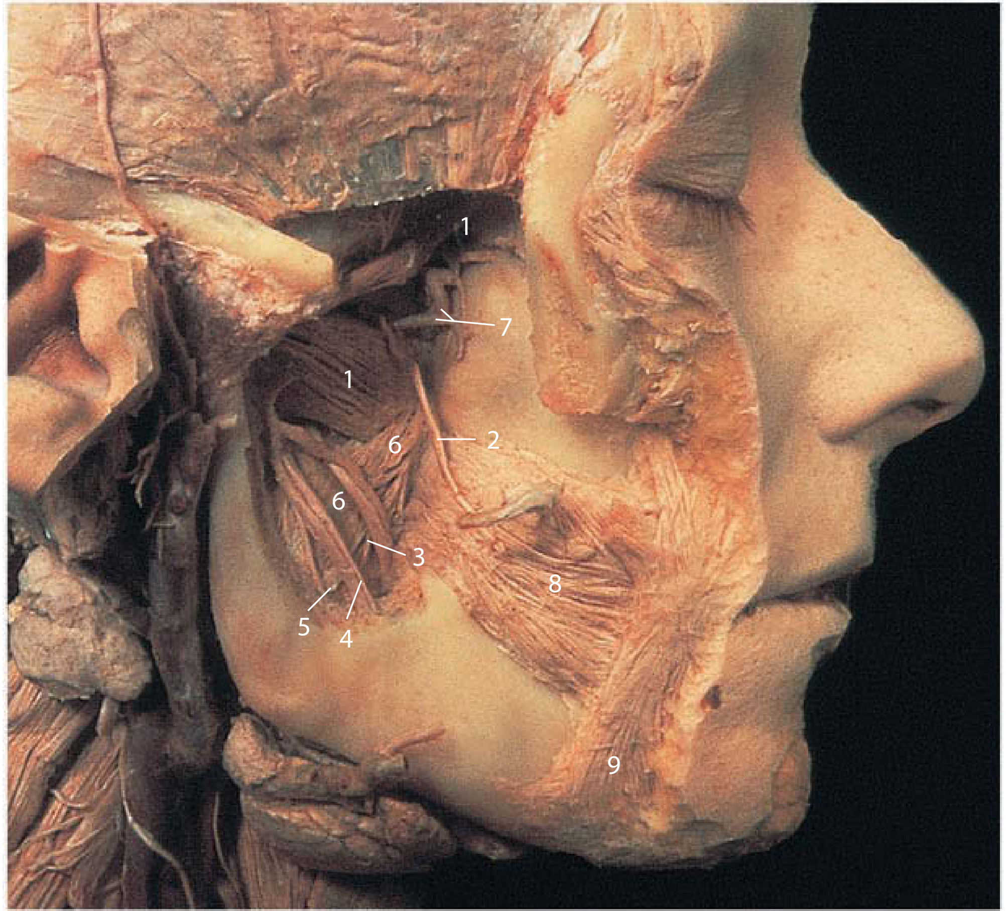
A real cadaveric dissection showing the infratemporal fossa. Matches your notes on Maxillary Artery Part 1 (Mandibular) - the inferior alveolar artery arising here - and Part 2 (Pterygoid) - masseteric, deep temporal, pterygoid, and buccal branches.
Key numbers: 1 = Lateral pterygoid (upper and lower heads), 2 = maxillary artery territory, 8 = buccinator muscle, 9 = buccal fat pad area.
Diagram 4 - Lingual Artery at the Floor of Mouth (Cadaveric Dissection)
Scott-Brown's Otorhinolaryngology, Figure 41.8 - Muscles of the Tongue and Structures on the Hyoglossus Muscle
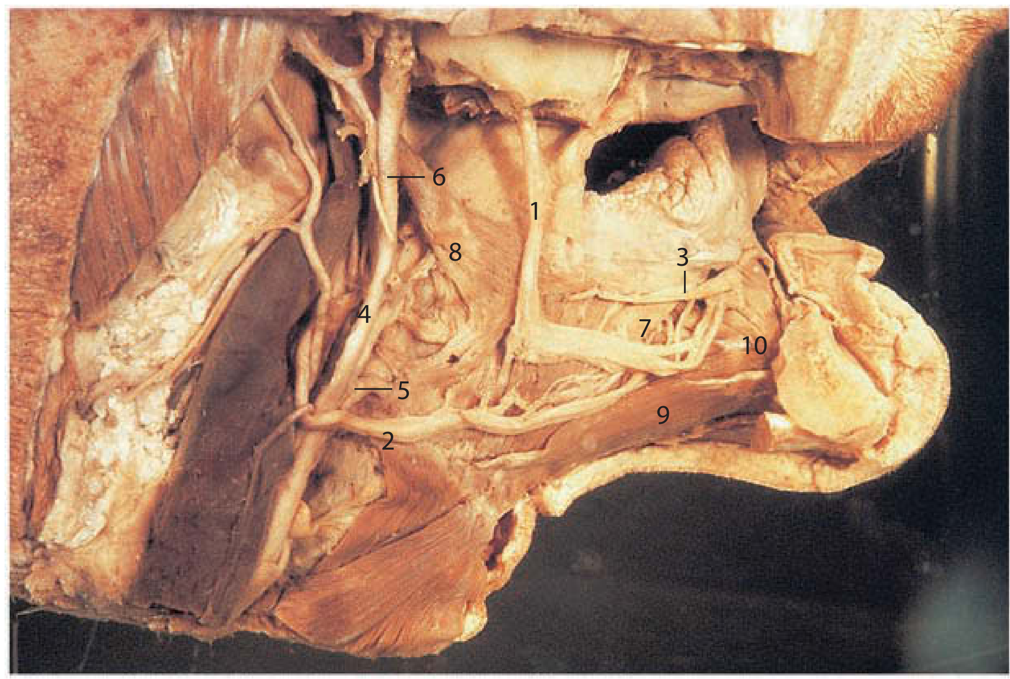
Real cadaveric dissection of the tongue and floor of mouth. Matches your notes on the lingual artery and its 4 branches. Shows the hyoglossus muscle as the key landmark, with the lingual artery running deep to it, and the hypoglossal nerve [XII] on its surface.
Quick Reference: Your Notes Mapped to Each Diagram
| Your Notes - Artery | Structure Supplied | Diagram to Use |
|---|---|---|
| Lingual a. - suprahyoid branch | Omohyoid, sternohyoid, thyrohyoid mx | Fig. 41.8 (Scott-Brown's) |
| Lingual a. - dorsal lingual a. | Post. 1/3 tongue, soft palate, tonsil, epiglottis | Fig. 41.8 |
| Lingual a. - deep lingual a. | Ventral surface of tongue | Fig. 41.8 |
| Lingual a. - sublingual a. | Sublingual gland, genioglossus, mylohyoid, mandible | Fig. 41.8 |
| Facial a. - ascending palatine | Soft palate, palatine tonsil | Fig. 8.70A (Gray's) |
| Facial a. - tonsillar branch | Palatine tonsil | Fig. 8.70A |
| Facial a. - submental a. | Chin and mylohyoid mx | Fig. 8.70A |
| Facial a. - glandular branches | Submandibular salivary gland | Fig. 8.70A |
| Facial a. - superior labial a. | Upper lip | Fig. 8.70A |
| Facial a. - inferior labial a. | Lower lip | Fig. 8.70A |
| Maxillary a. Part 1 - inf. alveolar a. | Mandible, lower teeth, chin (mental branch) | Fig. 41.5 (Scott-Brown's) |
| Maxillary a. Part 2 - buccal a. | Buccinator, cheek mucosa, overlying skin | Fig. 41.5 |
| Maxillary a. Part 3 - descending palatine a. | Hard palate (greater), soft palate (lesser) | Fig. 8.161 (Gray's) |
| Maxillary a. Part 3 - post. sup. alveolar a. | Upper premolar/molar teeth, maxillary sinus, gingiva | Fig. 8.161 |
| Maxillary a. Part 3 - infra-orbital a. + ant. sup. alveolar a. | Upper canine/incisor teeth, maxillary sinus | Fig. 8.161 |
Sources: Gray's Anatomy for Students (Figs. 8.70, 8.161); Scott-Brown's Otorhinolaryngology Head & Neck Surgery (Figs. 41.5, 41.8)
This is a shared conversation. Sign in to Orris to start your own chat.