
Diabetic foot
Reading File
Now I'll search the medical library for diabetic foot content across multiple dimensions in parallel.
Finding Sources
Finding Sources
Searching PubMed
"diabetic foot"[MeSH Terms] AND management
Excellent results. Now let me read the most comprehensive sources in parallel - Bailey & Love (surgery), Campbell's Orthopaedics, Goldman-Cecil Medicine, and Fischer's Mastery of Surgery.
Reading File
Reading File
Reading File
Reading File
Now let me get more content from Fischer's and Campbell's for classification, management, and fetch the clinical image.
Reading File
Reading File
Loading Image
Now let me get the classification systems and management sections from Campbell's and Fischer's.
Reading File
Reading File
Now let me get more on Wagner classification, Charcot foot, osteomyelitis, and the Bailey & Love surgical management section.
Finding Sources
Reading File
Reading File
Now let me get the Wagner classification table and treatment details from Campbell's, and Charcot neuroarthropathy.
Reading File
Finding Sources
Let me get the Wagner classification table and the Charcot foot section from Campbell's.
Reading File
Finding Sources
Now let me get the Wagner classification table itself, the Charcot section, and amputations section from Campbell's.
Finding Sources
Reading File
I now have comprehensive content. Let me get the Wagner table itself and Charcot/amputation sections from Sabiston, plus a web search for current guidelines.
Reading File
Reading File
Searching the Web
diabetic foot management guidelines 2024 2025 IWGDF classification treatment
I now have comprehensive, authoritative content from multiple textbooks and the latest IWGDF guidelines. Let me compile the full response.
Diabetic Foot
A diabetic foot encompasses any foot pathology that arises as a direct consequence of diabetes mellitus - primarily neuropathy, peripheral arterial disease (PAD), and infection. Globally, 15% of diabetic patients will develop a foot ulcer in their lifetime, with an annual incidence of ~2%. Up to one-fifth of these will progress to amputation. Notably, the 5-year mortality of diabetic foot complications rivals that of many cancers, underscoring the severity of this condition.
Pathophysiology
Three overlapping mechanisms drive diabetic foot disease:
1. Peripheral Neuropathy
The most pivotal contributor. Intracellular glucose accumulates and is shunted to sorbitol and fructose, impairing ATPase pumps and axonal transport. Free radical generation from mitochondria causes oxidative damage. Glycosylation of the vasa nervorum causes basement membrane thickening, leading to chronic ischemia of nerve tissue.
- Sensory neuropathy: Loss of light touch, temperature, and pain in a glove-and-stocking distribution. Patients cannot feel minor injuries or pressure ulcers.
- Autonomic neuropathy: Decreased sweating and skin lubrication - dry, fissured skin prone to breakdown.
- Motor neuropathy: Intrinsic muscle wasting leading to claw toes, hammer toes, and distal migration of the fat pad, creating abnormal plantar pressure points especially at the 1st and 5th metatarsal heads.
2. Peripheral Arterial Disease (PAD)
Glycosylation of endothelial basement membranes impairs oxygen diffusion. Atherosclerosis, arteriosclerosis, and calcinosis narrow the lumen, reducing perfusion - mainly affecting the tibial or femoral-popliteal distribution. Diabetic patients with PAD are 9 times more likely to develop a foot ulcer than those without PAD.
3. Immune Dysfunction
Hyperglycemia alters polymorphonuclear cell chemotaxis and impairs macrophage function. Epigenetic modifications lock macrophages in a persistent pro-inflammatory state, preventing progression from inflammation to the proliferation/remodeling phases of wound healing. This, combined with biofilm formation, makes wounds chronic and resistant to treatment.
Clinical Presentation
Ulcers most commonly develop under the 1st or 5th metatarsal head, the plantar surface, and the tips of toes (ischemic). Venous stasis ulcers may appear at the medial malleolus.
A key feature: the patient often does not remember the inciting injury due to loss of protective sensation. Wounds may be completely painless.
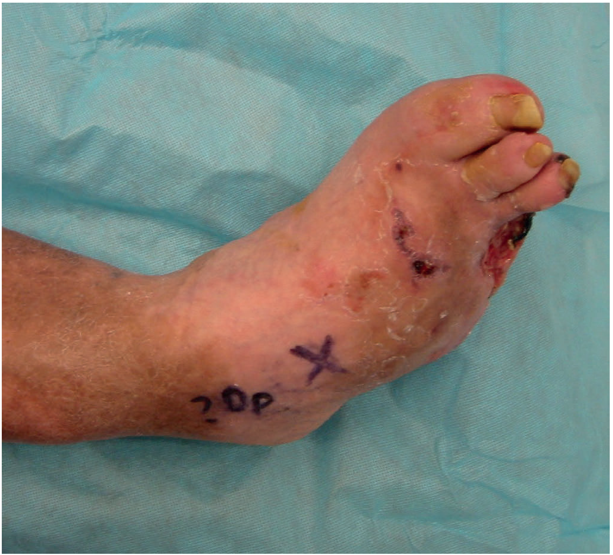
A severe diabetic foot infection with marked necrosis and tissue loss. (Bailey & Love's, 28th Ed.)
Common Signs
| Feature | Significance |
|---|---|
| Shiny, taut, hairless skin | Peripheral arterial disease |
| Dry, scaly skin | Autonomic neuropathy |
| Erythema, warmth, swelling | Infection or Charcot neuroarthropathy |
| Callus over bony prominences | Abnormal pressure - may overlie ulcer |
| Absent/diminished pulses | PAD - requires Doppler |
| Claw/hammer toes | Motor neuropathy, abnormal pressure |
Distinguishing Charcot from infection: Erythema and warmth from Charcot neuroarthropathy typically subside with limb elevation; infectious erythema does not.
Classification Systems
Wagner Classification (most widely used clinically)
| Grade | Description |
|---|---|
| 0 | No open lesion; high-risk foot (callus, deformity) |
| I | Superficial ulcer, not penetrating to tendon/capsule/bone |
| II | Deep ulcer to tendon, capsule, or bone |
| III | Deep ulcer with osteomyelitis or abscess |
| IV | Localized gangrene (toe/forefoot) |
| V | Extensive gangrene of entire foot |
Note: Wagner does not account for neuropathy or ischemia, limiting its ability to distinguish infectious from ischemic lesions.
IWGDF/IDSA Classification (preferred in guidelines)
| IWGDF Grade | Clinical Features |
|---|---|
| 1 / Uninfected | No local or systemic signs |
| 2 / Mild | ≥2 local signs (erythema >0.5 to <2 cm, warmth, swelling, pain, purulence); no systemic signs |
| 3 / Moderate | Erythema ≥2 cm AND/OR tissue deeper than skin/subcutaneous involved (tendon, muscle, joint, bone) |
| 4 / Severe | Any infection with systemic inflammatory response (fever, tachycardia, leukocytosis) |
The 2023 IWGDF/IDSA guidelines recommend hospitalization for all patients with severe (Grade 4) infection.
Investigations
| Test | Role |
|---|---|
| Semmes-Weinstein 10-g monofilament | Screen for loss of protective sensation (10 sites, or 4.5 g under 1st metatarsal heads) |
| Ankle-Brachial Index (ABI) | Normal 0.9-1.2; <0.5 = unlikely to heal without vascular intervention; >1.3 = non-compressible (calcified) |
| Toe pressures / Toe-Brachial Index | More reliable when ABI is non-compressible; >40 mmHg associated with wound healing |
| Plain radiograph | Detects osteomyelitis (may be normal early), foreign bodies, soft tissue gas, Charcot changes |
| MRI | Most sensitive modality for osteomyelitis and deep tissue involvement |
| CT scan | Detects deep abscesses |
| CRP / ESR | Elevated supports infection; normal does not exclude it |
| Probe-to-bone test | PPV 57%, NPV 96% for osteomyelitis |
| Bone biopsy culture | Gold standard for osteomyelitis organism identification |
Superficial ulcer swabs are unreliable - cultures should be taken from deep tissue after debridement.
Microbiology
- Mild infections: Predominantly Gram-positive cocci - S. aureus (including MRSA), Streptococcus spp.
- Moderate-severe infections: Polymicrobial - aerobic Gram-positive cocci + Gram-negative bacilli (including Pseudomonas) + anaerobes
- Severe/gangrenous: Pseudomonas over-represented; anaerobes warrant metronidazole addition
Management
1. Prevention and Systemic Control
- HbA1c target <7%; tight glucose control can halt and even partially reverse neuropathy
- Antihypertensives (ACE inhibitor preferred), statins
- Antiplatelet therapy (aspirin) if PAD is present
- Smoking cessation (addressed at every visit)
- Annual foot examination (more frequent if prior ulcer or deformity); patient education on daily self-inspection and foot hygiene
- Well-fitting cushioned footwear; no barefoot walking
- Callus management by a professional; nail trimming by a professional
2. Wound Offloading
The single most important intervention for neuropathic plantar ulcers:
- Total contact casting (TCC): Gold standard - extends above the ankle, reduces plantar pressure by up to 60%; not removable, so not ideal for frequent dressing changes
- Fixed ankle walking boot / removable walker: Alternative when frequent dressing access needed
- Below-ankle orthoses are inadequate for healing
3. Local Wound Care
Goals: moist environment, exudate control, surrounding tissue protection:
- Small ulcers: Moist saline or Vaseline-impregnated gauze, changed daily or twice daily
- Fibrinous/exudative wounds: Foams, hydrofibers, calcium alginate, hydrocolloids
- Dry wounds: Hydrogels
- Silver-impregnated dressings: For infection prevention
- Enzymatic debridement: Collagenase for selective non-viable tissue removal
- Avoid hydrogen peroxide and betadine - these damage viable tissue
4. Debridement
Convert a chronic wound to an acute wound by sharply removing all hypertrophic callus and non-viable tissue. Wound irrigation with saline by syringe reduces bacterial load. Ulcers communicating with bone require surgical debridement.
5. Negative Pressure Wound Therapy (NPWT)
Increases local blood flow, decreases edema, concentrates growth factors. Per a 2024 systematic review and meta-analysis (PMID: 39241769), NPWT increases healing rates compared to standard dressings in diabetic foot ulcers. Note: Cochrane Reviews (2018, 2023) still rate this evidence as low quality.
6. Antibiotic Therapy
| Severity | Regimen |
|---|---|
| Mild (IWGDF Grade 2) | Oral anti-Gram-positive: TMP-SMX 800/160 mg BD; or clindamycin 300 mg TDS; or cefalexin 500 mg QDS |
| Moderate-Severe (IWGDF Grade 3-4) | IV piperacillin-tazobactam 3.375 g q8h + vancomycin 15 mg/kg q12h (covers MRSA and Pseudomonas) |
- Duration: typically 1-2 weeks for soft tissue infection; 4-6 weeks for osteomyelitis
- Blood flow is often compromised, so longer courses may be needed
- Anaerobic cover (metronidazole) for abscesses and devitalized tissue
7. Vascular Intervention
Mandatory vascular assessment in any patient with poor peripheral pulses or ABI <0.5. Proximal angioplasty or bypass surgery can improve distal perfusion enough to allow foot-saving surgery.
8. Surgical Management
Wagner Grade III: Surgical debridement ± partial amputation + culture-specific antibiotics
Wagner Grade IV-V: Local or major amputation based on extent of infection
Wagner Grade IV-V: Local or major amputation based on extent of infection
Surgical offloading options include:
- Metatarsal head resection: Reduces abnormal pressure and ulcer recurrence
- Achilles tendon lengthening / gastrocnemius recession: Reduces forefoot pressure 25-50%; ulcer recurrence at 2 years reduced from 81% to 38% vs. casting alone
For amputation: resection must be adequate to remove all infected material; below-knee amputation may be preferable if extensive neuropathy extends to the ankle/hindfoot.
Charcot Neuroarthropathy
A destructive arthropathy caused by repetitive trauma to an insensate foot. Presents as a warm, swollen, erythematous foot - easily confused with infection. Key differentiation: Charcot erythema and warmth subside with elevation. Radiographs show progressive joint destruction, fragmentation, and collapse of the midfoot arch (rocker-bottom deformity). Management involves prolonged non-weight-bearing and total contact casting until bone consolidation occurs.
Osteomyelitis
- Most important risk factor: presence of a foot ulcer
- Probe-to-bone test: positive result + elevated inflammatory markers + abnormal plain X-ray = presumptive diagnosis
- MRI is the most sensitive confirmatory modality
- Bone biopsy for culture in complex cases
- Treatment: culture-specific antibiotics ± surgical debridement/resection
- Superficial swabs must not be used to guide therapy
Prognosis
- Up to one-third of patients with deep/limb-threatening infections eventually undergo amputation
- 5-year mortality of diabetic foot ulcers (30.5%) approaches or exceeds that of many common cancers
- Prevention, early detection, and prompt multidisciplinary management significantly improve outcomes
Key Sources:
- Bailey & Love's Short Practice of Surgery, 28th Ed. - Diabetic Foot Infection, p. 628
- Campbell's Operative Orthopaedics, 15th Ed. (2026) - Diabetic Foot, Ch. 91
- Fischer's Mastery of Surgery, 8th Ed. - Diabetic Foot Wound Management
- Sabiston Textbook of Surgery - Diabetic Foot Infections
- Rosen's Emergency Medicine - The Diabetic Foot
- IWGDF/IDSA Guidelines 2023 on Diagnosis and Treatment of Foot Infection
- ADA Standards of Care 2025 - Neuropathy and Foot Care
This is a shared conversation. Sign in to Orris to start your own chat.