
Ventilator emergency medical technician paramedical level
Reading File
The query is broad - "ventilator emergency" at paramedical/EMT level could cover many topics. Let me clarify before spending budget.
Asking for Preferences
What specific ventilator topic do you need at the EMT/paramedical level?
Searching sources...
Finding Sources
Finding Sources
Searching the Web
mechanical ventilator emergency paramedic EMT level guide modes troubleshooting weaning
Reading File
Reading File
Finding Sources
Reading File
Reading File
Reading File
Finding Sources
Reading File
Reading File
Reading File
Reading File
I now have comprehensive content from the textbooks. Let me compile the full response.
Mechanical Ventilator - Comprehensive Guide (EMT/Paramedical Level)
Sources: Roberts and Hedges' Clinical Procedures in Emergency Medicine; Rosen's Emergency Medicine; Tintinalli's Emergency Medicine; Fishman's Pulmonary Diseases and Disorders
1. BASIC PHYSIOLOGY YOU MUST KNOW
Minute Volume (VE)
VE = Tidal Volume (VT) x Respiratory Rate (f)
- Normal VE = 7-10 L/min
- VT = Alveolar Volume (VA) + Dead Space Volume (VDS)
- Anatomic dead space ~ 2.2 mL/kg lean body weight (trachea + larger airways)
- Alveolar ventilation determines PaCO2 (carbon dioxide in blood)
Key Pressure Concepts
| Pressure | Definition | Clinical Significance |
|---|---|---|
| Peak Airway Pressure | Pressure during active inspiration (dynamic) | Reflects compliance + airway resistance |
| Plateau Pressure | Pressure at end-inspiration with breath-hold (static) | Reflects lung compliance only; target <30 cm H2O |
| PEEP | Pressure at end of exhalation | Keeps alveoli open; prevents atelectasis |
| Auto-PEEP | Inadvertent PEEP from air trapping | Dangerous in asthma/COPD |
Peak-Plateau Gradient: Normal < 4 cm H2O. If elevated = increased airway resistance (bronchospasm, secretions).
P = V / C (Pressure = Volume / Compliance)
- Stiffer lungs (low compliance) = higher pressures for same volume
- Bigger volume = higher pressure
2. VENTILATOR SETTINGS (Key Parameters)
| Parameter | Typical Initial Setting | Notes |
|---|---|---|
| Tidal Volume (VT) | 6-8 mL/kg ideal body weight (IBW) | Never use actual weight; use IBW |
| Respiratory Rate (RR) | 10-14 breaths/min | Adjust based on blood pH/CO2 |
| FiO2 | Start at 100%, wean down | Target SpO2 > 92-95% |
| PEEP | 5 cm H2O (standard) | Increase if hypoxic |
| Flow Rate | 60 L/min | Increase for air hunger; prolongs expiration |
| I:E Ratio | 1:2 (normal) | Obstructive disease needs longer expiration (1:3 or 1:4) |
3. VENTILATOR MODES
A. Spontaneous / Support Modes
Pressure Support Ventilation (PSV)
- Patient triggers every breath; ventilator adds a set pressure boost
- Patient controls rate and tidal volume
- No set respiratory rate (backup apnea rate present)
- Used for: weaning, alert/breathing patients
CPAP (Continuous Positive Airway Pressure)
- Constant positive pressure throughout breathing cycle
- Patient breathes spontaneously against set pressure
- Used for: sleep apnea, mild respiratory failure, post-extubation support
BiPAP (Non-Invasive - NPPV)
- IPAP (inspiratory) + EPAP (expiratory) pressures set separately
- IPAP initial: 10-25 cm H2O | EPAP initial: 5-8 cm H2O
- PSV = IPAP - EPAP (the support given to each breath)
- Titrate FiO2 to maintain SpO2 86-92%
B. Mandatory (Machine-Controlled) Modes
Volume-Cycled Ventilation (VCV) - "Volume Control"
- Ventilator delivers a set tidal volume regardless of pressure needed
- Clinician sets: VT, RR, FiO2, PEEP, flow rate
- Advantage: guaranteed volume delivery
- Risk: pressure can spike if lungs become stiffer
Pressure-Cycled Ventilation (PCV) - "Pressure Control"
- Ventilator delivers gas to a set pressure; volume varies with compliance
- Clinician sets: pressure target, RR, FiO2, PEEP
- Advantage: safer pressures, better for stiff lungs
- Risk: VT drops if compliance worsens
Assist-Control (AC) Mode
- Every patient breath triggers a full machine-assisted breath
- Machine also delivers breaths if patient doesn't breathe at set rate
- Most common initial mode in emergency/ICU
- Can be used with VCV (AC-VCV) or PCV (AC-PCV)
SIMV (Synchronized Intermittent Mandatory Ventilation)
- Machine delivers set number of mandatory breaths synchronized with patient effort
- Patient can breathe spontaneously between mandatory breaths
- Add PSV on top of SIMV for patient comfort
C. Advanced/Specialty Modes
| Mode | Key Feature |
|---|---|
| APRV / Bilevel | Spends most time at high pressure; brief release for CO2 clearance; allows spontaneous breathing |
| PRVC / Volume Control+ | Closed-loop dual mode; adjusts pressure breath-to-breath to guarantee target VT |
| HFV (High-Frequency Ventilation) | Very high rates + tiny VT; mainly used in neonates and severe ARDS |
4. INITIATING MECHANICAL VENTILATION
Indications
- Respiratory or cardiac arrest
- Coma / altered consciousness
- Refractory hypoxemia (SpO2 < 90% despite O2)
- Severe respiratory acidosis
- Paradoxical breathing / impending fatigue
- Failure of non-invasive ventilation
RSI (Rapid Sequence Intubation) for Asthma/Bronchospasm
- Ketamine 1-2 mg/kg - preferred induction (bronchodilator + sympathomimetic)
- Propofol 1.5-2 mg/kg - alternative (bronchodilator but can cause hypotension)
- Succinylcholine 1.5 mg/kg OR Rocuronium 1 mg/kg for paralysis
- Use large ETT ≥ 8.0 mm to allow suctioning and bronchoscopy
Initial Settings for a Standard Adult Patient
Volume Control (VCV-AC):
- RR: 10-14 | VT: 7-8 mL/kg IBW | PEEP: 5 | FiO2: 100%
- Flow rate: 60 L/min | Waveform: Decelerating
Pressure Control (PCV-AC):
- RR: 12-16 | Pressure high: ~20 cm H2O | PEEP: 5 | FiO2: 100%
- Monitor VT obtained; adjust pressure to achieve ~7-8 mL/kg IBW
NPPV/BiPAP Initiation (Non-Invasive):
- Low-high approach: IPAP 10 / EPAP 5 / FiO2 100%, titrate IPAP up
- High-low approach: IPAP 20-25 / EPAP 5 / FiO2 100%, titrate down
- Reassess at 30 minutes; check ABG within 1-2 hours
5. VENTILATOR EMERGENCIES & TROUBLESHOOTING
The DOPES Mnemonic (Sudden Deterioration on Ventilator)
| Letter | Problem | Action |
|---|---|---|
| D | Displacement of ETT (esophageal, right main bronchus) | Check tube position, CXR, capnography |
| O | Obstruction (secretions, mucus plug, biting tube) | Suction, reposition head, bite block |
| P | Pneumothorax | Assess breath sounds, tracheal deviation, needle decompression |
| E | Equipment failure (circuit disconnect, power failure) | Check all connections; switch to BVM |
| S | Stacking / Auto-PEEP | Disconnect briefly (20-30 sec); reduce rate and increase expiration time |
Alarm Troubleshooting Table
| Alarm | Possible Causes | Interventions |
|---|---|---|
| High Pressure | Secretion/mucus plug, bronchospasm, biting tube, pneumothorax | Suction; bronchodilator; sedate; check breath sounds |
| Low Pressure / Apnea | Circuit disconnection, leak, cuff deflation, tracheostomy dislodgement | Check all circuit connections; inflate cuff; reposition tube |
| Low Power | Battery depleted | Plug into AC power outlet |
| Setting Error | Incorrect settings entered | Manually ventilate with BVM; transport patient + ventilator |
| Power Switchover | Shifted from AC to battery | Press "alarm silent" after confirming battery is operational |
Hypotension on Ventilator
- Auto-PEEP / air trapping - most common in asthma/COPD
- Action: Disconnect patient from ventilator for 20-30 seconds to allow full expiration; reduce RR; increase flow rate
- Tension pneumothorax
- Action: Needle decompression (2nd ICS MCL or 4th-5th ICS AAL)
- Decreased venous return from high PEEP/hyperinflation
- Volume depletion (give IV fluids)
- Sedative-induced vasodilation
Barotrauma
Caused by excessive airway pressures. Types: pneumothorax, pneumomediastinum, subcutaneous emphysema.
- Prevention: Keep plateau pressure < 30 cm H2O, tidal volume 6-8 mL/kg IBW
- Permissive hypercapnia (accept higher CO2 to keep pressures safe) - especially in asthma/ARDS
6. LUNG-PROTECTIVE VENTILATION (Critical Concept)
For ARDS, ALI, asthma, any ventilated patient:
| Target | Value |
|---|---|
| Tidal Volume | 6 mL/kg IBW (low stretch) |
| Plateau Pressure | < 30 cm H2O |
| PEEP | Titrate to oxygenation (usually 8-15 in ARDS) |
| SpO2 target | 88-95% (accept mild hypoxemia to avoid lung injury) |
| PaCO2 | Allow to rise (permissive hypercapnia) if needed |
Avoid: PaCO2 > 100 mmHg; plateau > 30; high tidal volumes (ventilator-induced lung injury).
7. WEANING FROM VENTILATOR
When to Consider Weaning
- Underlying cause of respiratory failure is resolving
- Patient is awake, following commands
- Hemodynamically stable (minimal/no vasopressors)
- FiO2 ≤ 40-50% and PEEP ≤ 5-8 cm H2O
- Adequate oxygenation (SpO2 > 92% on low settings)
Rapid Shallow Breathing Index (RSBI) = f/VT
- Most validated weaning predictor
- Measure during spontaneous breathing (no pressure support)
- RSBI < 100 = likely successful weaning
- RSBI > 100 = rapid, shallow breathing = likely weaning failure
Spontaneous Breathing Trial (SBT)
- Place patient on T-piece, low-level CPAP (5 cm H2O), or minimal PSV (5-8)
- Duration: 30 minutes to 2 hours
- Monitor: RR, SpO2, heart rate, BP, work of breathing, mental status
- If patient tolerates → proceed to extubation
- If patient fails → resume full ventilatory support; identify and treat cause
4 Methods for Weaning Trial
- T-tube trials - patient breathes through T-piece; oldest method
- SIMV - gradually reduce mandatory breath rate
- PSV - gradually reduce pressure support level
- CPAP - patient breathes spontaneously at low PEEP
Key fact: Weaning comprises ~40% of total ventilation duration. 20-30% of patients are difficult to wean. Weaning failure = failure of SBT OR need for reintubation within 48 hours of extubation.
8. QUICK REFERENCE - WAVEFORM SELECTION
| Waveform | Best For | Advantage |
|---|---|---|
| Decelerating (Ramp) | ARDS, standard patients | Better recruitment, lower peak pressure |
| Square | Asthma, COPD, head injury | Longer expiration time, lower mean airway pressure |
9. NON-INVASIVE VENTILATION (NIV/NPPV) KEY POINTS
- Best evidence: COPD exacerbation, cardiogenic pulmonary edema, immunocompromised patients
- Contraindications: respiratory arrest, inability to protect airway, excessive secretions, facial trauma, vomiting
- Mask fit is critical - check for leaks
- Reassess response within 30 minutes
- If no improvement or deterioration → intubate
Summary - EMT/Paramedic Priority Actions
- Any sudden deterioration - disconnect from ventilator, give BVM, use DOPES mnemonic
- High pressure alarm - suction first, then bronchodilator, then check for pneumothorax
- Low pressure alarm - check all circuit connections immediately
- Hypotension - think auto-PEEP (disconnect briefly) or tension pneumothorax
- Power failure - switch to BVM; never leave patient without ventilation
- Always use ideal body weight for tidal volume calculations, not actual weight
- Roberts and Hedges' Clinical Procedures in Emergency Medicine | - Tintinalli's Emergency Medicine | - Rosen's Emergency Medicine | - Fishman's Pulmonary Diseases and Disorders
Innum details aa besic to advanced All mode One by one Average student solli kudukura mathiri solli kudu
Searching sources...
Finding Sources
Finding Sources
Reading File
Finding Sources
Reading File
Reading File
Reading File
Reading File
I now have all the content needed for a full, student-friendly explanation. Let me write it out clearly in Tamil-English (Tanglish) style, simple language from basic to advanced.
🫁 VENTILATOR - BASIC TO ADVANCED (All Modes)
Average Student-ku Pura'njukura Mathiri - Simple Tamil-English Mix
FIRST THINGS FIRST - Ventilator Enna Panradhu?
Simple-a solli kuda:
"Patient-ku saaga vida maatom - machine breath kudukuthu"
Patient-ku tannoda breathe panna mudiyala? Appo ventilator machine - patient-kku forcefully air push pannuthu, CO2 vaanguthu, O2 kuduthu.
CORE CONCEPT - 3 PILLARS
VENTILATOR = VOLUME control + PRESSURE control + PATIENT effort
Yaar control panuradhu? Machine-a? Patient-a? Both-a?
- Atha based-a MODES decide panrom.
PART 1: BASIC SETTINGS (Settings Enna Enna Set Panurom?)
Ventilator switch-on panna first-a 6 settings set pannom:
| Setting | Meaning | Normal Value |
|---|---|---|
| VT - Tidal Volume | Oru breath-la poga venda air amount | 6-8 mL/kg IBW |
| RR - Rate | 1 minute-la machi breath count | 10-14/min |
| FiO2 | Oxygen percentage kodukurom | Start: 100%, Wean down |
| PEEP | Breath-ku apram lungs close aagaama vaikkum pressure | 5 cm H2O |
| Flow Rate | Air ethana speed-la pokudu | 60 L/min |
| I:E Ratio | Inhale time vs Exhale time ratio | 1:2 normal |
⚠️ IBW = Ideal Body Weight - Actual weight use panna KOODATHU. Height-based calculate panurom. Why? Heavy patient-ku lungs same size dhan. Actual weight use panna over-ventilation aagum.
PART 2: VENTILATOR MODES - ONE BY ONE
Modes = "Machine ethana control panum, Patient ethana control padum?"
MODE 1: CONTROLLED VENTILATION (CV)
aka IMV - Intermittent Mandatory Ventilation
Simple English:
Machine FULL control. Patient zero effort. Machine decides everything.
Analogy:
Patient-ai auto-pilot plane-la vacha mathiri. Pilot (patient) hands off. Machine alone fly pannuthu.
How it works:
- Machine-e decide pannum: Rate, Volume, everything
- Patient breathe panna try pannaluam - MACHINE LISTEN PANNADU
- Patient effort = 0 (or heavily sedated/paralyzed)
Used when:
- General anesthesia (OT-la)
- Heavily sedated ICU patients
- Completely paralyzed patients (neuromuscular blockade)
- Apnea (breathe panna marakirkavan)
Settings clinician sets:
- VT, RR, FiO2, PEEP - all of it
Advantage:
- Doctor 100% control pannan
Disadvantage:
- Patient-ku heavy sedation MUST (otherwise patient fight pannan with machine - "dyssynchrony")
- Patient effort use aagadu = muscle waste aagum
MODE 2: ASSIST-CONTROL (AC) Mode
MOST COMMON - Emergency-la Default Mode
Simple English:
Patient breath edukka try pannan - Machine help pannum. Patient breath edukka marandha - Machine alone pannum.
Analogy:
Auto-rickshaw back-a thallu pannum mathiri. Nee thallu pannan - auto thoothu varum. Nee thallu pannalana - auto dhan thallu pannikkum. But every breath = FULL support.
How it works:
- Patient inspiratory effort pannan (trigger) → Machine senses it → Full breath delivers
- Patient effort illana → Machine set rate-la breath delivers
- Every single breath = Full machine-assisted breath
Two Types of AC:
AC - Volume Control (AC-VC)
- Machine guarantees fixed VT (e.g., 500 mL every time)
- Pressure varies based on lung stiffness
- You set: VT, RR, PEEP, FiO2, Flow rate
- You watch: Peak pressure, Plateau pressure
Problem: Lung stiff aana → Pressure increase aagum → Barotrauma risk
AC - Pressure Control (AC-PC)
- Machine guarantees fixed Pressure (e.g., 20 cm H2O every time)
- Volume varies based on lung compliance
- You set: Driving pressure, RR, PEEP, FiO2
- You watch: Tidal Volume, Minute ventilation
Problem: Lung compliance change aana → VT change aagum → Under/over-ventilation risk
Used when:
- Newly intubated patients (ICU, ED)
- Critically ill, unstable patients
- Patients who need rest (full support needed)
Advantage:
- Minimum VE guaranteed
- Less sedation needed than pure controlled
- Patient can trigger extra breaths
Disadvantage:
- Patient fast breathe panan → Machine follows → Respiratory alkalosis (too much CO2 blown off)
- Asthma/COPD-la dangerous (air trapping = auto-PEEP)
Initial Settings for AC:
AC-VC: RR 10-14, VT 7-8 mL/kg IBW, PEEP 5, FiO2 100%, Flow 60 L/min
AC-PC: RR 12-16, Pressure 20 cm H2O, PEEP 5, FiO2 100%
MODE 3: SIMV - Synchronized Intermittent Mandatory Ventilation
Simple English:
Machine certain number of breaths kudukum. Middle-la patient sarakku breathe pannalaam - but alone (no help).
Analogy:
School-la attendance mathiri. Machine 10 breaths mandatory kudukum. Nee 5 extra breathe eduka want pana - edukalam, but without help. Own effort.
How it works:
- Clinician sets mandatory RR (e.g., 10/min)
- Machine delivers those 10 breaths - synchronized with patient effort
- Between those breaths → Patient can breathe spontaneously, but NO machine help (unless PSV added on top)
Two variations:
- SIMV-VC: Mandatory breaths = volume-targeted
- SIMV-PC: Mandatory breaths = pressure-targeted
PSV added to SIMV:
Usually SIMV + PSV together use panurom. Why? Spontaneous breaths-ku support kudukka. Otherwise patient extra work pannum.
Used when:
- Weaning process (slowly reducing machine support)
- Spontaneously breathing patients who need partial support
Advantage:
- Patient uses respiratory muscles = muscle strength maintain aaguthu
- Weaning tool as support slowly reduced
Disadvantage:
- Spontaneous breaths unsupported (unless PSV added) = patient works hard between mandatory breaths
- Complex monitoring needed
MODE 4: PSV - Pressure Support Ventilation
Simple English:
Patient breathe pannanum - machine boost kudukum. Patient breathe pannalana - machine pannadu.
Analogy:
Bike-la accelerator pidi mathiri. Nee pedal pannanum dhan - machine power add pannum. But pedal pannalana - nothing happens. 100% patient effort needed to start.
How it works:
- Patient makes inspiratory effort (trigger)
- Machine adds a preset pressure boost (e.g., PS 10)
- Breath ends when flow drops below threshold (~25% of peak)
- Patient controls: Rate, I-time, Flow demand
- Machine only controls: Pressure support level + PEEP + FiO2
Important:
- No set respiratory rate
- Backup apnea rate present in modern ventilators (safety net)
- VT depends on PS level + patient effort + lung compliance
Used when:
- Awake, breathing patients who need help
- Weaning from ventilator
- BiPAP (non-invasive) = essentially PSV
PSV Level Guide:
| PS Level | Meaning |
|---|---|
| PS 20+ | High support - near full assist |
| PS 10-15 | Medium support |
| PS 5-8 | Minimal support - almost ready to extubate |
| PS 0 (CPAP only) | No support - test if patient can breathe alone |
Advantage:
- Patient comfort - breathes naturally
- Respiratory muscles stay active
- Best weaning mode
Disadvantage:
- Patient apnea aana - no backup (danger! - that's why backup rate set panrom)
- VT not guaranteed
MODE 5: CPAP - Continuous Positive Airway Pressure
Simple English:
Constant pressure maintain panrom. Patient tannoda breathe pannum. Machine breath kudukadu - just "open door" kudukuthu.
Analogy:
Balloon slightly inflate vacha mathiri lungs vaikkurom. Deflate aagaama. Patient tannoda breathe pannum through that open balloon.
How it works:
- Same pressure (positive) maintained throughout - both inhale AND exhale
- No inspiratory pressure boost (unlike PSV)
- Patient does ALL the work
- Machine just maintains the set PEEP level
Used when:
- Weaning (SBT - Spontaneous Breathing Trial)
- Sleep apnea (non-invasive CPAP at home)
- Mild respiratory failure
- Post-extubation support
- RSBI test pannuvom - CPAP 5 + PS 0 la 3 minutes vaippom
Settings: Just CPAP level (usually 5) + FiO2
Difference CPAP vs PSV:
CPAP = Constant pressure, no boost during inhalation
PSV  = Pressure BOOST added during each inhalation
MODE 6: BiPAP - Bilevel Positive Airway Pressure
Simple English:
Inhale-la higher pressure, Exhale-la lower pressure. Non-invasive (mask, no tube).
Analogy:
Door-a open panna help panrom (IPAP), then slightly close but not full close (EPAP). Two levels of pressure.
Parameters:
- IPAP = Inspiratory Positive Airway Pressure (higher level)
- EPAP = Expiratory Positive Airway Pressure (lower level)
- Pressure Support = IPAP - EPAP (actual support given)
Initial Settings:
Low-High Approach: IPAP 10 / EPAP 5 / FiO2 100%
High-Low Approach: IPAP 20-25 / EPAP 5 / FiO2 100%

EPAP 8 if: Morbid obesity or auto-PEEP
Titration:
- Hypercapnia (CO2 high) → Increase IPAP (increase PS)
- Hypoxia (O2 low) → Increase EPAP + FiO2
- Fast RR / dyspnea → Increase IPAP
Used when:
- COPD exacerbation (BEST evidence)
- Cardiogenic pulmonary edema (BEST evidence)
- Immunocompromised patients (avoid intubation)
- Obesity hypoventilation
Contraindicated when:
- Respiratory arrest
- Vomiting / aspiration risk
- Cannot protect airway
- Facial trauma
- Excessive secretions
Reassess in: 30 minutes - ABG check 1-2 hours
MODE 7: APRV - Airway Pressure Release Ventilation
(Advanced Mode)
Simple English:
Lungs-ai HIGH pressure-la hold pannurom (oxygenation). Brief-a LOW pressure-la release panrom (CO2 out). Patient throughout breathe pannalaam.
Analogy:
Dam-la water (air) hold panrom. Short-a gate open panurom - flush aaguthu (CO2 out). Gate close - dam full again (oxygenation).
Parameters:
- P-high = High pressure level (oxygenation) - e.g., 25-35 cm H2O
- P-low = Low pressure (usually 0) - brief release
- T-high = Time at high pressure (4-6 seconds)
- T-low = Time at low pressure (0.2-0.8 sec for restrictive, 0.8-1.5 sec for obstructive)
Key points:
- Patient can breathe spontaneously throughout cycle
- Most time at P-high → oxygenation
- Brief release to P-low → CO2 clearance
- Less sedation needed (unlike inverse ratio ventilation)
Used when:
- Severe ARDS (refractory hypoxemia)
- Patients who failed conventional modes
- Open lung ventilation strategy
MODE 8: PRVC / Volume Control Plus
(Dual/Hybrid Mode - Advanced)
Simple English:
Machine guarantee-a oru volume kudukum, but pressure adjust panudu - lungs protect pannuvadha.
Analogy:
Smart AC mathiri - temperature target fix. But power automatically adjust pannum based on room condition.
How it works:
- Closed-loop system
- Each breath-ku compliance calculate pannum
- Next breath-la pressure adjust pannum to achieve target VT
- Volume guaranteed + Pressure kept as low as possible
Also called: PRVC, Auto-flow, VC+, Adaptive Pressure Ventilation
Advantage: Best of both worlds - VT guaranteed AND lung-protective
QUICK COMPARISON TABLE - All Modes at a Glance
| Mode | Who Controls Breath? | Volume Guaranteed? | Pressure Guaranteed? | Used For |
|---|---|---|---|---|
| CV (Controlled) | Machine 100% | Yes | No | OT, paralyzed patients |
| AC-VC | Both (machine dominant) | Yes | No | ICU initial, emergency intubation |
| AC-PC | Both (machine dominant) | No | Yes | High-pressure situations, ARDS |
| SIMV | Both (shared) | Mandatory-Yes, Spontaneous-No | Depends | Weaning, partial support |
| PSV | Patient (with boost) | No | No (PS is boost, not control) | Weaning, awake patients |
| CPAP | Patient alone | No | No | SBT, sleep apnea, mild failure |
| BiPAP | Patient (with 2-level support) | No | IPAP/EPAP set | COPD, Pulm edema, non-invasive |
| APRV | Patient + machine (unique cycle) | No | P-high/P-low set | Severe ARDS, refractory hypoxia |
| PRVC | Machine (smart) | Yes | Auto-adjusted | Lung protection + volume guarantee |
PART 3: PATIENT CONDITION → CORRECT MODE
Newly intubated, unstable patient?
→ AC-VC or AC-PC (full support needed)
COPD exacerbation, awake?
→ Try BiPAP first (avoid intubation)
ARDS patient?
→ AC-VC, low tidal volume (6 mL/kg), high PEEP, lung protective
Asthma severe, intubated?
→ AC-VC, low RR, high flow rate, permissive hypercapnia
Patient improving, trying to wean?
→ SIMV + PSV → Then PSV alone → Then CPAP → Then extubate
Sleep apnea?
→ CPAP (home device)
Ready to test if patient can breathe alone (SBT)?
→ CPAP 5 + PS 0 or T-piece
PART 4: WEANING STEP-BY-STEP
Step 1: Check readiness (ALL must be met)
- Patient awake, following commands ✓
- Hemodynamically stable ✓
- Cause of intubation resolving ✓
- FiO2 ≤ 40-50% ✓
- PEEP ≤ 5-8 cm H2O ✓
- RSBI < 105 ✓ (RR ÷ VT in Liters → e.g., 20 breaths / 0.5 L = 40 = good)
Step 2: Do RSBI test
- Put patient on CPAP 5 / PS 0 for 3 minutes
- Calculate RSBI = Respiratory Rate ÷ Tidal Volume (L)
- RSBI < 105 = likely can wean
- RSBI > 105 = 95% chance of failure - don't extubate
Step 3: SBT (Spontaneous Breathing Trial)
- CPAP 5 + PS 6-8 OR T-piece
- Watch 30-120 minutes
- STOP if: RR > 35, SpO2 < 90%, HR > 140, BP > 180 or < 90, agitation, distress
Step 4: Extubate if passed
Step 5: If failed → Back to full support, find reason
PART 5: EMERGENCY TROUBLESHOOTING
DOPES Mnemonic (Patient deteriorates on ventilator)
| Problem | Do this | |
|---|---|---|
| D | Displacement - tube moved out or into right lung | Check tube depth, auscultate, CXR |
| O | Obstruction - secretions, mucus plug | Suction immediately |
| P | Pneumothorax | Check breath sounds, trachea, needle decompress |
| E | Equipment failure - disconnect, power off | BVM bag the patient NOW |
| S | Stacking/Auto-PEEP | Disconnect 20-30 sec, slow rate, increase expiration |
Alarm Table
| Alarm | Why? | Fix |
|---|---|---|
| High Pressure | Secretions, bronchospasm, biting tube, pneumothorax | Suction → Bronchodilator → Sedate → Check for PTX |
| Low Pressure / Apnea | Circuit disconnected, cuff leak | Check all connections, inflate cuff |
| Low Battery/Power | Internal battery dead | Plug to AC power |
| Setting Error | Wrong setting entered | Manual BVM ventilation while fixing |
Hypotension on Vent - Think This Order:
- Auto-PEEP - disconnect briefly (most common in COPD/asthma)
- Tension pneumothorax - needle decompress
- High PEEP - reduce PEEP
- Volume depletion - IV fluids
- Sedation-induced - reduce sedation
EXAM SUMMARY BOX
MOST COMMON INITIAL MODE    → AC-VC
BEST MODE FOR WEANING       → PSV → CPAP
BEST NON-INVASIVE           → BiPAP
SEVERE ARDS MODE            → AC-VC (low VT) or APRV
PARALYZED PATIENT ONLY      → Controlled (CV/IMV)
SMART HYBRID MODE           → PRVC

RSBI < 105   = EXTUBATE ready
RSBI > 105   = 95% fail - don't extubate
Plateau P    < 30 cm H2O always
Tidal Vol    = 6-8 mL/kg IBW (never actual weight)
Sources: Roberts and Hedges' Clinical Procedures in Emergency Medicine | Pfenninger & Fowler's Procedures for Primary Care | Harrison's Principles of Internal Medicine 22E | Tintinalli's Emergency Medicine
First basic anatomy and physiology of ventilation
Searching sources...
Finding Sources
Finding Sources
Finding Sources
Finding Sources
Reading File
Reading File
Reading File
Finding Sources
Reading File
Reading File
Reading File
Reading File
Loading Image
Reading File
Reading File
Finding Sources
Reading File
Loading Image
Excellent! I now have comprehensive textbook content and key diagrams. Let me compile the full student-friendly anatomy and physiology guide.
🫁 ANATOMY & PHYSIOLOGY OF VENTILATION
Basic to Advanced - Average Student Purinja Mathiri
Sources: Costanzo Physiology 7e | Morgan & Mikhail's Clinical Anesthesiology 7e | Murray & Nadel's Respiratory Medicine
PART 1: BIG PICTURE - Enna Nadakuradhu?
Ventilation = Breathing process = Air in + Air out
Respiration = Ventilation + Gas Exchange + O2 delivery to cells
ATMOSPHERE
    ↓  (Air breathe in)
CONDUCTING AIRWAYS  (Transport panurom - NO gas exchange)
    ↓
ALVEOLI  (Gas exchange nadakudu - O2 in, CO2 out)
    ↓
BLOOD → HEART → WHOLE BODY
    ↑  (CO2 collect panni tirupi lungs-ku)
CELLS
PART 2: ANATOMY - Respiratory System Structure
TWO ZONES - Conducting Zone vs Respiratory Zone
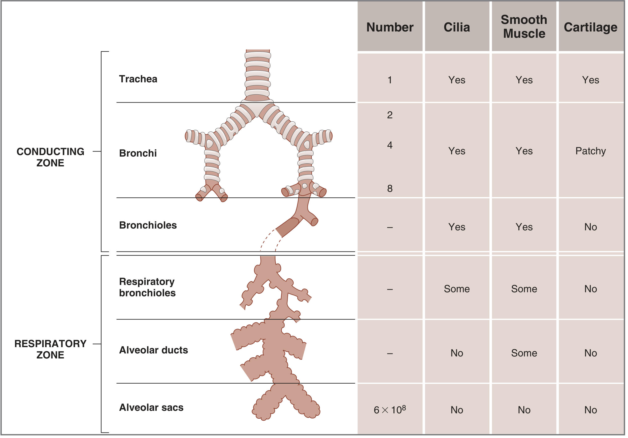
Conducting Zone - Respiratory Zone comparison (Costanzo Physiology)
ZONE 1: CONDUCTING ZONE (Air Highway)
Purpose: Air-a respiratory zone-ku transport panurom. Gas exchange ILLAI inga.
Extra jobs: Air warm pannurom + humidify pannurom + filter pannurom (dust, bacteria)
Path (top to bottom):
NOSE / MOUTH
    ↓
NASOPHARYNX / OROPHARYNX
    ↓
LARYNX (Voice box - epiglottis here)
    ↓
TRACHEA (Windpipe)
    ↓
RIGHT MAIN BRONCHUS + LEFT MAIN BRONCHUS (Carina-la split)
    ↓
BRONCHI (increasingly smaller)
    ↓
BRONCHIOLES
    ↓
TERMINAL BRONCHIOLES  ← End of conducting zone
Important points - One by One:
🔹 Trachea
- Length: 10-13 cm
- C-shaped cartilaginous rings (horseshoe shape) - front + sides protected; back = membranous wall
- Cricoid cartilage = narrowest part (adults): Men 17 mm, Women 13 mm
- Splits at CARINA → Right + Left main bronchus
🔹 Right vs Left Main Bronchus
| Right | Left | |
|---|---|---|
| Angle | More vertical (direct line with trachea) | More angled |
| Length | Shorter | Longer (~5 cm men, 4.5 cm women) |
| Why important? | Foreign body + ETT goes here more easily! | - |
⚠️ Clinical Pearl: ETT accidentally deep-a push panna → Right side-la goes (right mainstem intubation). Left lung ventilate aagadu. Left-side breath sounds disappear.
🔹 Airway Generations
- Trachea = Generation 0
- Right + Left bronchi = Generation 1
- Total 23 generations exist
- Each generation = smaller tubes, but MORE number = total area INCREASES
🔹 Airway Wall Layers
| Structure | Trachea | Bronchi | Bronchioles |
|---|---|---|---|
| Cilia | Yes | Yes | Yes |
| Smooth Muscle | Yes | Yes | Yes |
| Cartilage | Yes | Patchy | NO |
Cartilage = structural support. Bronchioles-la cartilage illai → Smooth muscle alone controls diameter → Bronchospasm possible here!
🔹 Smooth Muscle Control
- Sympathetic (β2 receptors): Bronchodilation - airways OPEN
- Adrenaline, Salbutamol (albuterol) work here
- Parasympathetic (Muscarinic receptors): Bronchoconstriction - airways CLOSE
- Atropine, Ipratropium block this
ZONE 2: RESPIRATORY ZONE (Gas Exchange Zone)
Path continues:
RESPIRATORY BRONCHIOLES (some alveoli here)
    ↓
ALVEOLAR DUCTS (fully lined with alveoli)
    ↓
ALVEOLAR SACS
    ↓
ALVEOLI ← Gas exchange happens here!
🔹 Alveoli - The Star of the Show
- Each lung: ~300 million alveoli (total body = 600 million)
- Size: ~200 micrometers diameter each
- Total surface area: ~70 square meters (size of a tennis court!)
- Walls: Ultra thin → allows rapid O2/CO2 diffusion
- Lined with Type I pneumocytes (gas exchange) + Type II pneumocytes (surfactant production)
🔹 Surfactant - Why Important?
Simple explanation:
Alveoli = bubbles. Bubbles want to collapse (surface tension). Surfactant is soap - reduces surface tension, prevents collapse.
Laplace's Law:
Pressure = 2 × Surface tension / Radius
- Small alveolus → surfactant MORE concentrated → tension LOW → won't collapse
- Large alveolus → surfactant LESS concentrated → tension HIGH → won't over-expand
- Net effect: All alveoli stay stable size
⚠️ Why PEEP is needed on ventilator? Surfactant loss (ARDS, prematurity) or low lung volume → alveoli collapse → PEEP open pannuthu alveoli-ai.
PART 3: PLEURA - Two Layers
LUNG
  ↓ (hugs the lung)
VISCERAL PLEURA
  [Intrapleural space - thin fluid layer, negative pressure]
PARIETAL PLEURA
  ↓ (lines chest wall)
CHEST WALL
Intrapleural pressure: Normally -5 cm H2O (negative = suction effect)
- This negative pressure keeps lungs expanded
- Lung-ku collapse aagathu pull pannuthu
⚠️ Pneumothorax: Air enters pleural space → negative pressure lost → lung collapses. Tension PTX: Pressure builds → mediastinum shifts → cardiac output drops → Emergency!
PART 4: MUSCLES OF BREATHING
Inspiration Muscles (Active process - energy use pannurom)
Primary muscle:
- Diaphragm (75% of work) - dome shape → contracts → flattens → chest volume increases
- Moves down 1.5 to 7 cm during breathing
- Nerve supply: Phrenic nerve (C3, C4, C5) - "C3, 4, 5 keeps the diaphragm alive"
Secondary/Accessory muscles (normal quiet breathing-la minimum use):
- External intercostal muscles - ribs up + out → chest expands
- Sternocleidomastoid (SCM) - elevates rib cage (distress-la use)
- Scalene muscles - prevents upper rib from collapsing inward
- Pectoralis muscles (arms fixed vacha) - chest expansion assist
⚠️ Accessory muscle use = respiratory distress sign! Patient-ku SCM, intercostal retractions visible = working hard to breathe. Intubation consider panum time.
Expiration Muscles (Normally PASSIVE - no energy needed!)
Quiet breathing-la expiration = elastic recoil alone does it. Muscles not needed.
Active expiration (exercise, disease):
- Abdominal muscles (rectus abdominis, obliques) - diaphragm-ai up push pannuthu
- Internal intercostal muscles - ribs down + in
Ventilator pearl: Ventilator expiration phase = 100% passive. Machine just opens expiratory valve → elastic recoil pushes air out. If expiration time not enough → air trap → auto-PEEP.
PART 5: MECHANICS OF BREATHING - How Air Moves
Boyle's Law Basis
Pressure × Volume = Constant Volume increase → Pressure decrease → Air flows IN Volume decrease → Pressure increase → Air flows OUT
Normal Inspiration
Brain says "breathe" → Phrenic nerve → Diaphragm contracts
    → Thoracic cavity volume INCREASES
    → Intrapleural pressure becomes MORE negative (-5 → -8 cm H2O)
    → Lungs expand (pulled by suction)
    → Intra-alveolar pressure drops BELOW atmospheric
    → Air flows IN (nose → alveoli)
Normal Expiration
Diaphragm relaxes
    → Thoracic cavity volume DECREASES
    → Elastic recoil of lungs kicks in
    → Intra-alveolar pressure RISES above atmospheric
    → Air flows OUT
    → (Passive - no muscle work!)
On Ventilator - REVERSED Mechanics!
| Normal Breathing | Mechanical Ventilation | |
|---|---|---|
| How air enters | Negative pressure (suction) | Positive pressure (push) |
| Diaphragm | Active - contracts | Passive/resting |
| Intrapleural pressure | More negative during inhalation | More positive during inhalation |
| Risk | None normally | Barotrauma, decreased venous return |
This is why ventilator is called Positive Pressure Ventilation - opposite of natural breathing!
PART 6: LUNG VOLUMES (Numbers to Know)
| Volume / Capacity | Abbreviation | Normal Value | What it means |
|---|---|---|---|
| Tidal Volume | VT | 500 mL | Each normal breath |
| Inspiratory Reserve Volume | IRV | 3000 mL | Extra air after normal inhale |
| Expiratory Reserve Volume | ERV | 1100 mL | Extra air forced out after normal exhale |
| Residual Volume | RV | 1200 mL | Air ALWAYS remaining - cannot breathe out |
| Total Lung Capacity | TLC | 6000 mL (6 L) | Maximum air lungs can hold |
| Vital Capacity | VC | 4700 mL | TLC - RV (what you can use) |
| Functional Residual Capacity | FRC | 2400 mL | ERV + RV (air at end of normal exhale) |
| Inspiratory Capacity | IC | 3500 mL | VT + IRV |
Memory tip:
FRC = Resting lung volume. PEEP increases FRC → keeps alveoli open. RV = Never goes out. Even after maximum forced exhale, 1.2 L remains.
PART 7: DEAD SPACE - Important Concept
Dead space = Air that's ventilated but NOT doing gas exchange
Types:
1. Anatomical Dead Space (~150 mL)
- Nose, trachea, bronchi, bronchioles (conducting zone)
- Air here doesn't reach alveoli for exchange
- Rule: ~2.2 mL/kg lean body weight
- Easy memory: 1 mL per pound body weight (150 lb → ~150 mL)
2. Alveolar Dead Space
- Alveoli that are ventilated but NOT perfused (no blood flow)
- Normally zero in healthy lungs
- Increases in: pulmonary embolism, low cardiac output
3. Physiological Dead Space = Anatomical + Alveolar
- Healthy person: ~150 mL (mostly anatomical)
- Sick patient: Can be much higher
Formula for Alveolar Ventilation:
VA (Alveolar ventilation) = (VT - VD) × Rate

Example:
VT = 500 mL, Dead space = 150 mL, Rate = 14
VA = (500 - 150) × 14 = 350 × 14 = 4900 mL/min
Ventilator impact: ETT adds dead space (tube itself). Tracheostomy reduces dead space (shorter path to lungs) → easier weaning.
PART 8: GAS EXCHANGE - O2 in, CO2 out
Where it happens: Alveolar-Capillary Membrane
Layers air must cross (extremely thin - <0.5 microns):
- Surfactant layer
- Alveolar epithelium (Type I cells)
- Basement membrane
- Capillary endothelium
Total = thinner than one cell! That's why exchange is so fast.
Partial Pressures - The Driving Force
Air molecules move from HIGH pressure → LOW pressure (diffusion)
| Location | PO2 | PCO2 |
|---|---|---|
| Atmospheric air | 160 mmHg | 0 mmHg |
| Alveolar air | 100 mmHg | 40 mmHg |
| Venous blood arriving at lung | 40 mmHg | 46 mmHg |
| Arterial blood leaving lung | 100 mmHg | 40 mmHg |
O2 movement: Alveoli (100) → Blood (40) → O2 diffuses INTO blood ✓
CO2 movement: Blood (46) → Alveoli (40) → CO2 diffuses OUT of blood ✓
The Golden Relationship - Alveolar Ventilation & CO2
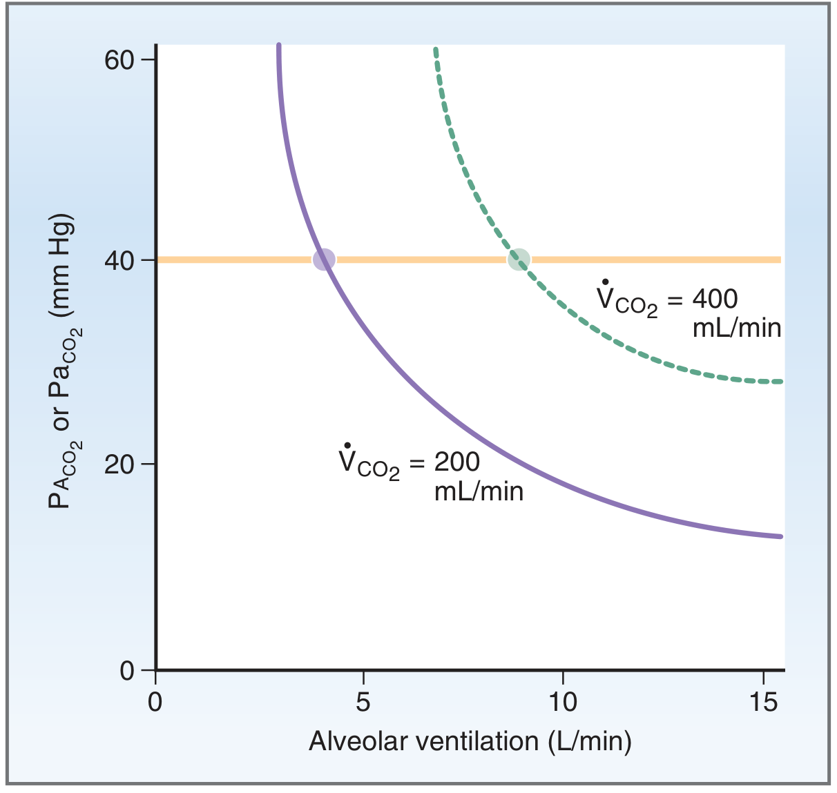
Inverse relationship: More ventilation = Less CO2. Less ventilation = More CO2.
PaCO2 = VCO2 × K / VA

Alveolar ventilation ↑ → PaCO2 ↓ (blows off CO2)
Alveolar ventilation ↓ → PaCO2 ↑ (CO2 builds up = hypercapnia)
This is why ventilator RR and VT control CO2!
- CO2 high (acidosis) → Increase RR or VT → blow off more CO2
- CO2 low (alkalosis) → Decrease RR or VT → retain more CO2
PART 9: COMPLIANCE - Stiffness of Lungs
Compliance = How easily lungs stretch
Compliance = Change in Volume / Change in Pressure

CL (Lung) = Normal: 150-200 mL/cm H2O
CW (Chest wall) = Normal: 200 mL/cm H2O
Total = 100 mL/cm H2O
Analogy:
New balloon = high compliance (easy to blow) Old balloon = low compliance (hard to blow, stiff)
| Condition | Compliance | Effect |
|---|---|---|
| ARDS, Pulm edema, Fibrosis | LOW (stiff) | High pressures needed to push same volume |
| Emphysema | HIGH (floppy) | Easy to inflate but hard to exhale - air traps |
| Normal | Normal | Normal pressures generate normal volumes |
Ventilator impact:
- Low compliance → plateau pressure rises → barotrauma risk
- That's why we monitor plateau pressure < 30 cm H2O
PART 10: AIRWAY RESISTANCE
Resistance = How hard it is for air to FLOW through airways
Normal: 0.5 to 2 cm H2O/L/s
Main contributors: Medium bronchi (before 7th generation)
Increased when:
- Bronchospasm (asthma - smooth muscle contracts)
- Secretions / mucus plug
- Mucosal edema (allergic, infection)
- Low lung volume (airway collapse)
Detected on ventilator by:
Peak Pressure - Plateau Pressure = Peak-Plateau Gradient Normal < 4 cm H2O HIGH gradient (>4) = increased resistance (bronchospasm, secretion)
PART 11: CONTROL OF BREATHING - Brain's Role
Three Brain Centers
CEREBRAL CORTEX
→ VOLUNTARY breathing (you decide when to breathe, speak, hold breath)

PONS (Pneumotaxic center)
→ Fine-tunes rhythm, controls respiratory rate

MEDULLA OBLONGATA (Main automatic center)
→ AUTOMATIC breathing (you don't think about it - happens 24/7)
→ DRG (Dorsal Respiratory Group) - Inspiration
→ VRG (Ventral Respiratory Group) - Expiration + Apneustic center
Chemical Control - What triggers breathing?
Primary trigger: CO2 (not O2!)
Central Chemoreceptors (Medulla):
- Detect CO2 / H+ in cerebrospinal fluid (CSF)
- CO2 crosses blood-brain barrier → forms H+ → stimulates breathing
- Main driver in normal people
Peripheral Chemoreceptors (Carotid + Aortic bodies):
- Carotid body - most important (IX nerve)
- Aortic body (X nerve)
- Detect: Low O2 (< 60 mmHg), High CO2, Low pH
- Backup system
Important: HYPOXIC DRIVE in COPD!
Normal people: CO2 is the main trigger for breathing
Chronic COPD patients:
- They ALWAYS have high CO2 (used to it - brain becomes desensitized)
- Their main trigger = LOW O2 (hypoxic drive)
- If you give too much O2 → PO2 rises → hypoxic drive removed → patient stops breathing!
- Therefore: COPD patients → Target SpO2 88-92% (not 100%)
PART 12: V/Q RATIO - Ventilation/Perfusion
V = Ventilation (air reaching alveoli)
Q = Perfusion (blood flow through lung capillaries)
Perfect match: V/Q = 1
- Both air and blood present → gas exchange perfect
V/Q Mismatch = Problem!
| Situation | V/Q | What happens |
|---|---|---|
| Normal | ~1 | Perfect exchange |
| Pulmonary Embolism | High (∞) | Alveolus ventilated, NO blood → Dead space ↑ |
| Atelectasis / Pneumonia | Low (0) | Blood flows, NO ventilation → Shunt → Hypoxia |
| ARDS | Very low V/Q | Massive shunt → Refractory hypoxia |
Shunt = O2 doesn't help! (blood bypasses ventilated areas)
Dead space = CO2 problem! (ventilation wasted)
Why PEEP helps in ARDS? PEEP opens collapsed alveoli → V/Q improves → O2 improves
SUMMARY CHART - All Key Numbers
| Parameter | Normal Value | Clinical Significance |
|---|---|---|
| Tidal Volume | 500 mL (7 mL/kg) | Ventilator: 6-8 mL/kg IBW |
| Respiratory Rate | 12-16/min | Ventilator: 10-14/min |
| Minute Ventilation | 7-10 L/min | VT × RR |
| PaO2 | 80-100 mmHg | < 60 = hypoxemia |
| PaCO2 | 35-45 mmHg | < 35 = alkalosis, > 45 = acidosis |
| pH | 7.35-7.45 | < 7.35 = acidosis, > 7.45 = alkalosis |
| SpO2 | 95-100% | COPD target 88-92% |
| FRC | 2400 mL | PEEP increases FRC |
| Dead Space | ~150 mL | 30% of tidal volume |
| Compliance (total) | 100 mL/cm H2O | Low in ARDS |
| Airway Resistance | 0.5-2 cm H2O/L/s | High in asthma/COPD |
FLOW CHART: Ventilation to Ventilator (How it connects)
PROBLEM: Patient can't breathe normally
         ↓
Why? Choose one:
         ↓
MUSCLES weak?         LUNGS stiff?          AIRWAY blocked?
(ALS, GBS,            (ARDS, Pulm edema,    (Asthma, COPD,
 Myasthenia)           Fibrosis)             Secretions)
         ↓                 ↓                      ↓
Diaphragm fails      Compliance LOW         Resistance HIGH
         ↓                 ↓                      ↓
No neg. pressure    Need high pressure      Need flow + time
to breathe          to open stiff lungs     for expiration
         ↓                 ↓                      ↓
VENTILATOR solves by POSITIVE PRESSURE
- Pushes air in (overcomes muscle failure)
- PEEP keeps alveoli open (overcomes stiffness)
- High flow rate + long expiration time (overcomes resistance)
Sources: Costanzo Physiology 7e | Morgan & Mikhail's Clinical Anesthesiology 7e | Murray & Nadel's Textbook of Respiratory Medicine | Fishman's Pulmonary Diseases and Disorders
This is a shared conversation. Sign in to Orris to start your own chat.