
Write about mammary gland under following headings a)extent B)relations c structure d blood supply e lymphatic drainage applied aspects 15mark answer mbbs
Reading File
Now let me search the medical library for comprehensive mammary gland content across all required headings simultaneously.
Finding Sources
Finding Sources
Finding Sources
Reading File
Reading File
Excellent content found. Now let me read the comprehensive breast anatomy sections from Gray's and Sabiston simultaneously.
Reading File
Reading File
Excellent detailed content. Now let me read the Fischer's Surgery anatomy section and search for blood supply/lymphatics specifically.
Reading File
Finding Sources
Finding Sources
Let me now read the detailed blood supply & lymphatics from histology and the thoracic wall sections.
Reading File
Reading File
Reading File
Now let me get the blood supply and histology/structure content specifically.
Finding Sources
Reading File
Now let me fetch the Thieme Atlas blood supply section and the detailed structure/histology from Sabiston and the applied aspects.
Reading File
Finding Sources
Reading File
I now have comprehensive content from multiple authoritative sources. Let me fetch a key anatomical image to include in the answer.
Loading Image
Loading Image
I now have all the content needed. Let me compile the comprehensive 15-mark MBBS answer.
MAMMARY GLAND
Sources: Gray's Anatomy for Students, Sabiston Textbook of Surgery, Fischer's Mastery of Surgery, Thieme Atlas of Anatomy, Histology: A Text and Atlas (Pawlina), Creasy & Resnik's Maternal-Fetal Medicine
a) EXTENT
The mammary gland is a modified sweat gland located within the superficial fascia of the anterior thoracic wall.
Vertical extent: From the 2nd rib superiorly to the 6th rib (or inframammary fold / upper border of the rectus sheath, corresponding to the 6th-7th rib) inferiorly.
Horizontal extent: From the lateral border of the sternum medially to the anterior axillary line (or medial border of latissimus dorsi) laterally.
Base: The base of the adult breast extends from the 2nd to 6th rib along the midclavicular line, directly overlying the pectoralis major, serratus anterior, and external oblique muscles.
Axillary Tail (Tail of Spence): The superolateral part of the gland extends along the inferior margin of the pectoralis major and passes around it through an opening in the deep fascia to enter the axilla. This is called the axillary process or tail of Spence. It rarely reaches the apex of the axilla. Ductal tissue can extend to the clavicle, anteriorly to the midline, and into the axilla.
Important note: The upper half of the breast, especially the upper outer quadrant, contains more glandular tissue than the rest of the breast. In 15% of cases, ducts extend beyond the inframammary fold.
b) RELATIONS
Anteriorly: Skin and subcutaneous tissue. The nipple-areolar complex (NAC) is situated at the level of the 4th intercostal space (varies with breast size).
Posteriorly (Deep surface): The breast lies on the retromammary space, a potential space containing loose areolar tissue. Deep to this are:
- Pectoralis major muscle (central portion)
- Serratus anterior (inferolateral)
- External oblique (lower part)
- Upper part of rectus sheath
This retromammary space allows the breast to move over the underlying pectoral fascia. When invaded by carcinoma, the breast becomes fixed to the chest wall.
Superiorly: Clavicle and subcutaneous tissue of the infraclavicular region.
Medially: Lateral border of the sternum, separated from the opposite breast by the intermammary cleft.
Inferolaterally: The axillary process/tail of Spence projects into the axilla.
Supporting structures: Cooper's ligaments (suspensory ligaments) are fibrous bands running between the deep fascia overlying the pectoralis major and the dermis of the overlying skin, providing structural support to the breast.
c) STRUCTURE
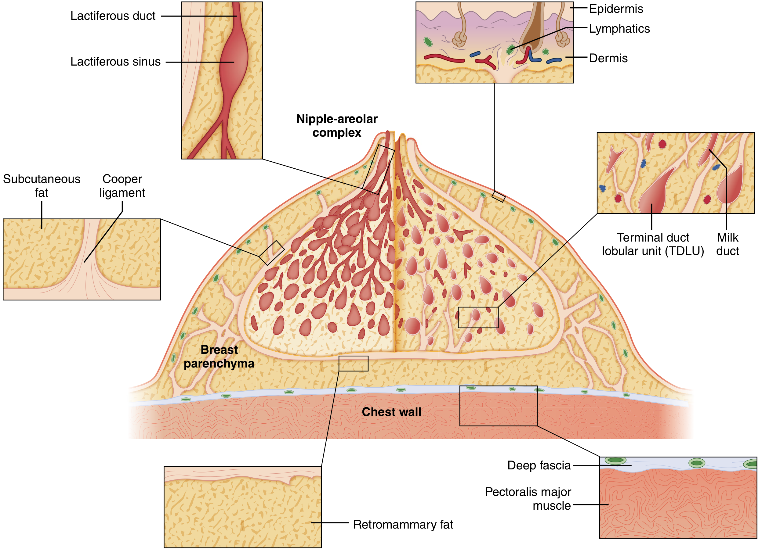
The mature breast is composed of three principal tissue types: (1) glandular epithelium, (2) fibrous stroma, and (3) adipose tissue.
Lobes and Ducts
- The glandular apparatus is arranged into 15-20 lobes, each drained by a lactiferous duct that opens at the nipple.
- Each lactiferous duct has a dilated segment just below the NAC called the lactiferous sinus (ampulla), then progressively branches into smaller interlobular ducts and finally terminates in the terminal ductules or acini.
- The lobes are separated by fibrous septa (interlobular stroma).
- The ducts are arranged radially, spreading outward from the nipple in an "inverted tree" configuration.
Terminal Duct Lobular Unit (TDLU)
- The TDLU consists of a terminal duct and its associated lobule (cluster of acini).
- The acini are the milk-forming glands of the lactating breast.
- The TDLU is invested in specialized loose intralobular connective tissue (containing capillaries and lymphocytes), clearly distinct from the denser interlobular stroma.
- The TDLU is the site of origin of most malignant breast tumors.
Nipple and Areola
- The nipple contains smooth muscle and numerous sensory nerve endings.
- The areola contains Montgomery's glands (modified sebaceous glands) which enlarge during pregnancy.
- Underlying the areola, the lactiferous sinuses act as temporary milk reservoirs.
Fascial Coverings
- Superficial layer of superficial fascia: Separates the breast from the overlying skin.
- Deep layer of superficial fascia: Separated from the pectoral fascia by the retromammary space.
- Cooper's ligaments: Fibrous suspensory bands passing from the deep fascia through the breast substance to fuse with the superficial fascia under the dermis.
Microscopic Structure
- Inactive breast: TDLUs have rudimentary acini without visible lumen, surrounded by dense stroma and adipose tissue.
- Lactating breast: Alveoli are large with columnar secretory cells; myoepithelial cells surround each alveolus.
- After menopause: Glandular tissue involutes, replaced largely by adipose tissue.
d) BLOOD SUPPLY
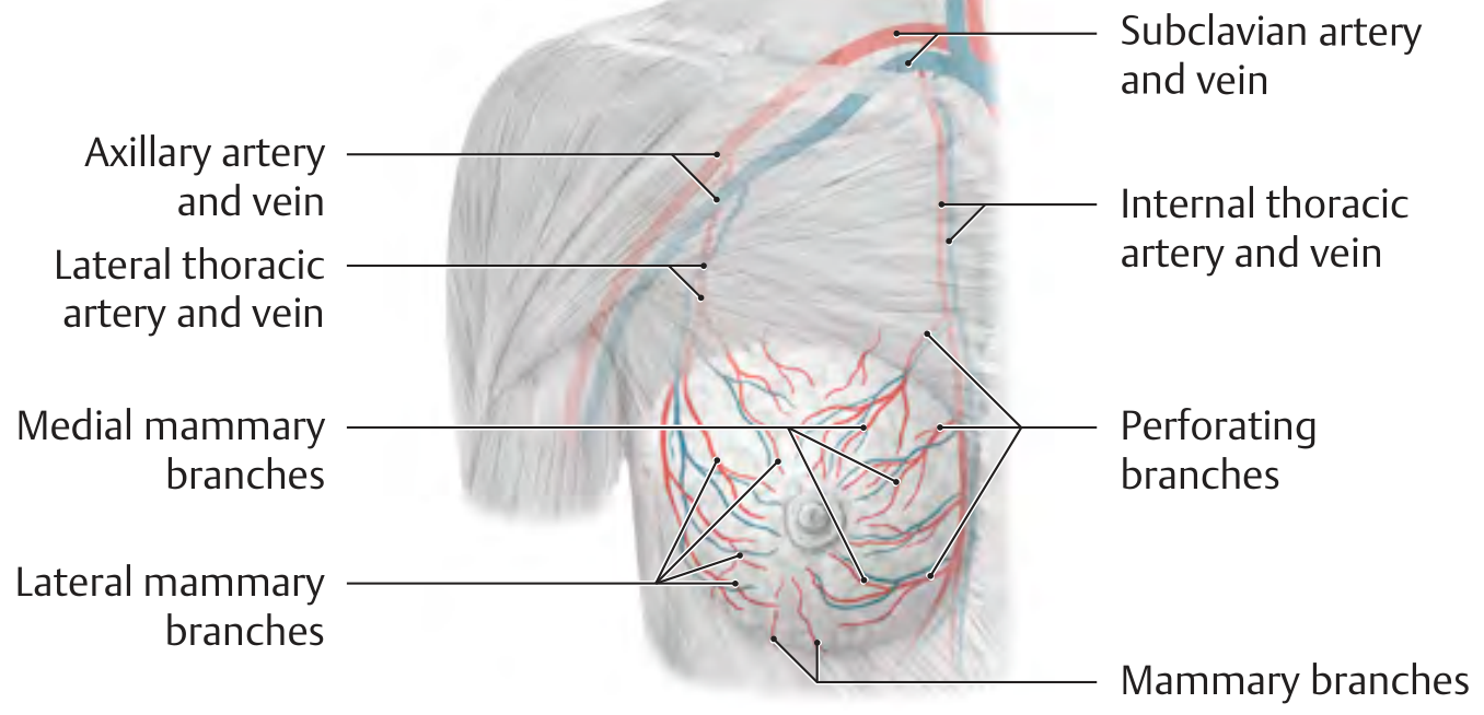
Arterial Supply (Three main sources)
| Source | Branches | Contribution |
|---|---|---|
| Internal thoracic (mammary) artery | Perforating branches (medial mammary branches) from 2nd-4th intercostal spaces | ~60% - main supply, supplies medial and central parts |
| Lateral thoracic artery (branch of axillary artery) | Lateral mammary branches | Supplies lateral and upper parts |
| Anterior intercostal arteries (2nd-5th) | Direct mammary branches | Supplies lower and central portions |
Additional minor contribution from the thoracoacromial artery (pectoral branch).
Branches pass primarily along the path of the alveolar ducts to reach capillary beds surrounding the alveoli.
Venous Drainage
Veins follow the course of the arteries and drain into:
- Internal thoracic (mammary) vein (medially) - drains into the brachiocephalic vein
- Axillary vein (laterally) - via lateral thoracic and other tributaries
- Posterior intercostal veins - which drain into the azygos/vertebral venous plexus
Clinical significance of venous drainage: The vertebral venous plexus (Batson's plexus) communicates with the intercostal veins. This provides a valve-less route for hematogenous metastasis of breast carcinoma directly to the vertebral column, skull, and brain, without passing through the lungs.
e) LYMPHATIC DRAINAGE
Routes of Drainage
1. Axillary Route (75%) - Most important
- Lymph from the lateral, central, and inferior parts of the breast drains to axillary lymph nodes.
- Axillary nodes are divided into 3 levels based on their relationship to the pectoralis minor muscle:
- Level I (Low axillary): Lateral to the lateral border of pectoralis minor - includes anterior/pectoral, posterior/subscapular, and lateral/humeral groups.
- Level II (Mid axillary): Behind pectoralis minor - includes central nodes and Rotter's (interpectoral) nodes (between pectoralis major and minor).
- Level III (Apical): Medial to the medial border of pectoralis minor - apical nodes; efferents form the subclavian trunk.
2. Internal Mammary (Parasternal) Route (~25%)
- Medial and central parts of the breast drain along perforating branches of the internal thoracic artery to parasternal lymph nodes (1st-4th intercostal spaces).
- These drain into the bronchomediastinal trunk or directly into the thoracic duct/right lymphatic duct.
3. Other Routes (Minor)
- Supraclavicular nodes: Via apical axillary nodes or directly from upper breast.
- Cross-drainage: To contralateral breast and axilla via subareolar plexus (Sappey's plexus).
- Inferior drainage: To abdominal lymphatics (diaphragmatic/subdiaphragmatic nodes).
- Posterior intercostal nodes: Via posterior intercostal lymphatics.
Subareolar (Sappey's) Plexus
A rich network of lymphatics beneath the areola that interconnects all quadrants of the breast. It is the basis for subareolar injection of radiotracer/blue dye in sentinel lymph node biopsy.
f) APPLIED ASPECTS
1. Carcinoma of the Breast
- Most common malignancy in women; most tumors (>50%) arise in the upper outer quadrant.
- TDLUs are the site of origin of most breast carcinomas (ductal carcinoma in situ, lobular carcinoma in situ, invasive ductal and lobular carcinomas).
Signs due to anatomical relations:
- Skin dimpling/retraction: Invasion of Cooper's ligaments tethers the skin, causing puckering ("peau d'orange" when edema is added).
- Nipple retraction: Fibrosis/invasion of lactiferous ducts.
- Peau d'orange (orange-peel appearance): Lymphedema of skin due to blockage of dermal lymphatics by tumor emboli; the skin is tethered by Cooper's ligaments at the pores, producing a dimpled appearance.
- Fixation to chest wall: Deep invasion involving the pectoral fascia or muscles obliterates the retromammary space.
- Fixation to skin: Superficial tumor extension.
- Edema of the arm: Axillary lymph node involvement blocks lymphatic and venous return from the upper limb.
Metastatic spread:
- Lymphatic spread: First to axillary nodes (most commonly Level I). Sentinel lymph node biopsy (SLNB) via subareolar injection maps the first draining node.
- Hematogenous spread: Via Batson's vertebral plexus to vertebrae, skull, and brain (explaining bone/brain metastases without pulmonary involvement first).
- Parasternal route: Medial quadrant tumors drain to parasternal nodes, may be missed in axillary dissection; explains recurrence after axillary clearance.
2. Lymphedema after Mastectomy/Axillary Dissection
Disruption of lymphatics during axillary clearance or radiotherapy results in lymphedema of the ipsilateral upper limb (brawny, non-pitting edema).
3. Accessory Breast Tissue / Polymastia / Polythelia
Failure of complete regression of the embryological mammary ridge (milk line, axilla to inguinal region) results in accessory nipples (polythelia) or accessory breast tissue (polymastia), most commonly in the axilla. These can enlarge during pregnancy.
4. Sentinel Lymph Node Biopsy (SLNB)
Based on the concept that breast lymph drains in an orderly fashion to the first (sentinel) node before proceeding to other axillary nodes. Injection of blue dye or Tc-99m colloid into the subareolar plexus identifies the sentinel node. If negative for metastases, full axillary dissection may be avoided, reducing morbidity.
5. Surgical Importance of Cooper's Ligaments
During mastectomy, flap dissection is carried out at the level of Cooper's ligaments. This plane is relatively avascular and helps preserve adequate skin flaps while removing all parenchyma.
6. Galactocele
A milk-containing cyst arising from obstruction of a lactiferous duct during or after lactation.
7. Fibroadenoma
The most common benign breast tumor, arising from the TDLU in young women. It is freely mobile ("breast mouse") because it is not attached to Cooper's ligaments.
8. Gynecomastia
Enlargement of the male breast due to proliferation of ductal tissue and stroma, associated with hormonal imbalance (increased estrogen/androgen ratio). Causes include puberty, cirrhosis, drugs (spironolactone, digoxin, cimetidine), and tumors.
References:
- Gray's Anatomy for Students (Surface anatomy of breast, Axillary process)
- Sabiston Textbook of Surgery, Chapter 68 (Breast Anatomy, Development and Physiology; Lymphatic drainage Fig. 68.4)
- Fischer's Mastery of Surgery, Chapter 59 (Boundaries of Mastectomy)
- Thieme Atlas of General Anatomy and Musculoskeletal System (Blood supply section D)
- Histology: A Text and Atlas - Pawlina (Blood Supply and Lymphatics; Involution)
- Creasy & Resnik's Maternal-Fetal Medicine (Nipple, areola, blood supply)
From definition to end
I need a quick clarification before diving in!
Asking for Preferences
Which topic should be covered from definition to end?
The answer to directly write in exam
The user wants a clean, exam-ready answer on the Mammary Gland - written exactly as you would write it in an MBBS exam paper, from definition to the end of applied aspects. I have all the content from the previous research. Let me write it directly.
MAMMARY GLAND
(15 Marks Answer)
DEFINITION
The mammary gland is a modified sweat (apocrine) gland present in the superficial fascia of the anterior thoracic wall. It is the characteristic feature of the class Mammalia and is the organ of milk production (lactation). In females it is well developed; in males it remains rudimentary throughout life.
a) EXTENT
The mammary gland is situated on the anterior thoracic wall between the skin and the deep fascia (pectoral fascia).
Vertical extent:
- Superiorly: 2nd rib
- Inferiorly: 6th rib (inframammary fold / upper border of the rectus sheath)
Horizontal extent:
- Medially: Lateral border of sternum
- Laterally: Anterior axillary line / mid-axillary line / medial border of latissimus dorsi
Base overlies:
- Pectoralis major (central and most of the area)
- Serratus anterior (inferolateral)
- External oblique (inferomedial)
- Upper part of rectus sheath
Axillary Tail (Tail of Spence):
The superolateral part of the gland extends around the lower border of pectoralis major, pierces the deep fascia through an opening called the foramen of Langer, and projects into the axilla. It rarely reaches the apex of the axilla.
Note: The upper outer quadrant contains the largest amount of glandular tissue and is the most common site of breast carcinoma.
b) RELATIONS
| Surface | Structure |
|---|---|
| Anterior | Skin, subcutaneous fat, superficial layer of superficial fascia |
| Posterior (deep surface) | Retromammary space (loose areolar tissue) → pectoral fascia → pectoralis major, serratus anterior, external oblique |
| Superior | Infraclavicular region; clavicle |
| Medial | Lateral border of sternum |
| Lateral/superolateral | Axillary tail projecting into axilla |
Retromammary Space:
A potential space between the deep layer of superficial fascia and the pectoral fascia. Contains loose areolar tissue that allows the breast to move freely over the chest wall. It is obliterated when carcinoma invades deeply, causing fixation of the breast.
Cooper's Ligaments (Suspensory Ligaments):
Fibrous bands that run from the deep fascia through the breast substance and attach to the overlying dermis. They maintain the shape and position of the breast. When invaded by carcinoma, they shorten and pull the skin inward, causing the characteristic skin dimpling.
c) STRUCTURE
Gross Structure
The breast consists of 3 principal tissue types:
- Glandular epithelium (parenchyma)
- Fibrous stroma (supporting framework)
- Adipose tissue (gives the rounded contour)
Lobes and Ducts:
- Gland is divided into 15-20 lobes arranged radially around the nipple
- Each lobe is drained by a lactiferous duct that opens on the nipple
- Just below the nipple, each duct dilates to form the lactiferous sinus (ampulla), which acts as a temporary milk reservoir during lactation
- Each lactiferous duct further branches into interlobular ducts → intralobular (terminal) ductules → acini (alveoli)
Terminal Duct Lobular Unit (TDLU):
- One lobule + its terminal duct = the basic functional secretory unit
- The acini are the milk-producing glandular units
- Surrounded by intralobular stroma (loose connective tissue with capillaries and lymphocytes)
- TDLU is the site of origin of most breast carcinomas
Nipple and Areola:
- Nipple: Projects at the level of 4th intercostal space; contains smooth muscle, numerous sensory nerve endings, and openings of lactiferous ducts
- Areola: Pigmented area around nipple containing Montgomery's glands (modified sebaceous glands that secrete to lubricate the nipple during lactation); these enlarge during pregnancy as small tubercles called Montgomery's tubercles
Fascial Coverings
- Superficial layer of superficial fascia: Between skin and gland
- Deep layer of superficial fascia: Between gland and retromammary space
- Cooper's ligaments: Connect the two layers, passing through the gland and inserting into the dermis
Microscopic Structure
- Inactive (non-lactating) breast: TDLUs contain rudimentary acini without lumina, surrounded by dense fibrous stroma
- Lactating breast: Alveoli are distended, lined by columnar secretory cells; surrounded by myoepithelial cells that contract under oxytocin to eject milk
- Postmenopausal breast: Glandular structures involute and are replaced by adipose tissue; no TDLU visible
d) BLOOD SUPPLY
Arterial Supply (Three main sources)
1. Internal Thoracic Artery (Internal Mammary Artery) - 60% (main supply)
- Gives perforating branches through the 2nd, 3rd, and 4th intercostal spaces
- These are called medial mammary branches
- Supply the medial and central portions of the breast
2. Lateral Thoracic Artery (branch of 2nd part of axillary artery)
- Gives lateral mammary branches
- Supplies the lateral and upper portions of the breast
3. Anterior Intercostal Arteries (2nd-5th)
- Give direct mammary branches
- Supply the lower and peripheral portions
Minor contributions: Thoracoacromial artery (pectoral branch), subscapular artery.
Venous Drainage
Veins follow the arteries and drain into:
- Internal thoracic vein (medially) → brachiocephalic vein
- Axillary vein (laterally) → subclavian vein
- Posterior intercostal veins → azygos vein → Batson's vertebral venous plexus
Clinical significance: Batson's plexus is valve-less and communicates with the vertebral venous system. This provides a route for hematogenous metastasis of breast carcinoma directly to the vertebral bodies, skull, and brain, bypassing the lungs.
Nerve Supply
- Anterior and lateral cutaneous branches of 2nd to 6th intercostal nerves
- Carry sensory and sympathetic fibers
- Secretory function is primarily hormonal (prolactin, oxytocin), not neural
e) LYMPHATIC DRAINAGE
The lymphatics of the breast originate in the interlobular connective tissue and drain via two main plexuses:
- Subareolar (Sappey's) plexus - a rich network beneath the areola connecting all quadrants
- Deep plexus - along the lactiferous ducts
Routes of Drainage
1. Axillary Route - 75% (most important)
Lymph from lateral, central, and upper parts drains to axillary nodes, which are grouped into 3 levels by their relationship to pectoralis minor:
| Level | Position | Groups Included |
|---|---|---|
| Level I | Lateral to pectoralis minor | Anterior (pectoral), Posterior (subscapular), Lateral (humeral) groups |
| Level II | Behind pectoralis minor | Central group + Rotter's (Interpectoral) nodes |
| Level III | Medial to pectoralis minor | Apical nodes |
Efferent vessels from apical nodes form the subclavian trunk → right lymphatic duct (right side) or thoracic duct (left side).
Spread of carcinoma follows the axillary levels in order: Level I → Level II → Level III.
2. Internal Mammary (Parasternal) Route - 25%
- Medial and central quadrants drain along perforating vessels of the internal thoracic artery to parasternal nodes in the 1st-4th intercostal spaces
- These drain into the bronchomediastinal trunk or directly into the thoracic duct
- Important in medial quadrant tumors - missed by axillary dissection alone
3. Other Routes (Minor)
- Supraclavicular nodes: Via apical nodes or directly from upper breast
- Contralateral breast and axilla: Via subareolar plexus cross-connections
- Abdominal lymphatics: Inferior drainage through diaphragm to subdiaphragmatic nodes
- Posterior intercostal nodes: Via posterior intercostal lymphatics
f) APPLIED ASPECTS
1. Carcinoma of the Breast
Most common malignancy in women. Most arise in the upper outer quadrant (most glandular tissue). Site of origin: TDLU (ductal and lobular carcinomas).
Physical signs explained anatomically:
| Sign | Anatomical Basis |
|---|---|
| Skin dimpling | Carcinoma invades and shortens Cooper's ligaments, tethering the overlying skin |
| Peau d'orange (orange-peel skin) | Tumor emboli block dermal lymphatics causing skin edema; the pores are tethered by Cooper's ligaments creating the dimpled appearance |
| Nipple retraction | Fibrosis/invasion of the lactiferous ducts pulls nipple inward |
| Fixation to chest wall | Deep invasion obliterates the retromammary space and invades pectoral fascia/muscles |
| Fixation to skin | Superficial invasion of overlying skin |
| Arm edema (lymphedema) | Axillary lymph node involvement obstructs lymphatics of the upper limb |
Metastatic spread:
- Lymphatic: To axillary nodes (most common), parasternal nodes (medial tumors)
- Hematogenous: Via Batson's plexus → vertebrae (commonest bone site), skull, brain, liver, lungs
2. Sentinel Lymph Node Biopsy (SLNB)
- Based on the principle that breast lymph drains in an orderly fashion to the first (sentinel) node before reaching other axillary nodes
- Blue dye or Tc-99m sulfur colloid is injected into the subareolar (Sappey's) plexus
- If sentinel node is negative for metastases, full axillary clearance is avoided, preventing morbidity (lymphedema)
3. Lymphedema of Arm after Mastectomy
Disruption of axillary lymphatics (during axillary clearance or radiotherapy) causes chronic lymphedema of the ipsilateral upper limb - brawny, non-pitting edema.
4. Polymastia / Polythelia
Due to incomplete regression of the embryological mammary ridge (milk line) - which normally extends from axilla to inguinal region.
- Polythelia: Accessory nipples along the milk line
- Polymastia: Accessory breast tissue, most commonly in the axilla; enlarges during pregnancy
5. Fibroadenoma
Most common benign breast tumor in young women; arises from TDLU. It is freely mobile ("breast mouse") because it is not attached to Cooper's ligaments or the skin.
6. Gynecomastia
Enlargement of the male breast due to proliferation of ductal tissue. Caused by increased estrogen/androgen ratio (puberty, cirrhosis, drugs: spironolactone, digoxin, cimetidine, anabolic steroids, anti-androgens).
7. Galactocele
A milk-containing retention cyst caused by obstruction of a lactiferous duct during or after lactation.
8. Mastitis / Breast Abscess
Infection (usually Staphylococcus aureus) enters through cracks in the nipple during lactation. The fibrous septa separating the lobes limit pus spread, creating loculated abscesses. Incision for drainage should be radial (not circumareolar) to avoid cutting across lobular septa and lactiferous ducts.
9. Poland Syndrome
Congenital unilateral absence of the breast, pectoralis major and minor muscles, and sometimes ribs - due to failure of development.
Memory Aid for Arterial Supply: "ILA" - Internal thoracic (medial mammary), Lateral thoracic (lateral mammary), Anterior intercostals (direct mammary branches)
Memory Aid for Lymph Node Levels: "Lateral - Behind - Medial" to pectoralis minor = Levels I, II, III
Brachial plexus
Reading File
Finding Sources
Reading File
Reading File
Finding Sources
Reading File
Finding Sources
Reading File
Finding Sources
Finding Sources
Reading File
Reading File
Reading File
Now let me get the key brachial plexus diagram and the branches table.
Loading Image
Loading Image
Reading File
I now have all the content needed for a comprehensive, exam-ready brachial plexus answer. Let me write it.
BRACHIAL PLEXUS
(Complete Exam Answer - MBBS)
DEFINITION
The brachial plexus is a somatic nerve plexus formed by the anterior rami (ventral rami) of the C5, C6, C7, C8, and T1 spinal nerves. It supplies all the muscles and skin of the upper limb (except the trapezius and an area of skin over the medial arm supplied by the intercostobrachial nerve - T2).
Pre-fixed plexus: Major contribution from C4 (C4-C8, T1) Post-fixed plexus: Major contribution from T2 (C5-T1, T2)
FORMATION AND PARTS
The parts of the brachial plexus from medial to lateral are:
Roots → Trunks → Divisions → Cords → Branches
Memory aid: "Read The Damn Cadaveric Book"

1. ROOTS (Anterior Rami: C5, C6, C7, C8, T1)
- The roots emerge between the anterior scalene and middle scalene muscles in the neck
- They lie posterior to the subclavian artery in the neck
- Each root receives gray rami communicantes from the sympathetic trunk carrying postganglionic sympathetic fibers
- The roots and trunks pass through the posterior triangle of the neck and cross over the 1st rib to enter the axilla
Branches from Roots:
| Nerve | Root | Muscle Supplied |
|---|---|---|
| Dorsal scapular nerve | C5 | Rhomboid major, Rhomboid minor, Levator scapulae |
| Long thoracic nerve (Nerve of Bell) | C5, C6, C7 | Serratus anterior |
| Contribution to phrenic nerve | C5 | Diaphragm |
2. TRUNKS
The three trunks pass laterally over rib I into the axilla:
| Trunk | Formed by | Position |
|---|---|---|
| Superior (Upper) trunk | C5 + C6 | Superior position |
| Middle trunk | C7 alone | Middle position |
| Inferior (Lower) trunk | C8 + T1 | Lies on rib I, posterior to subclavian artery |
Branches from Trunks (Only from Superior Trunk):
| Nerve | Roots | Muscle Supplied |
|---|---|---|
| Suprascapular nerve | C5, C6 | Supraspinatus, Infraspinatus |
| Nerve to subclavius | C5, C6 | Subclavius |
Erb's point: The point on the superior trunk where the suprascapular nerve and nerve to subclavius arise - at the junction of C5 and C6. Located 2.5 cm above the clavicle, at the posterior border of the sternocleidomastoid. Important site for nerve blocks and injuries.
3. DIVISIONS
Each trunk divides into an anterior and a posterior division - giving 6 divisions total.
- No peripheral nerves arise directly from the divisions
- Anterior divisions → supply anterior (flexor) compartments of arm and forearm
- Posterior divisions → supply posterior (extensor) compartments
4. CORDS
The three cords are named by their relationship to the 2nd part of the axillary artery (around which they are arranged):
| Cord | Formation | Position to Axillary Artery | Root Values |
|---|---|---|---|
| Lateral cord | Anterior divisions of Superior + Middle trunks | Lateral | C5, C6, C7 |
| Medial cord | Anterior division of Inferior trunk | Medial | C8, T1 |
| Posterior cord | All 3 posterior divisions | Posterior | C5, C6, C7, C8, T1 |
5. BRANCHES (TERMINAL AND COLLATERAL)

Branches from Lateral Cord (C5-C7):
| Nerve | Function |
|---|---|
| Lateral pectoral nerve | Pectoralis major (and minor via communication) |
| Musculocutaneous nerve | All anterior compartment arm muscles (biceps brachii, brachialis, coracobrachialis); skin of lateral forearm (as lateral cutaneous nerve of forearm) |
| Lateral root of median nerve | Joins medial root to form median nerve |
Branches from Medial Cord (C8, T1):
| Nerve | Function |
|---|---|
| Medial pectoral nerve | Pectoralis major and minor |
| Medial cutaneous nerve of arm | Skin of medial arm |
| Medial cutaneous nerve of forearm | Skin of medial forearm |
| Ulnar nerve | Most intrinsic hand muscles; flexor carpi ulnaris; medial half of flexor digitorum profundus; skin of medial 1.5 fingers |
| Medial root of median nerve | Joins lateral root to form median nerve |
Branches from Posterior Cord (C5-T1):
| Nerve | Function |
|---|---|
| Upper (Superior) subscapular nerve | Subscapularis (upper part) |
| Thoracodorsal nerve (nerve to latissimus dorsi) | Latissimus dorsi |
| Lower (Inferior) subscapular nerve | Subscapularis (lower part), Teres major |
| Axillary nerve | Deltoid, Teres minor; skin over deltoid (regimental badge area) |
| Radial nerve | All posterior compartment muscles of arm and forearm; skin of posterior arm, forearm, and dorsal lateral hand |
M-Shaped Arrangement in Axilla (Memory aid):
The musculocutaneous nerve, lateral root of median, median nerve, medial root of median, and ulnar nerve form an "M" shape over the 3rd part of the axillary artery. This is used to identify components during surgery.
SUMMARY TABLE: ALL BRANCHES
| From | Nerve | Roots | Supplies |
|---|---|---|---|
| Roots | Dorsal scapular | C5 | Rhomboids |
| Roots | Long thoracic | C5, C6, C7 | Serratus anterior |
| Superior trunk | Suprascapular | C5, C6 | Supraspinatus, Infraspinatus |
| Superior trunk | Nerve to subclavius | C5, C6 | Subclavius |
| Lateral cord | Lateral pectoral | C5-C7 | Pectoralis major |
| Lateral cord | Musculocutaneous | C5-C7 | Anterior arm |
| Lateral cord | Lateral root of median | C5-C7 | (contributes to median nerve) |
| Medial cord | Medial pectoral | C8, T1 | Pectoralis major & minor |
| Medial cord | Med. cutaneous of arm | C8, T1 | Medial arm skin |
| Medial cord | Med. cutaneous of forearm | C8, T1 | Medial forearm skin |
| Medial cord | Ulnar | C8, T1 | Hand intrinsics, medial forearm |
| Medial cord | Medial root of median | C8, T1 | (contributes to median nerve) |
| Posterior cord | Upper subscapular | C5, C6 | Subscapularis |
| Posterior cord | Thoracodorsal | C6, C7, C8 | Latissimus dorsi |
| Posterior cord | Lower subscapular | C5, C6 | Subscapularis, Teres major |
| Posterior cord | Axillary | C5, C6 | Deltoid, Teres minor |
| Posterior cord | Radial | C5-T1 | Posterior arm & forearm |
APPLIED ASPECTS / INJURIES TO BRACHIAL PLEXUS
1. ERB'S PALSY (Upper Brachial Plexus Injury - C5, C6)
Cause: Most common brachial plexus injury
- Obstetric: Excessive lateral flexion of neck away from shoulder during difficult delivery (shoulder dystocia)
- Adults: Motorcycle accident causing depression of shoulder and lateral neck traction
Muscles affected (C5, C6 paralyzed):
- Deltoid (abduction lost)
- Biceps (flexion and supination lost)
- Supraspinatus, Infraspinatus (abduction, external rotation lost)
- Brachioradialis (weak elbow flexion in neutral)
Characteristic Posture: "Waiter's tip" / "Bellman's tip" / "Porter's tip" deformity
- Arm: hangs at side, adducted and medially rotated
- Forearm: extended and pronated
- Wrist: flexed
- Hand: normal (unaffected)
Sensory loss: Over lateral arm and forearm (C5, C6 dermatomes)
Prognosis: Best prognosis of all brachial plexus injuries; most infants recover fully.
2. KLUMPKE'S PALSY (Lower Brachial Plexus Injury - C8, T1)
Cause: Less common
- Upward traction of arm (grabbing a branch during a fall from a height)
- Thoracic outlet syndrome
- Pancoast's tumour (apical lung carcinoma)
- Cervical rib
Muscles affected (C8, T1 paralyzed):
- All intrinsic hand muscles (thenar, hypothenar, interossei, lumbricals)
- Long finger flexors (medial half of flexor digitorum profundus)
Characteristic posture: "Claw Hand" (main en griffe)
- Hyperextension of MCP joints
- Flexion of IP joints
- Wasting of thenar and hypothenar eminences
Sensory loss: Medial aspect of arm, forearm, and hand (C8, T1 dermatomes)
Horner's Syndrome (associated if T1 injured proximal to sympathetic chain):
- Miosis (pupil constriction)
- Ptosis (drooping of upper eyelid)
- Enophthalmos (sunken eye)
- Anhydrosis (loss of sweating on face)
Prognosis: Poor prognosis.
3. TOTAL BRACHIAL PLEXUS PALSY (C5 - T1)
- Complete flail, insensate upper limb
- Worst prognosis
- Causes: High-velocity trauma, Pancoast's tumour invading entire plexus
4. LONG THORACIC NERVE INJURY (C5, C6, C7)
- Serratus anterior paralysis
- "Winging of the scapula" - medial border of scapula lifts off chest wall during forward pushing/flexion
- Causes: Axillary surgery, carrying heavy loads on shoulder, radical mastectomy
5. THORACIC OUTLET SYNDROME
- Compression of lower trunk (C8, T1) and subclavian vessels between the clavicle and first rib
- Features: Klumpke-type symptoms (ulnar-sided hand weakness + sensory loss) + vascular symptoms (pallor, coldness, absent radial pulse on raising arm)
- Cause: Cervical rib, fibrous band, scalene muscle hypertrophy
- Test: Adson's test (pulse disappears with head rotation + deep inspiration)
6. PANCOAST'S SYNDROME (Apical Lung Tumour)
- Apical lung carcinoma (usually non-small cell) invades the lower brachial plexus
- Produces Klumpke-type palsy + Horner's syndrome
- Hoarseness may occur if recurrent laryngeal nerve is involved
- In advanced cases, total brachial plexus invasion → flail limb
Summary of Injury Types:
| Injury | Roots | Cause | Deformity | Prognosis |
|---|---|---|---|---|
| Erb's palsy | C5, C6 | Lateral neck traction (obstetric / motorcycle) | Waiter's tip | Best |
| Klumpke's palsy | C8, T1 | Upward arm traction / Pancoast / cervical rib | Claw hand + Horner's | Poor |
| Total palsy | C5-T1 | High velocity trauma / tumour | Flail limb | Worst |
Sources: Gray's Anatomy for Students; Neuroanatomy through Clinical Cases (Blumenfeld); Schwartz's Principles of Surgery; Miller's Review of Orthopaedics; Campbell's Operative Orthopaedics
This is a shared conversation. Sign in to Orris to start your own chat.