
Stroke in pediatrics
"pediatric stroke"[MeSH Terms] AND management
"stroke" AND "children" AND "treatment"
pediatric stroke MRI brain ischemic infarct child
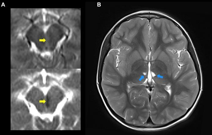
Diagnostic axial MRI brain scans demonstrating acute and chronic ischemic manifestations in a pediatric patient with Deficiency of Adenosine Deaminase 2 (DADA2). Panel A (left) displays an acute infarct in the left paramedial midbrain. The top image is a Diffusion-Weighted Imaging (DWI) sequence showing a focal area of hyperintensity (yellow arrow), and the bottom image is a corresponding Apparent Diffusion Coefficient (ADC) map showing signal hypointensity (yellow arrow), confirming true diffusion restriction characteristic of an acute stroke. Panel B (right) is a T2-weighted axial image at the level of the basal ganglia and thalamus. Blue arrows point to bilateral, small, hyperintense focal lesions in the thalamus, representing chronic lacunar infarcts. The clinical context highlights the tendency of this inflammatory vasculopathy to cause recurrent strokes in deep brain structures, including the midbrain, thalamus, and pons, often at a very young age.
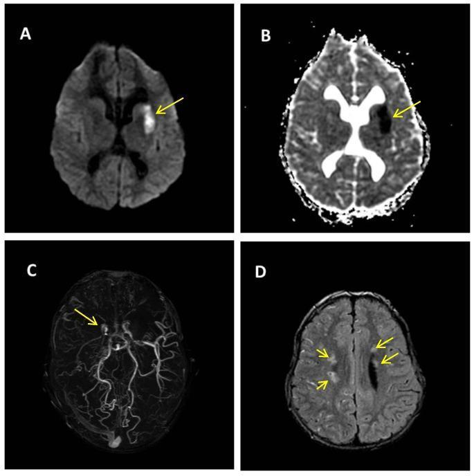
A multi-panel brain MRI demonstrating acute ischemic stroke and vascular stenosis in a pediatric patient. Panel (A) Diffusion-Weighted Imaging (DWI) and Panel (B) Apparent Diffusion Coefficient (ADC) map show an area of restricted diffusion (hyperintense on DWI, hypointense on ADC) in the left lenticular nucleus, anterior limb of the internal capsule, and the head and body of the caudate nucleus, consistent with an acute infarct. Panel (C) Time-of-Flight (TOF) Magnetic Resonance Angiography (MRA) reveals bilateral stenosis of the carotid siphon apices, most prominent on the right, with attenuated flow in the ipsilateral middle cerebral artery (MCA). Panel (D) Fluid-Attenuated Inversion Recovery (FLAIR) sequence demonstrates multiple punctate hyperintense signal alterations in the bilateral semioval centers, representing chronic ischemic changes or small vessel disease. Yellow arrows highlight the acute lesion (A, B), the vascular narrowing (C), and the white matter signal changes (D). The findings are highly suggestive of a progressive vasculopathy such as Moyamoya disease.
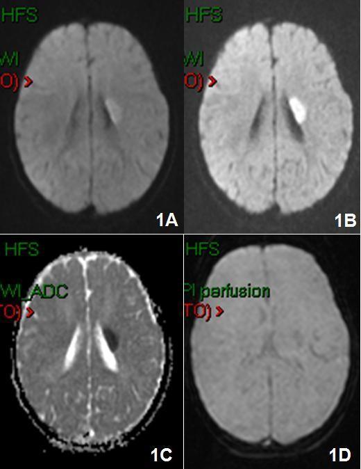
This diagnostic image is a four-panel axial brain MRI composite demonstrating a hyperacute ischemic stroke in a pediatric patient. Panels 1A and 1B show Diffusion-Weighted Imaging (DWI) sequences, highlighting a prominent, hyperintense (bright), ovoid-shaped lesion approximately 2 cm in diameter. The abnormality is localized to the left hemisphere, specifically involving the left lenticular nucleus with extension into the internal capsule. Panel 1C is an Apparent Diffusion Coefficient (ADC) map, which reveals corresponding signal hypointensity in the same region, confirming true restricted diffusion and distinguishing the acute infarct from T2 shine-through. Panel 1D presents a Perfusion-Weighted Imaging (PWI) sequence, showing an area of decreased signal intensity in the left basal ganglia region, indicative of reduced cerebral blood flow. This multi-parametric MRI study effectively illustrates the classic imaging features of early ischemic tissue damage and the associated hemodynamic changes within the deep gray matter and white matter tracts of the brain.
Stroke in Pediatrics
Overview
- Incidence: 2.6 to 13 per 100,000 children per year in the general pediatric population (Greater Cincinnati study: 4.4/100,000/year)
- Neonates are the highest-risk subgroup: arterial ischemic stroke affects 1 in 4000-10,000 neonates; symptomatic ICH affects 1 in 100 full-term neonates
- Overall ratio in children: 50% ischemic, 50% hemorrhagic - but 80% ischemic in perinatal stroke
Classification
1. Arterial Ischemic Stroke (AIS)
2. Hemorrhagic Stroke
3. Cerebral Venous Thrombosis (CVT)
4. Perinatal Stroke
Risk Factors
Cardiac
- Congenital heart defects (most common in childhood AIS)
- Right-to-left shunts, valvular disease
- Cardiomyopathy, endocarditis, arrhythmias
- History of cardiac surgery
Hematologic / Coagulation
- Sickle cell disease - the most important risk factor; sickling causes thrombosis and endothelial injury, sometimes with associated moyamoya syndrome. Children with SCD have stroke rates approaching those of older adults
- Hemophilia (ICH in 7% of infants ≤2 years)
- ITP, TTP, iron deficiency anemia, thrombocytosis
- Inherited thrombophilias: protein C/S deficiency, antithrombin deficiency, factor V Leiden, prothrombin 20210A, MTHFR variants, antiphospholipid antibodies, lipoprotein(a)
Cerebral Arteriopathy
- Moyamoya disease/syndrome - progressive stenosis of distal ICAs with collateral formation; associated with SCD, Down syndrome, NF1
- Focal cerebral arteriopathy (including post-varicella arteriopathy - VZV)
- Arterial dissection (cervico-cephalic, including carotid and vertebral)
- CNS vasculitis
Vascular Malformations
- AVMs (may be part of neurocutaneous syndromes: Sturge-Weber, Osler-Weber-Rendu, Wyburn-Mason, Klippel-Trenaunay)
- Cavernous malformations (can be genetic/familial)
- Intracranial aneurysms (rare; carotid bifurcation most common site in children)
Infections
- Meningitis, encephalitis
- Varicella (post-VZV focal cerebral arteriopathy)
- HIV, TB, syphilis, mastoiditis
Metabolic / Genetic
- Homocystinuria, Fabry disease, organic acidurias
- MELAS (Mitochondrial Encephalomyopathy, Lactic Acidosis, Stroke-like episodes)
- Menkes disease, hyperlipidemia
Drugs / Other
- L-asparaginase (chemotherapy), oral contraceptives, cocaine
- Trauma (including non-accidental)
- Dehydration
Clinical Presentation
| Age Group | Typical Presentation |
|---|---|
| Neonate (perinatal) | Seizures, altered consciousness, feeding difficulties, early hand preference |
| Infant/Young child | Seizures, altered consciousness, hemiparesis (less focal) |
| Older child/Adolescent | More adult-like: hemiparesis, hemianopsia, aphasia, ataxia |
- Seizures at onset occur in ~20% of pediatric stroke cases, especially in children under 6 years
- MCA territory: upper extremity hemiplegia (contralateral)
- ACA territory: lower extremity weakness
- Posterior circulation: ataxia, nystagmus, vertigo, hemiparesis, hemianopsia
- In adults, focal deficits are the rule; in children, headache and seizure are far more common presenting features
Stroke Mimics (Must Exclude)
- Migraine (including hemiplegic migraine)
- Seizure ± postictal (Todd) paralysis
- Functional neurologic disorder
- Intracranial mass lesion
- CNS infection (encephalitis, abscess)
- Drug toxicity (e.g., methotrexate)
- Posterior Reversible Encephalopathy Syndrome (PRES)
- Metabolic abnormality (hypoglycemia, hyperammonemia)
- Demyelinating lesion (ADEM, MS)
Diagnostic Evaluation
Imaging
| Modality | Role |
|---|---|
| Non-contrast CT head | First-line; rules out hemorrhage; may miss ischemia within 24h |
| MRI + DWI | Gold standard for AIS - detects ischemia within hours; DWI bright for ~2 weeks in older children, but may pseudonormalize within days in neonates |
| MRA | Detects vasculopathy (moyamoya, dissection, vasculitis of large vessels) |
| CT angiography/venography | Rapid vascular imaging |
| Transcranial Doppler | Screening for SCD patients; elevated velocities = increased stroke risk (screening should begin in infancy) |
| Conventional angiography | Gold standard for moyamoya, vasculitis, dissection; 6-vessel study required for moyamoya |
| Cranial ultrasound | Useful for neonatal IVH/PVL; <50% sensitivity for arterial stroke in neonates |
Laboratory Workup
- CBC with differential and platelets
- PT/INR, aPTT
- Protein C, protein S, antithrombin levels
- Activated protein C resistance
- Plasminogen, fibrinogen, homocysteine
- Antiphospholipid antibody screen
- Lipoprotein(a), lipid panel
- Genetics: factor V Leiden, prothrombin 20210A, MTHFR
- Hemoglobin electrophoresis (if SCD suspected)
- Inflammatory markers, toxicology screen
- Metabolic screen (if metabolic etiology suspected)
Cardiac Workup
- ECG - arrhythmia, structural abnormality
- Echocardiogram - intracardiac shunts, clots, structural lesions
Management
Acute Stabilization (ABC First)
- Airway, Breathing, Circulation - avoid hypoxemia and hypotension
- Seizure control - anticonvulsants promptly
- Normoglycemia - correct hypo- and hyperglycemia
- Normothermia - aggressive fever management (fever worsens ischemic injury)
- ICP monitoring - especially in hemorrhagic stroke
- Blood pressure: allow permissive hypertension in ischemic stroke (to maintain CPP); in hemorrhagic stroke, reduce BP slowly to avoid worsening CPP
Reperfusion Therapies
- IV tPA (alteplase): Consider within 4.5-hour window in AIS; data limited in children but utilization has increased significantly in the last decade. Requires referral to a stroke center with pediatric expertise
- Mechanical thrombectomy: Consider within 24-hour window; increasingly used in pediatric centers
- Transfer emergently to a tertiary center with pediatric stroke capability
Specific Therapies
| Etiology | Treatment |
|---|---|
| Hemorrhagic stroke / AVM | Neurosurgical intervention - decompression, blood evacuation, AVM resection/embolization |
| Sickle cell disease | Exchange transfusion or simple transfusion (target HbS <30%) - reduces circulating HbS; consult hematology + neurology |
| CVT | Anticoagulation (LMWH or unfractionated heparin, even in the presence of hemorrhagic transformation) |
| AIS (non-SCD) | Aspirin (antithrombotic); anticoagulation for cardioembolic/dissection etiology |
| Moyamoya | Surgical revascularization (indirect: EDAS, pial synangiosis; or direct bypass) |
| VZV arteriopathy | Acyclovir + corticosteroids |
Prevention of Recurrence
- Chronic transfusion program for SCD (most established prevention strategy)
- Antiplatelet therapy (aspirin) for arteriopathy
- Anticoagulation for thrombophilia/cardioembolic
- Risk factor-directed therapy (treat cardiac lesions, thrombophilias)
MRI Examples in Pediatric Stroke
Special Considerations
Sickle Cell Disease
- Highest pediatric stroke risk (~11% lifetime risk of clinical stroke by age 20)
- TCD screening is standard of care starting at age 2; velocities >200 cm/s indicate high stroke risk
- Chronic transfusion reduces recurrence from ~70% to ~10%
- The STOP trial established transfusion as prevention
Neonatal/Perinatal Stroke
- Often presents with seizures in the first days of life; diagnosis confirmed on MRI
- Etiology often unclear; maternal-fetal factors implicated
- DWI may pseudonormalize within days (unlike in older children)
- Long-term outcome varies; many develop hemiplegia, learning difficulties, or epilepsy
Moyamoya Disease
- Progressive occlusion of the distal ICAs with characteristic "puff of smoke" collaterals on angiography
- MRI: diminished flow voids in distal ICA, proximal MCA/ACA; prominent basal ganglia collateral voids
- Management is surgical revascularization; aspirin is used as a bridge
Prognosis & Outcomes
- Mortality: Lower than adults, but morbidity is high (developing brain affected)
- Recurrence: ~20-40% risk overall; highest in arteriopathy, cardiac disease, SCD, and thrombophilia
- Neurologic sequelae: Hemiplegia (most common), cognitive/learning difficulties, epilepsy, behavioral disorders
- Neuroplasticity in younger children can mitigate some deficits, but recovery is incomplete in many
- Bradley and Daroff's Neurology in Clinical Practice, Chapter 68 (pp. 1465-1472)
- ROSEN's Emergency Medicine, Chapter 169 (p. 3296)
- The Harriet Lane Handbook, 23rd ed., Chapter 20 (pp. 724-726)
Neonatal sepsis
"neonatal sepsis" AND management
neonatal sepsis clinical features signs newborn
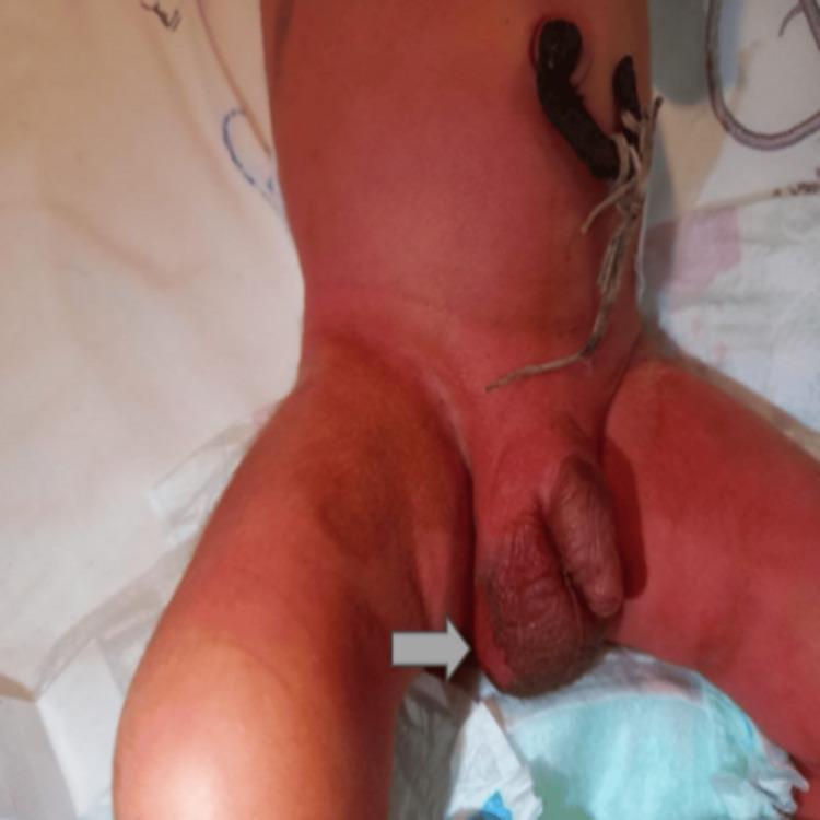
Clinical photograph of a newborn's abdomen and genital region, demonstrating potential portals of entry for neonatal infection. The umbilical cord stump appears dark and thickened, coated with a black traditional medicinal substance (henna mixture). The surrounding periumbilical skin and lower abdomen exhibit diffuse erythema. The scrotum is notably enlarged and edematous, particularly on the right side, which features a well-demarcated, dark, ulcerative lesion (indicated by a white arrow) suggestive of localized necrosis or gangrenous changes. The overall clinical appearance is highly suggestive of neonatal tetanus or omphalitis with secondary scrotal involvement, highlighting the risks associated with non-sterile traditional cord care practices. The image serves as a teaching tool for recognizing signs of neonatal sepsis and inflammatory skin lesions in infants.
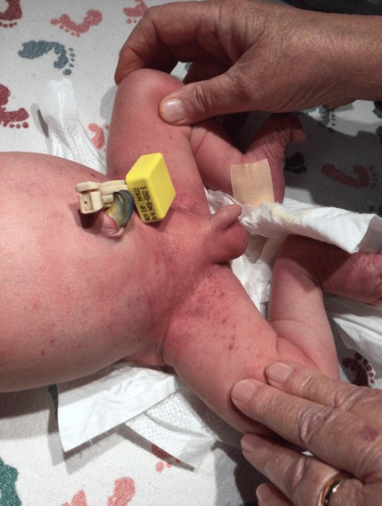
This clinical photograph displays the lower abdomen and perineal region of a newborn infant. A non-blanching petechial rash is visible, characterized by numerous pinpoint, dark red to purple macules. The lesions are most densely concentrated within the inguinal folds and the perineal area, with scattered distribution extending onto the lower torso and medial aspects of the thighs. The infant is positioned on a white clinical diaper featuring a footprint pattern. Key neonatal features include a stump of the umbilical cord secured with a yellow and tan plastic umbilical clamp and a tan adhesive bandage on the right ankle. An examiner's gloved hands are visible, retracting the legs to demonstrate the extent of the eruption in the skin folds. This presentation is significant for neonatal dermatology and hematology, often requiring investigation for congenital infections (TORCH), neonatal alloimmune thrombocytopenia, or sepsis.
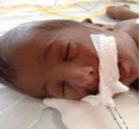
Clinical photograph of a neonate in a supine position, demonstrating dysmorphic facial features and generalized cutaneous findings. The infant exhibits widespread, dark hyperpigmentation across the entire face and scalp. Significant dysmorphic features include a flattened nasal bridge, small and low-set ears, and retrognathia (receding lower jaw). The infant is shown in a clinical setting with medical adhesive and tubing visible near the mouth, indicative of neonatal intensive care. These visual signs, particularly the generalized hyperpigmentation in a newborn, are highly suggestive of primary adrenal insufficiency or congenital adrenal hypoplasia/agenesis, where elevated ACTH levels lead to increased melanin production. This image serves as an educational example of the clinical presentation of neonatal endocrine disorders and associated craniofacial anomalies.
Neonatal Sepsis
Definition
- Fever = rectal temperature ≥38°C (100.4°F)
- Hypothermia = rectal temperature <36.5°C (97.7°F)
- Neonates have ~twice the risk of serious bacterial infection compared with infants aged 4-8 weeks
Classification: Early-Onset vs. Late-Onset
| Feature | Early-Onset Sepsis (EOS) | Late-Onset Sepsis (LOS) |
|---|---|---|
| Timing | First 7 days of life (some use ≤72h in NICU settings) | After 7 days (up to 3 months) |
| Onset | Fulminant, rapid deterioration | More gradual |
| Source | Maternal/perinatal - vaginal flora acquired at/before birth | Environmental - nosocomial (NICU lines, ventilators), community |
| Association | Maternal fever, GBS-positive swabs, prolonged rupture of membranes (PROM), chorioamnionitis, fetal distress | Less associated with maternal risk factors |
| Common complications | Septic shock, neutropenia, pneumonia | Meningitis (more common in LOS) |
| Premature infants | At risk for both types due to immature immune system | VLBW neonates especially vulnerable (catheters, ventilators) |
Etiology / Causative Organisms
Early-Onset Sepsis
- Group B Streptococcus (GBS) / Streptococcus agalactiae - most common cause of EOS and early-onset bacterial meningitis
- Escherichia coli - second most common, especially in preterm
- Listeria monocytogenes - longer incubation, presents as late-onset but can cause EOS; associated with maternal gastroenteritis
- Klebsiella species, Haemophilus influenzae
- Gram-positive cocci (other beta-hemolytic streptococci)
Late-Onset Sepsis
- Coagulase-negative Staphylococci (CoNS) - most common in NICU/VLBW infants (associated with central lines)
- Staphylococcus aureus (including MRSA)
- Gram-negative enteric rods: E. coli, Klebsiella, Pseudomonas, Enterobacter
- Candida species - significant in very premature infants on broad-spectrum antibiotics
- Herpes simplex virus (HSV) - must always consider; can present as late-onset sepsis-like illness
Risk Factors
Maternal / Perinatal
- Prolonged/premature rupture of membranes (PROM >18h)
- Chorioamnionitis (intra-amniotic infection)
- Maternal GBS colonization / GBS bacteriuria
- Maternal fever during labor
- Preterm delivery (<37 weeks)
- Group B Streptococcus-positive vaginal swab
- Previous sibling with invasive GBS disease
- Young maternal age, Black race
- Inadequate intrapartum antibiotic prophylaxis (IAP)
Neonatal
- Prematurity and very low birth weight (VLBW <1500g)
- Invasive procedures: central venous catheters, endotracheal intubation
- Prolonged hospitalization (NICU)
- Male sex
- Galactosemia (predisposes to E. coli sepsis)
- Immune deficiencies
- Skin disruption, omphalitis
Clinical Presentation
Signs and Symptoms of Neonatal Sepsis
| System | Features |
|---|---|
| Temperature | Hypothermia (more common than fever in neonates) or hyperthermia |
| CNS | Lethargy, poor tone, irritability, seizures, bulging fontanelle |
| Respiratory | Apnea, tachypnea, grunting, nasal flaring, retractions |
| Cardiovascular | Poor perfusion, mottling, prolonged CRT, hypotension, tachycardia |
| GI/Feeding | Poor feeding, vomiting, gastric distension, diarrhea |
| Skin | Jaundice (early/prolonged), petechiae, rashes, sclerema |
| Metabolic | Hypoglycemia, metabolic acidosis |
Key clinical pearl: Apnea and bradycardia in a neonate with no obvious respiratory cause must raise suspicion for sepsis. Temperature instability (especially hypothermia) is often the earliest sign - premature neonates especially lose heat rapidly and cannot generate it by shivering (rely on brown fat thermogenesis).
Diagnosis
Blood Culture
- Gold standard - positive culture of blood (or other sterile body fluid)
- Always obtain before starting antibiotics
- Minimum 1 mL blood volume recommended
Complete Blood Count (CBC)
| Finding | Significance |
|---|---|
| Leukopenia | More concerning than leukocytosis in neonates |
| Neutropenia | Bone marrow failure in severe sepsis |
| I/T ratio ≥0.2 | Immature:total neutrophil ratio - sensitive indicator of neonatal sepsis |
| Thrombocytopenia | Associated with sepsis; gram-negative sepsis consumes more platelets (42% vs. 17% gram-positive); severe thrombocytopenia ≤50×10⁹/L = higher mortality |
C-Reactive Protein (CRP)
- High specificity and positive predictive value (both >90%) for neonatal infection
- Most commonly trended biomarker in NICUs
- Normal CRP has negative predictive value >95% (helpful to rule out infection)
- Serial CRP more useful than a single value
Lumbar Puncture (LP) / CSF Analysis
- Lower threshold for LP in neonates than older infants
- Mandatory when meningitis is suspected or before empiric antibiotics in high-risk infants
- Do not defer antibiotics if LP would be delayed or is contraindicated by clinical instability
- Meningitis is more common with LOS than EOS
Other Studies
- Urine culture (catheter specimen) - UTI is a major source of neonatal sepsis
- Blood glucose (hypoglycemia common)
- ABG or VBG (metabolic acidosis, respiratory failure)
- Chest X-ray (pneumonia, air leak syndromes)
- Procalcitonin - rising utility as a biomarker; serial trending useful
- Toxicology screen if relevant
Neonatal Early-Onset Sepsis (EOS) Calculator
- Validated multivariate risk tool (Kaiser Permanente) combining maternal GBS status, gestational age, duration of ROM, intrapartum fever, and infant clinical status
- Provides estimated risk and recommended clinical action (observation vs. labs vs. empiric antibiotics)
Management
1. Supportive Care
- Airway and respiratory support: supplemental O₂, CPAP, or mechanical ventilation for respiratory distress/apnea
- Circulatory support: IV fluid resuscitation (10-20 mL/kg NS bolus) for poor perfusion; vasopressors (dopamine/dobutamine) if fluid-refractory shock
- Glucose control: correct hypoglycemia with D10W bolus + maintenance dextrose; monitor glycemia frequently
- Temperature regulation: radiant warmers, incubators, forced-air heating - avoid hypothermia
- Seizure management: phenobarbital first-line
- Monitor for DIC: especially in gram-negative sepsis (thrombocytopenia, coagulation abnormalities)
2. Empiric Antibiotic Therapy
| Scenario | Regimen | Notes |
|---|---|---|
| EOS (presumed) | Ampicillin 50 mg/kg + Gentamicin 2.5 mg/kg | Covers GBS, Listeria (ampicillin), E. coli and gram-negatives (gentamicin) |
| Suspected gram-negative meningitis | Ampicillin + Cefotaxime or Ceftazidime 50 mg/kg | Better CNS penetration than gentamicin |
| LOS / NICU-acquired | Vancomycin + Gentamicin (or piperacillin-tazobactam) | Covers CoNS, MRSA; broaden gram-negative cover |
| Suspected HSV | Add IV Acyclovir 60 mg/kg/day (÷ q8h) | All ill-appearing neonates; maternal HSV history; CSF with lymphocytes + RBCs |
| Suspected Candida | Add Amphotericin B or Fluconazole | VLBW, prolonged antibiotics, abdominal surgery |
⚠️ AVOID CEFTRIAXONE in neonates - displaces bilirubin from albumin and can cause kernicterus. Use cefotaxime instead.
3. Duration of Therapy
| Infection | Duration |
|---|---|
| Bacteremia without meningitis | 10-14 days |
| Gram-positive meningitis (GBS) | 14-21 days |
| Gram-negative meningitis | 21 days (or minimum 14 days after CSF sterilization) |
| Culture-negative clinical sepsis | 48-72h if cultures negative and clinical improvement; individualize |
4. Specific Organisms
- GBS: Penicillin G or ampicillin (drug of choice)
- Listeria: Ampicillin (+ gentamicin for synergy in meningitis)
- MRSA: Vancomycin
- HSV: Acyclovir (high-dose IV, 60 mg/kg/day)
Prevention
GBS Intrapartum Antibiotic Prophylaxis (IAP)
- GBS-positive vaginal/rectal swab at 35-37 weeks
- GBS bacteriuria in current pregnancy
- Previous infant with invasive GBS disease
- Unknown GBS status with any of: preterm labor <37 wks, ROM >18h, intrapartum fever >38°C
Other Preventive Measures
- Hand hygiene in NICU
- Central line bundle care (CLABSI prevention)
- Minimal invasive procedures in VLBW infants
- Probiotics (some evidence in preterm for reducing LOS)
- Breast milk/colostrum (passive immunity, reduces NEC and infection)
- Fluconazole prophylaxis in VLBW neonates <1000g in high-prevalence NICUs
Complications
| Complication | Details |
|---|---|
| Meningitis | 15-20% of neonatal sepsis cases; more common in LOS; risk of hydrocephalus, deafness, neurodevelopmental impairment |
| Septic shock | Multi-organ failure; DIC; more common in EOS |
| Neutropenia | Bone marrow failure in overwhelming sepsis |
| NEC (necrotizing enterocolitis) | Associated with gram-negative organisms in premature infants |
| Intraventricular hemorrhage | Complication in premature infants with sepsis |
| Death | Mortality 10-30% in VLBW neonates with LOS; higher in gram-negative and fungal sepsis |
Differential Diagnosis (Sepsis Mimics in Neonates)
- Congenital heart disease (ductal-dependent lesions presenting as shock - give prostaglandin E1)
- Inborn errors of metabolism (organic acidemias, urea cycle defects)
- Congenital adrenal hyperplasia (CAH) - salt-wasting crisis
- Intracranial hemorrhage
- Abdominal catastrophe (malrotation/volvulus, NEC)
- TORCH infections (congenital CMV, toxoplasmosis, rubella, HSV)
- Neonatal abstinence syndrome
Recent Evidence (PubMed 2024-2026)
- Tintinalli's Emergency Medicine, Chapter 116 (pp. 773-775)
- Mulholland & Greenfield's Surgery, Chapter 97 (pp. 5301-5302)
- Miller's Anesthesia, 10th ed., Box 75.5
- Robbins Pathologic Basis of Disease - Perinatal Sepsis
- Red Book 2021 (AAP) - Management of Neonates at Risk for GBS Disease
- Creasy & Resnik's Maternal-Fetal Medicine, Chapter 51