How pkdl is spread

Reading File
Searching the Web

PKDL post kala-azar dermal leishmaniasis transmission spread sandfly

Finding Sources
Searching Images

PKDL post kala-azar dermal leishmaniasis skin lesions
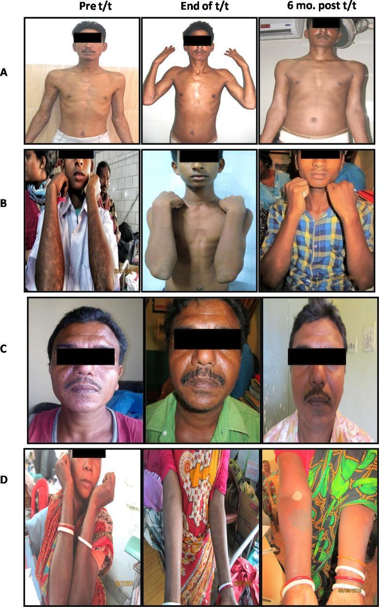
Clinical photograph series illustrating the progression of Post-Kala-Azar Dermal Leishmaniasis (PKDL) in four subjects (A-D) across three temporal stages: baseline (Pre t/t), treatment completion (End of t/t), and six months follow-up (6 mo. post t/t). The visual displays two distinct morphological patterns of PKDL: polymorphic and macular. Subjects A and C demonstrate polymorphic lesions, characterized by various skin manifestations including facial erythema, papules, and nodules. Subjects B and D display macular PKDL, marked by hypopigmented or hyperpigmented macules and patches primarily distributed across the extremities and face. The comparative grid demonstrates the efficacy of Miltefosine (A, B) and Liposomal Amphotericin B (LAmB) (C, D) in reducing lesion density, improving skin tone uniformity, and resolving dermal inflammation. This diagnostic comparison serves to educate on the clinical presentation and therapeutic response of dermal leishmaniasis, a neglected tropical disease. The images focus on the face, torso, and upper limbs to highlight characteristic lesion morphology and their gradual resolution over the follow-up period.
Clinical photograph series illustrating the progression of Post-Kala-Azar Dermal Leishmaniasis (PKDL) in four subjects (A-D) across three temporal stages: baseline (Pre t/t), treatment completion (End of t/t), and six months follow-up (6 mo. post t/t). The visual displays two distinct morphological patterns of PKDL: polymorphic and macular. Subjects A and C demonstrate polymorphic lesions, characterized by various skin manifestations including facial erythema, papules, and nodules. Subjects B and D display macular PKDL, marked by hypopigmented or hyperpigmented macules and patches primarily distributed across the extremities and face. The comparative grid demonstrates the efficacy of Miltefosine (A, B) and Liposomal Amphotericin B (LAmB) (C, D) in reducing lesion density, improving skin tone uniformity, and resolving dermal inflammation. This diagnostic comparison serves to educate on the clinical presentation and therapeutic response of dermal leishmaniasis, a neglected tropical disease. The images focus on the face, torso, and upper limbs to highlight characteristic lesion morphology and their gradual resolution over the follow-up period.

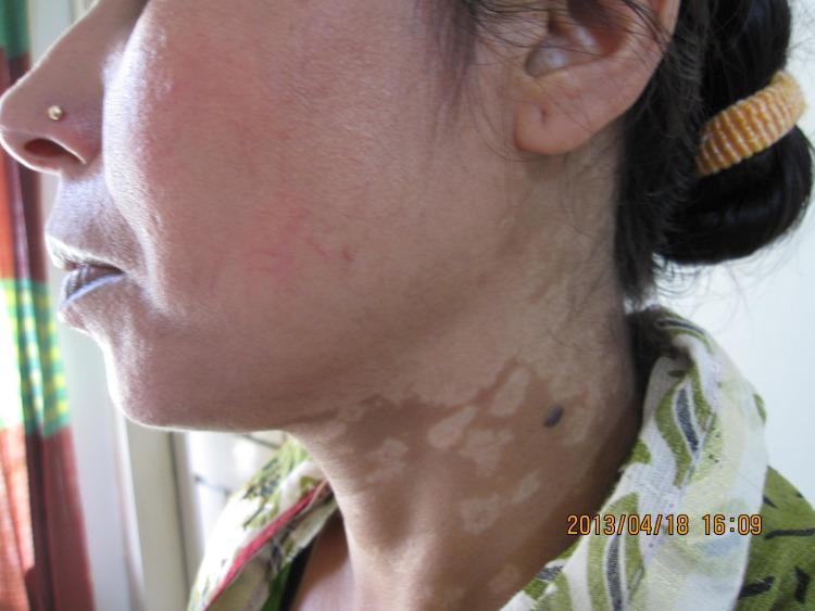
A clinical photograph of the face and neck of a patient from Bangladesh illustrating post-kala-azar dermal leishmaniasis (PKDL). The primary dermatological finding is a widespread, confluent macular rash characterized by hypopigmented to depigmented patches. These pale, irregularly shaped lesions are prominently visible across the cheek and extend inferiorly onto the neck. The lesions vary in size, with some smaller macules coalescing into larger, irregularly bordered areas. Additionally, a single, dark, well-demarcated circular nodule or papule is noted on the lateral aspect of the neck, situated within the hypopigmented region. This visual presentation represents the macular form of PKDL, which is a common cutaneous complication following visceral leishmaniasis (Kala-azar) in Southeast Asian populations. The image serves as a diagnostic reference for identifying characteristic pigmentary changes and distribution patterns of Leishmania donovani-induced skin disease.
A clinical photograph of the face and neck of a patient from Bangladesh illustrating post-kala-azar dermal leishmaniasis (PKDL). The primary dermatological finding is a widespread, confluent macular rash characterized by hypopigmented to depigmented patches. These pale, irregularly shaped lesions are prominently visible across the cheek and extend inferiorly onto the neck. The lesions vary in size, with some smaller macules coalescing into larger, irregularly bordered areas. Additionally, a single, dark, well-demarcated circular nodule or papule is noted on the lateral aspect of the neck, situated within the hypopigmented region. This visual presentation represents the macular form of PKDL, which is a common cutaneous complication following visceral leishmaniasis (Kala-azar) in Southeast Asian populations. The image serves as a diagnostic reference for identifying characteristic pigmentary changes and distribution patterns of Leishmania donovani-induced skin disease.

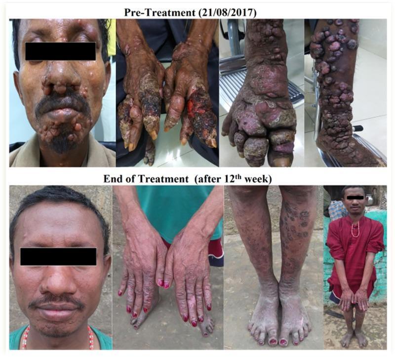
A series of clinical photographs presenting a comparison of a male patient with Post-Kala-azar Dermal Leishmaniasis (PKDL) before and after a 12-week treatment period. The 'Pre-Treatment' row shows extensive dermatological manifestations including multiple nodular lesions concentrated on the face (chin, nose, and cheeks), severely ulcerated and crusting lesions on the dorsal hands and fingers, and densely packed papulo-nodular eruptions on the lower extremities and feet, demonstrating a high disease burden. The 'End of Treatment' row illustrates significant clinical improvement across all anatomical regions. Facial nodules have resolved, leaving smoother skin with residual post-inflammatory hyperpigmentation. The ulcerated lesions on the hands have healed, showing improved skin integrity. The lower limbs exhibit a marked reduction in the size and quantity of papules, with the remaining lesions appearing flattened and less inflamed. This visual documentation serves as an educational tool for identifying the nodular and ulcerative subtypes of PKDL and monitoring the therapeutic response in a tropical medicine context.

A series of clinical photographs presenting a comparison of a male patient with Post-Kala-azar Dermal Leishmaniasis (PKDL) before and after a 12-week treatment period. The 'Pre-Treatment' row shows extensive dermatological manifestations including multiple nodular lesions concentrated on the face (chin, nose, and cheeks), severely ulcerated and crusting lesions on the dorsal hands and fingers, and densely packed papulo-nodular eruptions on the lower extremities and feet, demonstrating a high disease burden. The 'End of Treatment' row illustrates significant clinical improvement across all anatomical regions. Facial nodules have resolved, leaving smoother skin with residual post-inflammatory hyperpigmentation. The ulcerated lesions on the hands have healed, showing improved skin integrity. The lower limbs exhibit a marked reduction in the size and quantity of papules, with the remaining lesions appearing flattened and less inflamed. This visual documentation serves as an educational tool for identifying the nodular and ulcerative subtypes of PKDL and monitoring the therapeutic response in a tropical medicine context.

Your credits are exhausted. You can purchase a top-up or subscribe to a plan to continue using Orris. If you're already subscribed, your daily credits will reset tomorrow.
This is a shared conversation. Sign in to Orris to start your own chat.