
Can we control the position(retrocecal ,pelvic, splenic,etc)of appendix
positions of appendix retrocecal pelvic diagram anatomy
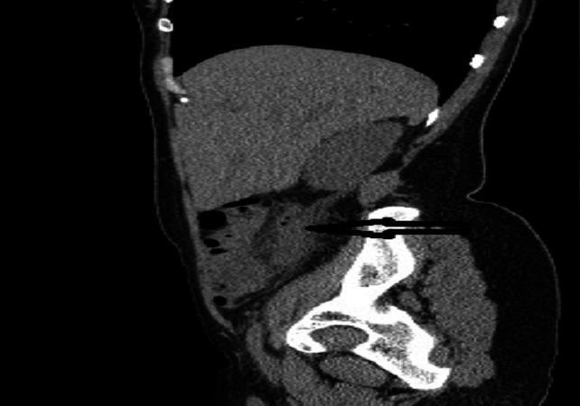
This diagnostic image is a sagittal view of an abdominal-pelvic CT scan (TDM). It demonstrates key radiological findings indicative of acute retrocecal appendicitis. A prominent feature is the distended and thickened vermiform appendix, indicated by a horizontal black arrow. The appendix measures approximately 12 mm in diameter, exceeding the standard threshold for inflammation. It is positioned posteriorly to the cecum, confirming a retrocecal anatomical variant. Furthermore, there is a visible loss of the normal low-density appearance of the surrounding mesenteric fat, characterized as periappendiceal fat stranding or infiltration. This increased attenuation in the fat neighboring the thickened appendix is a secondary sign of localized inflammatory response. The image serves as a clinical example for medical students and radiology residents to identify primary and secondary signs of appendicitis in unconventional anatomical positions using cross-sectional imaging.
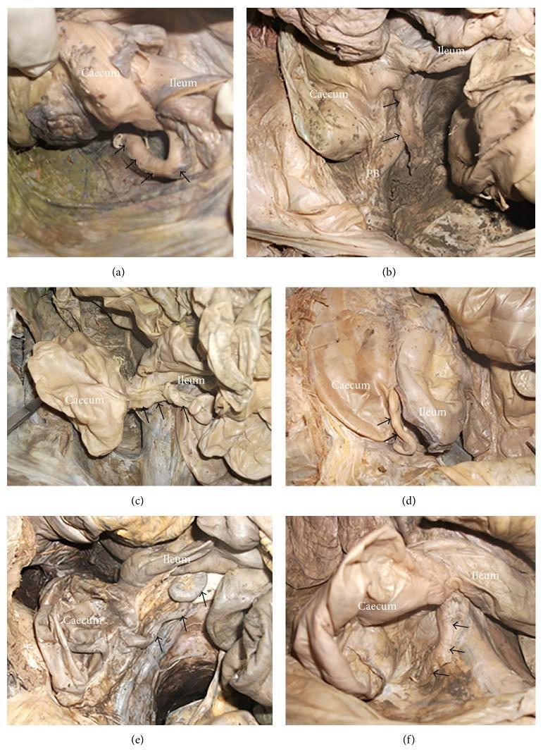
A series of six anatomical photographs (a-f) from human cadaveric dissections illustrating the various anatomical positions of the vermiform appendix relative to the cecum and terminal ileum. Each panel identifies the cecum and ileum as primary landmarks with arrows pointing to the appendix. (a) Retrocecal: the appendix is positioned posterior to the cecum. (b) Pelvic: the appendix descends over the pelvic brim (PB). (c) Preileal: the appendix is situated anterior to the terminal ileum. (d) Subileal: the appendix is located inferior or deep to the ileum. (e) Postileal: the appendix is positioned posterior to the terminal ileum. (f) Subcecal: the appendix is located inferior to the fundus of the cecum. These images provide essential clinical context for understanding surgical variations and diagnostic challenges in appendicitis and lower gastrointestinal procedures.
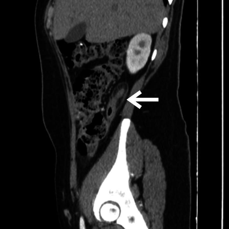
This diagnostic image is a contrast-enhanced computerized tomography (CT) scan of the abdomen and pelvis in a sagittal view. A white arrow points to a dilated and thickened vermiform appendix located in a retrocecal position. The primary visible pathology is appendiceal dilation and wall thickening, which are hallmark radiological signs of acute appendicitis. Additionally, the scan demonstrates subtle increased attenuation and striation within the periappendicular fat, representing inflammatory stranding. Anatomical landmarks include the kidney, liver, and pelvic bones, providing orientation for the right lower quadrant pathology. This image is used as an educational resource to illustrate the diagnostic imaging criteria for appendicitis, specifically emphasizing how sagittal reconstructions help visualize the longitudinal course and surrounding tissue changes of the appendix in complex or retrocecal presentations.
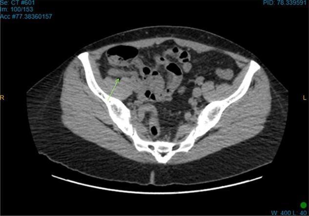
This diagnostic image is an axial computed tomography (CT) scan of the lower abdomen and pelvis, demonstrating diagnostic findings of acute appendicitis. The view captures the cross-sectional anatomy of the iliac bones (bright white, high-density structures) and pelvic musculature. A green arrow points specifically to a thickened, tubular structure in the right iliac fossa, located in a retrocecal position, which is consistent with an inflamed appendix. Pathological features include increased density and a hazy, smudged appearance of the adjacent mesenteric fat, known as fat stranding, which indicates local inflammatory infiltration. Several air-filled bowel loops are visible within the abdominal cavity. This imaging is characteristic of an emergency surgical presentation, used to differentiate between causes of right lower quadrant pain, such as acute appendicitis or appendiceal diverticulitis.
Can We Control the Position of the Appendix?
Embryological Basis (Why We Cannot Control It)
- The degree of cecal descent
- The extent of peritoneal fixation
- The configuration of the cecum
- Appendiceal length
- Associated adhesions
- Body habitus
Known Positions and Their Frequencies
| Position | Frequency | Description |
|---|---|---|
| Retrocecal | ~60-65% (most common) | Posterior to the cecum, intraperitoneal |
| Pelvic (descending) | ~30-31% | Descends over the pelvic brim |
| Subcecal | ~2.3% | Below the fundus of the cecum |
| Pre-ileal | ~1% | Anterior to the terminal ileum |
| Retroileal (postileal) | ~0.4% | Posterior to the terminal ileum |
| Retroperitoneal | ~7% | Posterior to peritoneum |
| Retrocolic | Rare | Posterior to ascending colon |
Why This Matters Clinically
Retrocecal Appendix
- Rigidity is often absent (the gas-distended cecum shields it from anterior pressure)
- Tenderness may be absent anteriorly - called a "silent appendix"
- Deep tenderness present in the loin
- Rigidity of quadratus lumborum
- Psoas sign positive (hip flexion for pain relief, or pain on hyperextension)
Pelvic Appendix
- Complete absence of abdominal rigidity
- Tenderness over McBurney's point often lacking
- Tenderness in the rectovesical pouch or pouch of Douglas on rectal examination
- May cause early diarrhoea (contact with rectum)
- May cause urinary frequency (contact with bladder) - more common in children
- Obturator sign may be positive
Summary
- The position of the appendix is determined at birth by embryological development (gut rotation, cecal descent, peritoneal fixation).
- There is no way - surgical, pharmacological, or otherwise - to alter this position in a living individual (short of appendectomy itself).
- The variation in position is clinically important because it changes the presentation of appendicitis and other appendiceal pathology, making diagnosis harder in atypical positions.