
Lamellar cataract as a thory question and a case
Your credits are exhausted. You can purchase a top-up or subscribe to a plan to continue using Orris. If you're already subscribed, your daily credits will reset tomorrow.
Lamellar cataract as a thory question and a case For pg ophthalmogy From AK khurama Kanski Dukes
Reading File
Finding Sources
Finding Sources
Finding Sources
Finding Sources
Finding Sources
Now I have the key file paths. Let me read the congenital cataract section from Kanski (which will cover lamellar cataract) and the main cataract block, plus search for images.
Reading File
Reading File
Searching Images
lamellar cataract zonular cataract lens opacity
Let me read more of the Kanski congenital cataract section to find the lamellar/zonular cataract content:
Reading File
I need to find the section on types/morphology of congenital cataract within Kanski. Let me search more specifically:
Finding Sources
Finding Sources
Reading File
Finding Sources
Excellent! I found the morphology section. Line 7847 onwards has key content. Let me read more of this section:
Reading File
I now have the Kanski content. Let me also fetch the image referenced (Fig. 10.27D for lamellar cataract) and search for more relevant images:
Finding Sources
Searching Images
congenital cataract morphology types nuclear lamellar polar
Let me look for the actual figure 10.27D image in the textbook blocks:
Finding Sources
Let me find the figure 10.27 in the textbook by reading around the morphology section:
Reading File
I found the figure. Let me fetch that image:
Loading Image
I have all the information I need. Let me now compile the comprehensive answer.---
Lamellar (Zonular) Cataract - PG Ophthalmology
THEORY QUESTION
Definition
Lamellar cataract (also called zonular cataract) is a type of congenital/developmental cataract in which the opacity affects a specific lamella (shell/zone) of the lens, both anteriorly and posteriorly, surrounding a clear nucleus with clear cortex outside. It is the most common type of visually significant congenital cataract.
Synonyms
- Zonular cataract (because it involves a zone/lamella of the lens)
- Perinuclear cataract
Aetiology / Causes
The opacity represents a temporary disturbance of lens metabolism during a critical period of lens fiber formation. Any insult during that specific phase will affect that particular zone.
Hereditary:
- Autosomal dominant (AD) - most common inherited form
- Can occur as an isolated finding
Metabolic causes (most important for PG):
| Cause | Mechanism |
|---|---|
| Hypocalcaemia (hypoparathyroidism, rickets) | Most classic cause of lamellar cataract |
| Galactosaemia | Galactitol accumulation (but more commonly produces "oil-droplet" nuclear opacity) |
| Galactokinase deficiency | Similar mechanism |
| Hypoglycaemia | Neonatal |
| Vitamin D deficiency | Rickets |
| Lowe syndrome | Amino acid metabolism disorder |
Intrauterine infections: Rubella, toxoplasmosis, CMV, varicella
PG High-Yield: Lamellar cataract is classically associated with neonatal tetany due to hypocalcaemia (hypoparathyroidism). The lens zone being laid down at the time of the hypocalcaemic insult becomes opaque.
Morphology / Slit-Lamp Appearance
- Discrete disc-shaped opacity affecting a specific lamella of the lens
- The nucleus within is clear
- The cortex outside is clear
- "Riders" - radial extensions of opacity projecting outward from the equatorial edge of the opacity like spokes; these are pathognomonic of lamellar cataract
- Typically bilateral and symmetrical
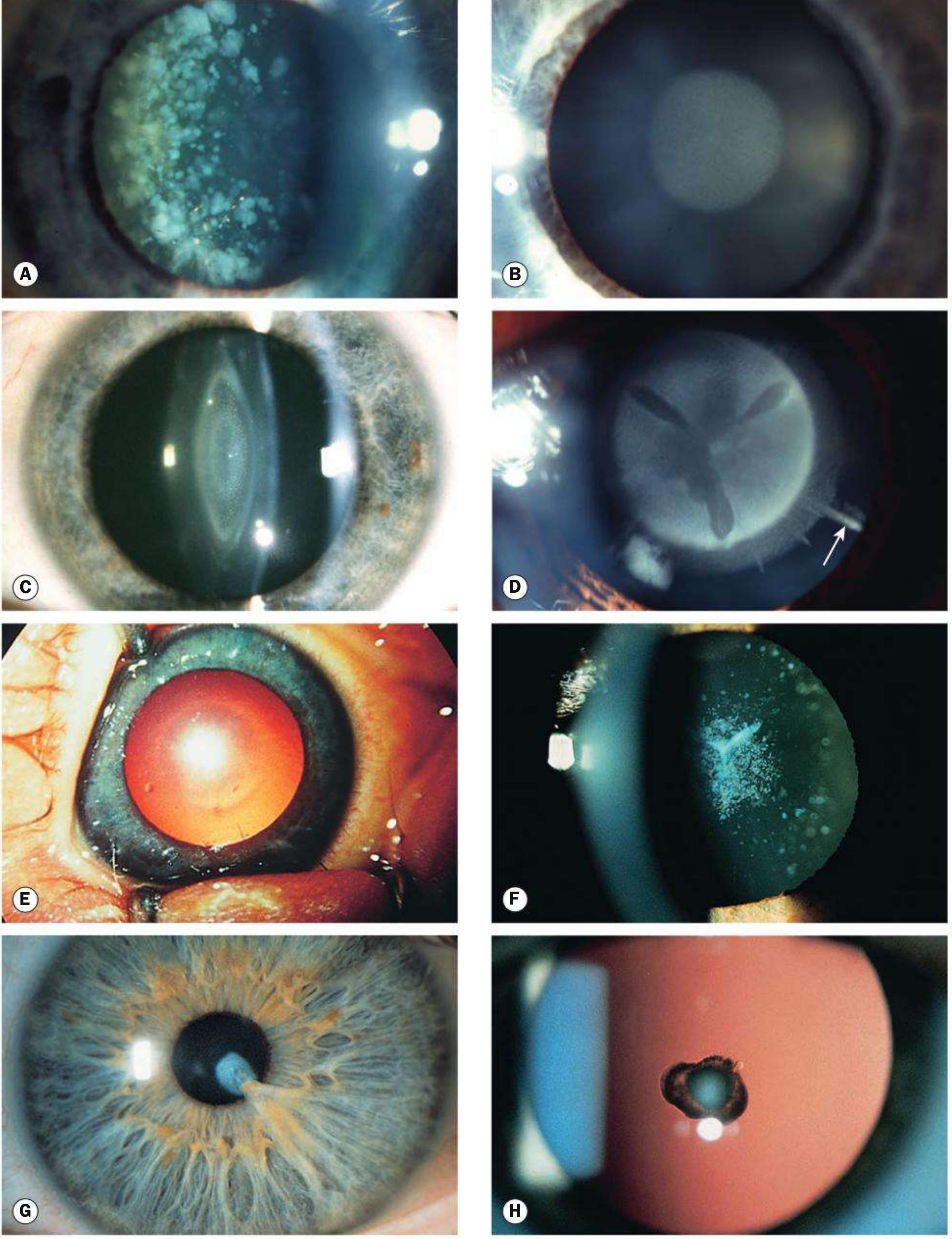
Kanski's Clinical Ophthalmology, 10th ed. - Fig. 10.27. Panel D shows lamellar cataract with riders (arrow).
Visual Significance
- Partially visually significant - the clear nucleus and clear outer cortex allow some vision
- Vision is typically worse in bright light (pupil constricts, the opacity now fills the pupillary aperture) - this is the "paradoxical pupil" phenomenon or photophobia
- Vision is better in dim light (pupil dilates, allowing light through the clear cortex peripherally)
- This behavior is characteristic and clinically important
Genetics
- AD inheritance - most common
- Can also be sporadic (isolated) or secondary to metabolic/systemic disorders
- Gene mutations involved: GJA3 (connexin 46), GJA8 (connexin 50), CRYAA, CRYGD
Differential Diagnosis of Lamellar Cataract Morphology
| Type | Key Feature |
|---|---|
| Lamellar/Zonular | Clear nucleus + clear outer cortex + riders |
| Nuclear | Confined to embryonic/fetal nucleus; dense/pulverulent |
| Coronary (supranuclear) | Deep cortex around nucleus like a crown |
| Anterior polar | At anterior pole, may project into AC |
| Posterior polar | At posterior pole, risk of capsular rupture at surgery |
| Oil-droplet | Central; characteristic of galactosaemia |
| Sutural | Follows Y-suture pattern |
Management
When to operate:
- Bilateral dense cataracts: Surgery between 4-10 weeks of age to prevent deprivation amblyopia
- Bilateral partial (lamellar): May not require surgery urgently; monitor closely. Dilating drops (cyclopentolate/atropine) may help by dilating the pupil to allow vision through clear outer cortex
- Unilateral dense cataract: Surgery within first 6-10 weeks
Surgical approach:
- Lensectomy + anterior vitrectomy (in infants)
- Posterior capsulorhexis + vitrectomy (to prevent posterior capsular opacification, which is nearly universal if the posterior capsule is left intact)
- IOL implantation increasingly performed even in young children
Post-operative:
- Aggressive amblyopia therapy (patching of the fellow eye)
- Optical rehabilitation: contact lenses preferred for unilateral aphakia in infants; spectacles for bilateral aphakia in older children
Systemic workup for suspected metabolic cause:
- Serum calcium, phosphorus, PTH
- Urine reducing substances (after milk feeding) - for galactosaemia
- Urine amino acid chromatography - for Lowe syndrome
- Fasting blood glucose
- TORCH screen (intrauterine infections)
CLINICAL CASE (PG Viva / Long Case Format)
Case Presentation:
A 3-year-old boy is brought by his parents with complaints of bilateral defective vision and squinting since 1 year of age. Parents note the child seems to see better in dim light (e.g., evenings) and worse in bright sunlight. There is no family history of similar complaints. The child had a history of neonatal convulsions on day 3 of life, treated and resolved.
On examination:
- Visual acuity: 6/36 (right), 6/24 (left) by Snellen (age-appropriate testing)
- Convergent squint (esotropia) right eye
- Nystagmus absent
- Pupils: Equal, reactive; both equal in size
- Red reflex: Diminished bilaterally, with a central grey disc-shaped opacity visible
- Slit-lamp examination: Bilateral lens opacities - discrete, disc-shaped zones involving the perinuclear lamella, with clear nucleus centrally and clear cortex peripherally. Radial "rider" extensions noted at the equatorial margins (arrow-like projections). Opacity measures ~4.5 mm in each eye.
- Fundus: Normal (viewed indirectly through dilated pupil past the clear cortex)
Diagnosis: Bilateral Lamellar (Zonular) Cataract
Viva Questions and Model Answers
Q1. What is the most likely diagnosis and how did you arrive at it?
Bilateral lamellar cataract. The slit-lamp shows a discrete disc-shaped opacity in a specific lamella of the lens with clear nucleus inside and clear cortex outside, and the classic radial "rider" extensions at the equator. Bilateral symmetrical involvement, better vision in dim light (when the pupil dilates to expose clear cortex), and the history of neonatal convulsions (suggesting hypocalcaemia) all support this.
Q2. What is the pathognomonic feature?
"Riders" - radial spoke-like extensions at the equatorial margin of the lamellar opacity.
Q3. What is the pathophysiology of this cataract in the context of neonatal hypocalcaemia?
The lens grows by adding new fiber layers (lamellae) around the nucleus throughout life. An insult (e.g., hypocalcaemia) affecting the lens epithelium during a specific developmental period causes opacity limited to the zone of fibers being laid down at that time. The period of insult determines which lamella is affected. Since the insult is transient, only the affected zone is opaque; earlier and later fibers (nucleus and outer cortex) remain clear.
Q4. Why does this child see better in dim light?
The opacity is in a central/perinuclear lamella. In bright light, the pupil constricts and the opacity fills the entire pupillary aperture, blocking vision. In dim light, the pupil dilates, allowing light to pass through the clear cortex peripherally, improving vision. This is a characteristic feature of lamellar cataract and should prompt dilation drops as a temporizing measure.
Q5. What investigations would you order?
- Serum calcium, phosphorus, PTH - to confirm hypocalcaemia/hypoparathyroidism
- Urine reducing substances after milk feeding - to exclude galactosaemia
- TORCH serology - to exclude intrauterine infection
- Urine amino acid chromatography - to exclude Lowe syndrome
- Fasting blood glucose
- Chromosome analysis if dysmorphic features present
Q6. What is the management?
- Partial bilateral lamellar cataracts may be monitored if vision is only mildly affected; mydriatics (atropine drops) help by dilating the pupil
- If visually significant (>3 mm opacity or VA significantly reduced): bilateral lensectomy + posterior capsulorhexis + anterior vitrectomy; IOL implantation considered (age-appropriate target refraction)
- Post-operative: aggressive amblyopia therapy (patching), optical correction (contact lenses/spectacles), and regular follow-up for glaucoma (up to 20% risk by 5 years post-surgery)
- Treat underlying metabolic cause (calcium supplementation for hypoparathyroidism)
Q7. What are complications of cataract surgery in children?
- Posterior capsular opacification (near universal if posterior capsule retained) - hence posterior capsulorhexis + vitrectomy done primarily
- Secondary membrane formation across pupil
- Glaucoma (open-angle in up to 20% by 5 years; angle-closure early post-op due to pupillary block)
- Amblyopia (main long-term threat to vision)
- Retinal detachment (rare, late)
- Lens epithelial proliferation / Soemmering ring
Q8. What is the critical timing for surgery in bilateral dense cataract?
Between 4 and 10 weeks of age. Surgery before 4 weeks risks higher glaucoma incidence; surgery after 10 weeks risks irreversible deprivation amblyopia.
Summary Box (High-Yield PG Points)
| Feature | Detail |
|---|---|
| Most common visually significant congenital cataract | Lamellar/Zonular |
| Pathognomonic sign | Riders (radial equatorial extensions) |
| Classic metabolic cause | Hypocalcaemia (hypoparathyroidism, rickets) |
| Characteristic symptom | Better vision in dim light (worse in bright light) |
| Inheritance | AD (most common hereditary type) |
| Management (bilateral dense) | Surgery 4-10 weeks; posterior capsulorhexis + vitrectomy |
| Key post-op complication | Glaucoma, amblyopia, PCO |
| Temporizing measure | Atropine/cyclopentolate mydriasis |
Sources: Kanski's Clinical Ophthalmology: A Systematic Approach, 10th ed. (pp. 352-354, Fig. 10.27); AK Khurana principles (congenital cataract classification and management); Goldman-Cecil Medicine (congenital cataract overview).
This is a shared conversation. Sign in to Orris to start your own chat.