
Micro practical agar plate example tablewith their use base on 2 nd year mbbs level with agar photo pdf send fast
agar plate culture media microbiology blood agar MacConkey
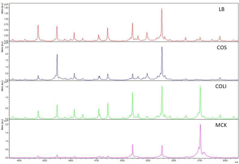
A multi-panel comparison chart of Matrix-Assisted Laser Desorption/Ionization Time-of-Flight (MALDI-TOF) mass spectra used in clinical microbiology. The image displays the spectral profiles of an Escherichia coli (E. coli) isolate grown on four different solid agar media: Luria Broth (LB, red), Columbia agar with sheep blood (COS, blue), Coliformen agar (COLI, green), and MacConkey agar (MCK, pink). The x-axis represents the mass-to-charge ratio (m/z) in the 4000–6000 range, while the y-axis indicates relative intensity. The LB spectrum shows the highest peak density and resolution, serving as the reference for reproducibility. In contrast, the COS, COLI, and MCK spectra demonstrate variations in peak presence and signal intensity, illustrating how different growth media influence the proteomic fingerprint of bacterial isolates. This diagnostic imaging comparison highlights the importance of standardized culture protocols for accurate microbial characterization and subspecies-level identification beyond simple species detection.
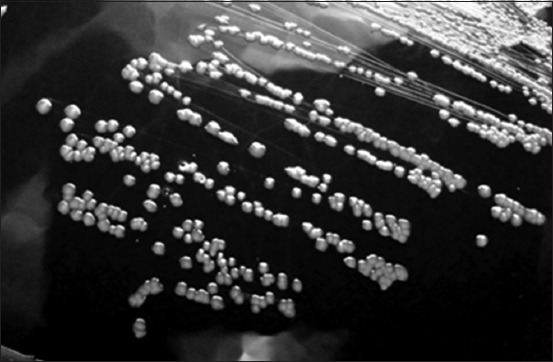
**Modality:** Microbiology culture photograph (grayscale). **Specimen:** *Corynebacterium pseudotuberculosis* (formerly *Corynebacterium ovis*) isolated on blood agar medium. **Morphological Features:** The image displays a streak plate demonstrating isolated bacterial colonies. The colonies are characterized by a distinct circular morphology with an entire margin. They exhibit a convex elevation and appear opaque with a grayish-white coloration against the dark background of the agar. The surface texture of the colonies appears slightly dry or waxy, which is characteristic of this genus. **Distribution:** Streaking pattern shows confluent growth in the primary sectors transitioning to well-defined, discrete individual colonies in the peripheral sectors, allowing for the observation of specific colony macro-morphology. **Diagnostic Context:** These visual features are consistent with the growth characteristics of *Corynebacterium* species, typically found in veterinary clinical microbiology samples. Key differentiating features include the specific convex profile and circular uniformity of the colonies on enriched media.
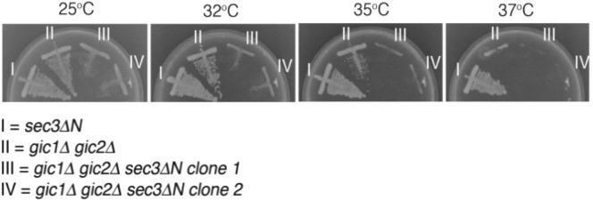
A comparison chart consisting of four clinical microbiology photographs showing yeast culture growth on YPD agar plates at different temperatures: 25°C, 32°C, 35°C, and 37°C. Each plate displays four different strains of Saccharomyces cerevisiae (labeled I-IV) via a streak-plate method to evaluate synthetic growth defects and temperature sensitivity. Strain I (sec3∆N single mutant) shows robust, consistent growth across all temperatures. Strain II (gic1∆ gic2∆ double mutant) demonstrates normal growth at 25°C but displays a progressive reduction in colony density as temperature increases, with minimal viability at 37°C. Strains III and IV (two independent clones of the sec3∆N gic1∆ gic2∆ triple mutant) exhibit a severe synthetic growth defect, characterized by significantly reduced colony density at 25°C and a complete loss of viability at temperatures of 32°C and above. This diagnostic image illustrates the essential parallel roles of Sec3 and Gic proteins in maintaining cell viability and polarized secretion, particularly under thermal stress.
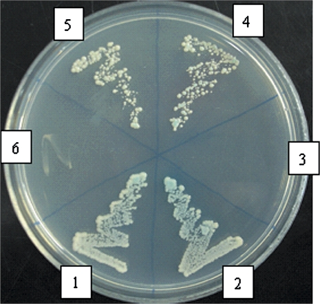
**Imaging Modality:** Laboratory photograph of a microbiology culture plate (Petri dish). **Analysis Method:** Yeast Two-Hybrid (Y2H) assay utilizing a β-galactosidase overlay activity reporter system. **Description:** The image shows a circular agar plate divided into six sectors, labeled 1 through 6, containing *Saccharomyces cerevisiae* colonies. The medium is a selective synthetic dropout agar (SD/-Ade/-His/-Leu/-Trp). **Visual Findings:** - **Positive Protein-Protein Interaction:** Sectors 1, 2, 4, and 5 demonstrate robust yeast growth with a distinct blue chromogenic shift, indicating activation of the *LacZ* reporter gene and successful nutrient prototrophy. Sector 2 serves as the positive control. - **Negative/Lack of Interaction:** Sector 3 (negative control) and Sector 6 show no significant yeast growth or color change, indicating a failure of the protein domains to interact and reconstitute the transcription factor. - **Experimental Significance:** The assay maps the interaction between sspg1d and IPG-1. The positive results in sectors 4 and 5 versus the negative result in sector 6 suggest that the C-terminal domain of IPG-1 is essential for the protein-protein interaction, while the N-terminal deletion does not abolish binding. **Key Features:** Blue colony pigmentation, selective growth patterns, and comparative sectoral analysis for molecular binding validation.

This clinical diagram illustrates a systematic inoculation pattern used in dental microbiology studies. The visual depicts a top-down view of a circular agar culture medium within a Petri dish. A continuous, serpentine, or meandering path is shown spread across the entire surface of the medium, representing the application of dental impression tray adhesive using a cap-attached brush. The path is characterized by a uniform thickness and a textured, coarse stroke appearance, consistent with a brush-applied liquid sample. This illustration demonstrates the standardized method for inoculating culture media to test for cross-contamination of oral microorganisms (bacteria and fungi) between patients via reusable adhesive application brushes. The clinical relevance focuses on infection control and hygiene protocols within prosthetic dentistry and dental laboratory procedures.
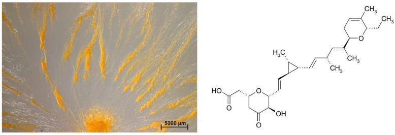
A dual-panel educational image showcasing the microbiological morphology of a myxobacterium and its chemical metabolic profile. The left panel is a clinical photograph of a Sorangium sp. strain Soce 1014 culture on a VY/2-agar plate. It demonstrates a classic swarming growth pattern, with a dense, orange-pigmented central origin and filamentous, radial streaks extending outwards across the translucent agar. These orange multicellular aggregates represent coordinated bacterial movement and high cell density. A 5000 µm scale bar is included for size reference. The right panel displays the skeletal chemical structure of Ambruticin A, a significant secondary metabolite and antifungal agent produced by this strain. The diagram details the molecular architecture including a cyclopropane ring, tetrahydropyran rings, and hydroxyl groups. This visual combination illustrates the relationship between bacterial macroscopic swarming behavior and the production of bioactive natural products, relevant to pharmaceutical microbiology and drug discovery.
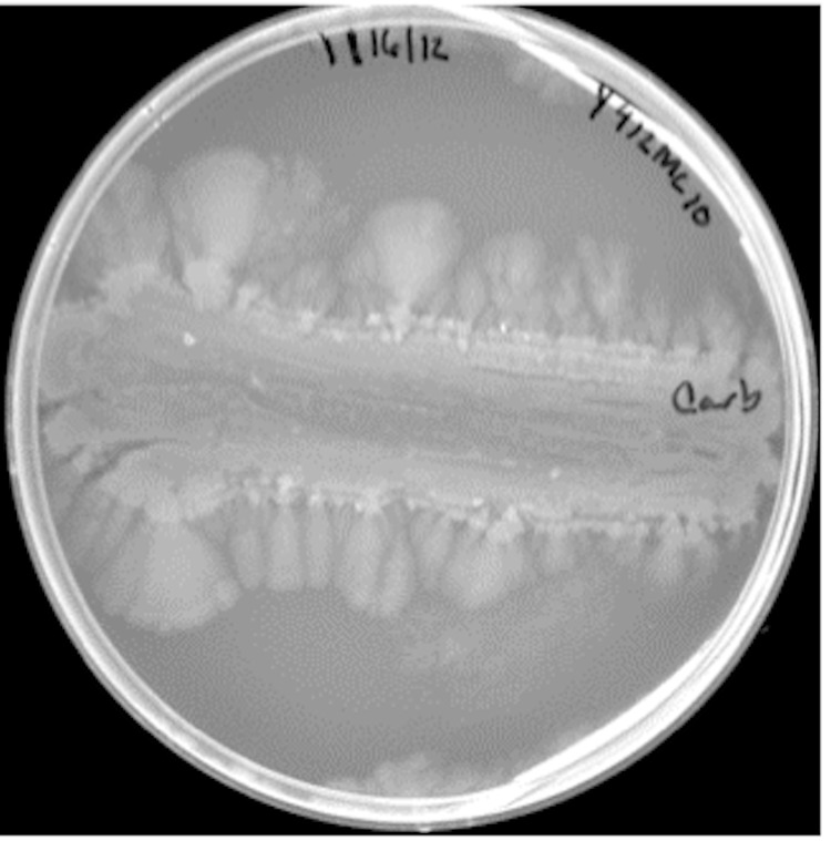
**Modality:** Microbiology culture photograph. **Specimen/Entity:** *Paenibacillus* sp. (strain Y412MC10). **Culture Conditions:** Growth on Yeast Extract-Tryptone (YT) agar medium, following 168 hours of incubation at 37°C. **Morphological Features:** The image displays a macro-view of a Petri dish containing a bacterial culture with a distinctive growth pattern. A horizontal streak serves as the primary inoculation site, showing central clearing or lysis. From the primary streak, there is significant lateral spreading of the culture toward the periphery of the plate. **Growth Pattern:** The organism exhibits characteristic colonial expansion and motility typical of *Paenibacillus* species. The leading edges of the culture demonstrate a dendritic or lobate-like spreading pattern, transitioning from a dense primary inoculum to feathered, translucent fan-like extensions. **Diagnostic Features:** Key visual indicators include high peripheral motility (swarming/spreading) and the progressive thinning or clearing of the central biomass over a prolonged incubation period. These features are critical for identifying motile, pattern-forming Gram-positive bacilli within a laboratory setting.
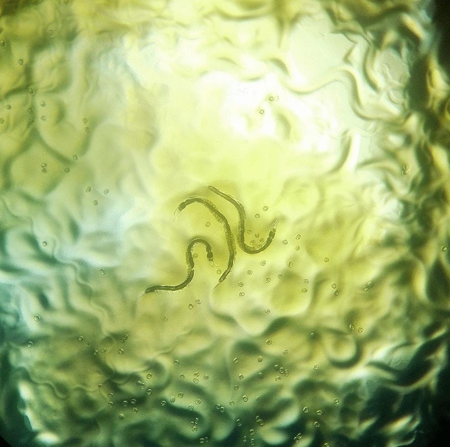
This diagnostic image shows a macroscopic view of an agar plate culture used for the detection of Strongyloides stercoralis, a human pathogenic nematode. The culture medium has a translucent, yellowish-green appearance with a highly irregular, wrinkled surface texture. These surface furrows and grooves represent characteristic 'larval tracks,' which are formed as the motile larvae migrate across the agar surface, often carrying bacteria along their path that then grow into visible trails. Centrally located are two slender, elongated adult worms exhibiting a curved morphology. Scattered throughout the medium are numerous small, circular, translucent structures consistent with parasite ova. This agar plate culture technique is a highly sensitive diagnostic method for identifying strongyloidiasis in clinical stool samples, as the visualization of these tracks and parasite life stages (larvae, adults, and eggs) provides definitive evidence of infection.
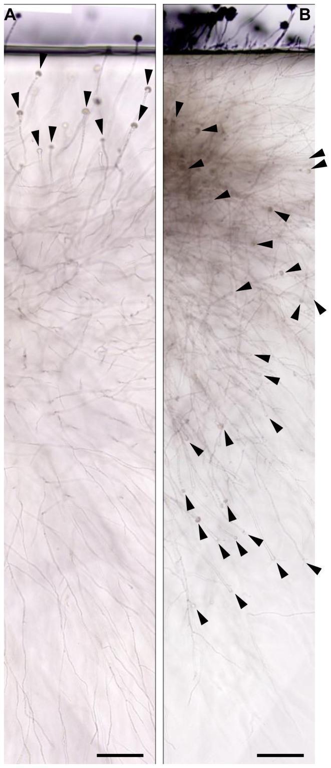
This diagnostic microbiological image demonstrates the oxygen-dependent development of Aspergillus fumigatus conidiophores within a gel-embedded substrate. The image is a comparative study using the sandwiched culture method on glucose minimal agar (GMM) media after 72 hours of incubation. Panel A illustrates fungal colonies grown in ambient air, showing that conidiophores (indicated by black arrowheads) are primarily restricted to the upper agar layer near the agar-air interface. Panel B depicts colonies grown in oxygen-saturated agar, revealing a significantly higher density of conidiophores distributed throughout the colony matrix, including deeper layers. The visual components include translucent vegetative hyphae and distinct, developing conidiophores characterized by stalks and vesicles. In Panel B, these structures appear more developed and abundant, suggesting that oxygen concentration serves as both a prerequisite and a directional cue for conidiogenesis in an embedded environment. Scale bars represent 100 µm. This material is pedagogically relevant for medical mycology and microbiology, illustrating the environmental regulation of fungal morphology and reproductive differentiation.
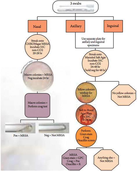
A clinical microbiology flowchart detailing the systematic isolation and identification of Methicillin-resistant Staphylococcus aureus (MRSA) from three anatomical sites: nasal, axillary, and inguinal. The nasal pathway utilizes CHROMagar MRSA, where mauve colonies indicate presumptive MRSA, followed by a confirmatory coagulase plasma test shown in test tubes. The axillary and inguinal pathways use Mannitol Salt Agar (MSA) for initial screening. Yellow colonies on MSA (indicating mannitol fermentation) are subcultured onto Blood Agar Plates for 18-24 hours. Final identification of MRSA requires a battery of tests confirming Gram-positive cocci (GPC) morphology, positive coagulase production, and oxacillin resistance via screening agar. The diagram incorporates photographs of culture plates and biochemical test results to illustrate the diagnostic criteria. This algorithm is designed for laboratory medicine and infectious disease training to demonstrate standard microbial culture protocols and differential media selection based on sample source.
blood agar hemolysis alpha beta gamma colony morphology bacteria
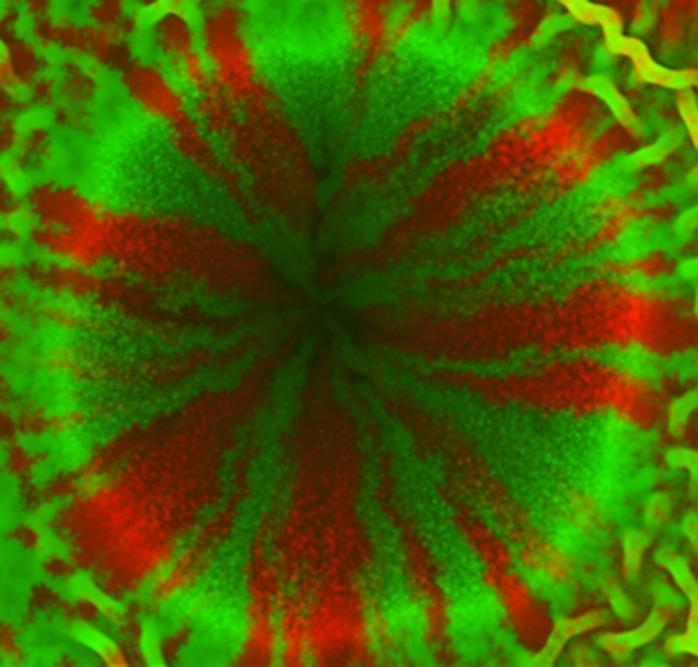
This fluorescence microscopy image demonstrates a time-lapse study of bacterial colony dynamics, specifically highlighting inward-growing growth patterns on an agar surface. Two identical strains of rod-shaped bacteria, likely Escherichia coli or Bacillus subtilis, are visualized using fluorescent protein markers: Red Fluorescent Protein (RFP) and Green Fluorescent Protein (GFP). The image captures an 'aster' structure, characterized by a distinct +1 topological defect. Within the central domain, the bacteria exhibit high radial alignment, forming alternating wedge-like sectors of red and green colonies that converge toward a central dark point devoid of signal. This radial organization is driven by flow-induced alignment and nonlinear velocity profiles occurring during inward growth into a confined circular area. The periphery shows a transition to less ordered, higher-density arrangements with mottled color distributions and filamentous tendrils, reflecting the initial mixed seeding. This visual material is used in microbiology and biophysics education to teach concepts of active matter, bacterial population segregation, and the physical forces governing colony morphology.
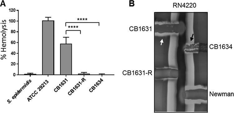
Educational medical graphic illustrating staphylococcal hemolytic activity through quantitative and qualitative assays. Panel A is a bar graph comparing the percentage of rabbit erythrocyte hemolysis among different bacterial strains. S. aureus ATCC 29213 (positive control) shows ~100% hemolysis, while S. epidermidis (negative control) and daptomycin-resistant strains CB1631-R and CB1634 show minimal activity. The daptomycin-susceptible strain CB1631 exhibits intermediate hemolysis (~60%). Statistical significance is indicated by asterisks (****, P < 0.0001). Panel B presents a blood agar plate showing a CAMP-like hemolytic assay. Strains are streaked perpendicular to S. aureus RN4220, which produces a central vertical band of beta-hemolysis. Visible clearing zones at the intersections differentiate toxin types: a white arrow highlights alpha-hemolysis in CB1631, while black arrows indicate delta-hemolysis in CB1631, CB1634, and the Newman strain. CB1631-R shows markedly diminished clearing. This visual demonstrates the attenuation of virulence factors (hemolysins) associated with the development of daptomycin resistance in MRSA clinical isolates.
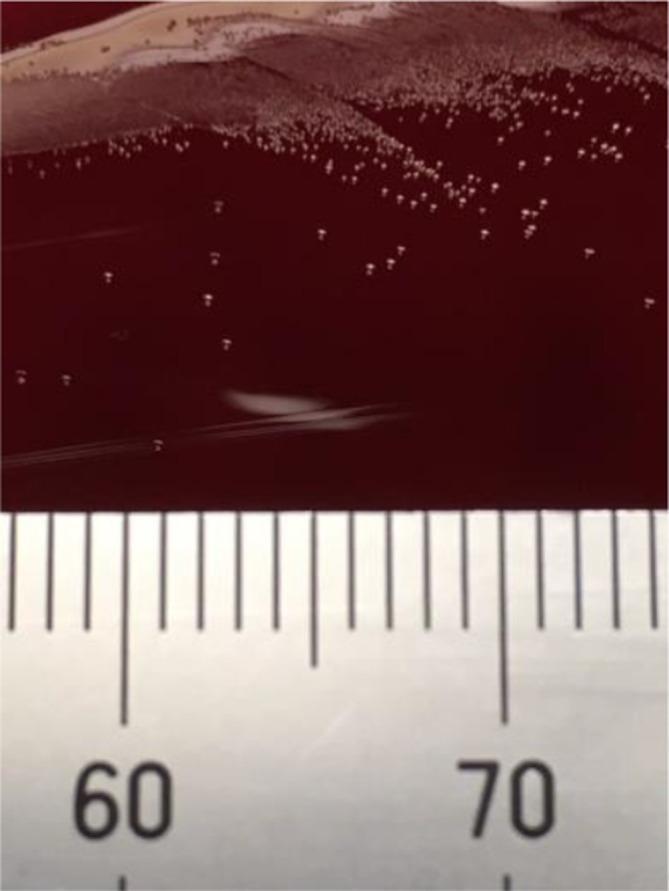
A diagnostic laboratory photograph showing bacterial colonies of Finegoldia sanguinis cultured on a Brucella blood agar plate. The image depicts numerous small, pinpoint, round colonies with a white to off-white appearance, contrasting against the deep red background of the agar medium. The colony distribution exhibits a density gradient: there is a high-density cluster (confluence) in the upper section of the plate, transitioning to more isolated, discrete colonies toward the center, while the lower portion remains relatively clear of growth. Along the bottom edge of the frame, a calibrated metric ruler with millimeter markings (labeled '60' and '70') is included to provide a scale for morphology measurement. This visual demonstrates the typical growth characteristics and colonial morphology of F. sanguinis, an anaerobic gram-positive coccus, which is clinically significant in various infections, including urinary tract and soft tissue involvements.
**Modality:** Microbiology culture photograph (grayscale). **Specimen:** *Corynebacterium pseudotuberculosis* (formerly *Corynebacterium ovis*) isolated on blood agar medium. **Morphological Features:** The image displays a streak plate demonstrating isolated bacterial colonies. The colonies are characterized by a distinct circular morphology with an entire margin. They exhibit a convex elevation and appear opaque with a grayish-white coloration against the dark background of the agar. The surface texture of the colonies appears slightly dry or waxy, which is characteristic of this genus. **Distribution:** Streaking pattern shows confluent growth in the primary sectors transitioning to well-defined, discrete individual colonies in the peripheral sectors, allowing for the observation of specific colony macro-morphology. **Diagnostic Context:** These visual features are consistent with the growth characteristics of *Corynebacterium* species, typically found in veterinary clinical microbiology samples. Key differentiating features include the specific convex profile and circular uniformity of the colonies on enriched media.
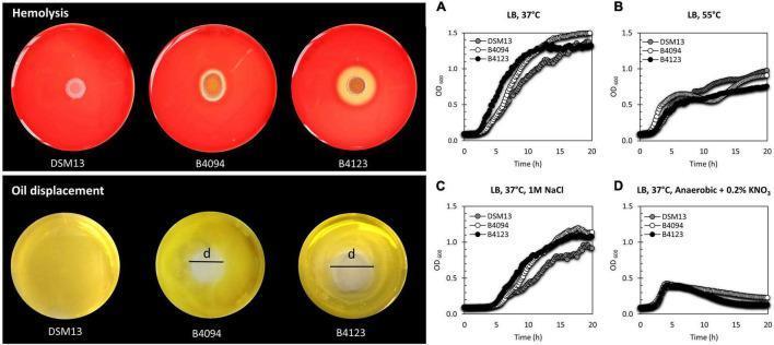
This composite educational graphic illustrates the phenotypic and growth characteristics of three bacterial strains (DSM13T, B4094, and B4123), focusing on biosurfactant production and clinical relevance. Top Left (Hemolysis Assay): Three Columbia blood agar plates demonstrate varying hemolytic activity. DSM13T shows no clearing; B4094 shows a small clearing zone; B4123 displays a large, prominent zone of beta-hemolysis, indicating erythrocyte lysis by the biosurfactant lichenysin. Bottom Left (Oil Displacement Assay): Visual evidence of surfactant activity. DSM13T shows no displacement, whereas B4094 and B4123 show clear displacement zones (labeled 'd'), reflecting their ability to repel oil from the water's surface. Right (Growth Curves): Four line graphs (A-D) plot Optical Density (OD600) over 20 hours under different environmental stressors: (A) standard LB at 37°C, (B) high heat (55°C), (C) high salinity (1M NaCl), and (D) anaerobic conditions with nitrate. The data illustrates how these food isolates adapt to conditions relevant to human infection or food safety pathways.
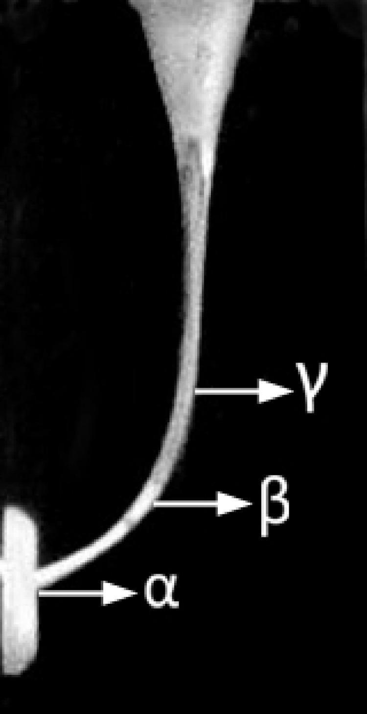
This diagnostic image displays a monochrome view of a simulated root canal structure, likely used for endodontic research or education. The visual demonstrates the morphology of a curved canal, tapering from a wider coronal aspect toward a narrow apical region. Three critical anatomical reference points are identified using Greek letters: Gamma (̸) indicates the most coronal portion of the curvature, Beta (̲) marks the middle section of the canal along the outer curvature, and Alpha (̱) denotes the apical end near the termination point. The canal exhibits a distinct lateral deflection, characteristic of natural root canal anatomy. This material serves as a model for evaluating dental instrumentation techniques, canal preparation efficiency, and the maintenance of canal centering during biomechanical preparation.
MacConkey agar gram negative bacteria selective differential media
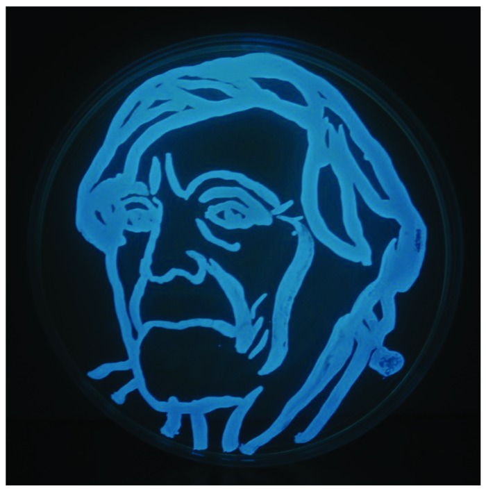
**Imaging Modality:** Microbiological clinical photograph; dark-field bioluminescence imaging. **Specimen and Media:** A circular petri dish containing marine microbiological agar. **Observed Organism:** *Photobacterium leiognathi*, a Gram-negative, bioluminescent bacterium. **Visual Features:** The image displays a controlled inoculation of luminous bacteria across the agar surface, arranged in a deliberate figurative pattern representing a human portrait. The bacteria exhibit intense, intrinsic cyan-blue bioluminescence against a pitch-black background. The growth pattern follows precise, thick-stroked lines with varying opacity, indicating localized concentrations of bacterial colonies. **Characteristic Findings:** The luminescence is self-generated by the bacteria via the luciferase enzyme system, allowing for visualization without external excitation light sources or fluorescent staining. **Clinical/Scientific Context:** This image demonstrates the application of microbial bioluminescence in "agar art." In a clinical or diagnostic context, such bioluminescent properties are key identifying features of certain marine Vibrionaceae and are utilized in molecular biology as reporter genes for gene expression and metabolic activity assays.
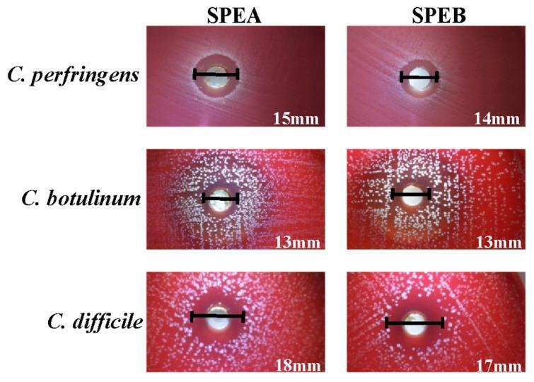
This diagnostic image showcases a series of agar well diffusion assays used for antimicrobial susceptibility testing of three clinically significant Gram-positive anaerobic bacteria: Clostridium perfringens, Clostridium botulinum, and Clostridioides difficile. The image displays a 3x2 grid of Petri dishes with blood agar medium, comparing the efficacy of two saffron petal extracts (SPEA and SPEB). Each panel illustrates an inhibition halo (zone of inhibition) surrounding a central well containing the extract. The diameter of each halo is marked with a black scale bar and measured in millimeters: for C. perfringens, SPEA (15mm) and SPEB (14mm); for C. botulinum, both SPEA and SPEB show 13mm; and for C. difficile, SPEA (18mm) and SPEB (17mm). The background shows white bacterial colonies against the red agar, with varying densities of growth. This comparison demonstrates the differential antibacterial activity of phenolic-rich extracts, where a larger halo indicates greater bacterial sensitivity, a key concept in identifying potential natural treatments for Clostridiaceae family infections.
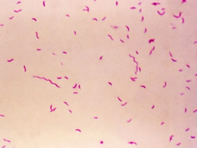
Bright-field light microscopy of a Gram-stained bacterial smear reveals numerous small, slender, curved Gram-negative rods with a characteristic gull-wing appearance consistent with Campylobacter species. The image captures bacteria oriented individually and in small clusters against a pale pink counterstain, illustrating Bacillary morphology typical of enteric pathogens. Gram staining shows purple/blue rods (crystal violet retained by cell wall) with a light pink background from the counterstain (safranin), enabling discrimination from Gram-positive organisms. The organisms appear slender and curved, with a single polar flagellum suggested by motility or alignment in the smear. Specimen type is a bacterial smear obtained from a gastrointestinal sample (feces or culture isolate), prepared for diagnostic microbiology. The imaging modality is bright-field microscopy at high magnification (approximately 1000x with oil immersion), following Gram staining to highlight bacterial cell wall structure. Clinically, detection of Campylobacter species supports infectious gastroenteritis; in the IPSID (immunoproliferative small intestinal disease) context, Campylobacter involvement has been proposed as a pathogenic trigger mirroring Helicobacter pylori's role in gastric MALT lymphoma. Differential diagnoses include Helicobacter, Vibrio, and other curved Gram-negative bacteria. This image serves educational and diagnostic utility for microbiology, clinical pathology, gastroenterology, and infectious disease research.
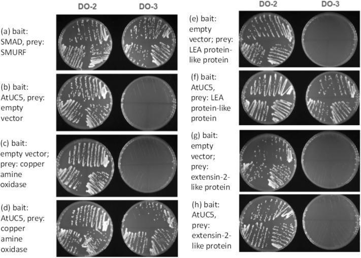
This composite diagnostic image displays results from a pairwise yeast two-hybrid (Y2H) analysis, a common molecular biology technique used to investigate protein-protein interactions (PPIs). The panel compares yeast growth on two different selective agar media: DO-2 (lacking tryptophan and leucine, selecting for the presence of both bait and prey plasmids) and DO-3 (lacking tryptophan, leucine, and histidine, selecting for a physical interaction between bait and prey proteins). Results are organized into rows (a-h). Row (a) serves as a positive control using the human TGF-β/Smad pathway proteins SMAD and SMURF, showing robust growth on both media. Rows (b, c, e, g) serve as negative controls using empty vectors, demonstrating growth on DO-2 but no growth on DO-3. Rows (d, f, h) test the interaction between the plant protein AtUC5 (uclacyanin 5) and various stress-related proteins. Significant growth on DO-3 in rows (d) and (f) indicates positive protein-protein interactions between AtUC5 and copper amine oxidase, as well as LEA protein-like protein. Row (h) shows minimal growth on DO-3, indicating a lack of significant interaction with extensin-2-like protein.
mkdir -p /tmp/workspace/agar-plates && ls /tmp/workspace/agar-plates/
~/agar-plates/generate_pdf.py
#!/usr/bin/env python3
"""Generate a comprehensive Microbiology Agar Plates PDF for 2nd Year MBBS students."""
import urllib.request
import os
from reportlab.lib.pagesizes import A4
from reportlab.lib import colors
from reportlab.lib.units import cm, mm
from reportlab.platypus import (
SimpleDocTemplate, Table, TableStyle, Paragraph, Spacer,
Image, PageBreak, HRFlowable, KeepTogether
)
from reportlab.lib.styles import getSampleStyleSheet, ParagraphStyle
from reportlab.lib.enums import TA_CENTER, TA_LEFT, TA_JUSTIFY
from reportlab.platypus import FrameBreak
from io import BytesIO
# ── Download images ──────────────────────────────────────────────────────────
IMG_DIR = "/tmp/workspace/agar-plates/imgs"
os.makedirs(IMG_DIR, exist_ok=True)
image_urls = {
"mrsa_flowchart": "https://cdn.orris.care/cdss_images/pmc_clinical_VQA_e99f60f6425c105d498ba9923e52984a16d8c0942eb1dcf5b2ac681c34943399.jpg",
"hemolysis": "https://cdn.orris.care/cdss_images/pmc_clinical_VQA_ed2c87110cd718e73b2aba82cd117a3896d97fbdd52a0c2cdf5dfb8726045b82.jpg",
}
downloaded = {}
for name, url in image_urls.items():
dest = os.path.join(IMG_DIR, f"{name}.jpg")
try:
req = urllib.request.Request(url, headers={"User-Agent": "Mozilla/5.0"})
with urllib.request.urlopen(req, timeout=15) as r:
with open(dest, "wb") as f:
f.write(r.read())
downloaded[name] = dest
print(f"Downloaded: {name}")
except Exception as e:
print(f"Failed {name}: {e}")
# ── Document setup ────────────────────────────────────────────────────────────
OUTPUT = "/tmp/workspace/agar-plates/Microbiology_Agar_Plates_2ndYr_MBBS.pdf"
doc = SimpleDocTemplate(
OUTPUT,
pagesize=A4,
rightMargin=1.5*cm, leftMargin=1.5*cm,
topMargin=2*cm, bottomMargin=2*cm,
title="Microbiology Agar Plates – 2nd Year MBBS",
author="Orris Medical Education"
)
W, H = A4
PAGE_W = W - 3*cm # usable width
# ── Styles ────────────────────────────────────────────────────────────────────
base_styles = getSampleStyleSheet()
TITLE_STYLE = ParagraphStyle(
"Title", parent=base_styles["Title"],
fontSize=20, textColor=colors.HexColor("#1a237e"),
spaceAfter=6, leading=26, alignment=TA_CENTER
)
SUBTITLE_STYLE = ParagraphStyle(
"Subtitle", parent=base_styles["Normal"],
fontSize=11, textColor=colors.HexColor("#5c6bc0"),
spaceAfter=4, alignment=TA_CENTER
)
SECTION_STYLE = ParagraphStyle(
"Section", parent=base_styles["Heading2"],
fontSize=13, textColor=colors.white,
backColor=colors.HexColor("#1565c0"),
spaceBefore=12, spaceAfter=4, leading=18,
leftIndent=4, rightIndent=4, borderPad=4
)
SUBSECTION_STYLE = ParagraphStyle(
"SubSection", parent=base_styles["Heading3"],
fontSize=11, textColor=colors.HexColor("#0d47a1"),
spaceBefore=8, spaceAfter=3, leading=15
)
BODY_STYLE = ParagraphStyle(
"Body", parent=base_styles["Normal"],
fontSize=9.5, leading=14, spaceAfter=4,
alignment=TA_JUSTIFY
)
CAPTION_STYLE = ParagraphStyle(
"Caption", parent=base_styles["Normal"],
fontSize=8, textColor=colors.HexColor("#546e7a"),
alignment=TA_CENTER, italics=True, leading=11
)
FOOTER_STYLE = ParagraphStyle(
"Footer", parent=base_styles["Normal"],
fontSize=7.5, textColor=colors.HexColor("#78909c"),
alignment=TA_CENTER
)
HEADER_CELL = ParagraphStyle(
"HeaderCell", parent=base_styles["Normal"],
fontSize=9, textColor=colors.white, alignment=TA_CENTER,
fontName="Helvetica-Bold", leading=12
)
CELL_STYLE = ParagraphStyle(
"CellStyle", parent=base_styles["Normal"],
fontSize=8.5, leading=12, alignment=TA_LEFT
)
KEY_STYLE = ParagraphStyle(
"KeyPoint", parent=base_styles["Normal"],
fontSize=9, leading=13, leftIndent=10,
textColor=colors.HexColor("#1a237e")
)
# ── Helper colours ────────────────────────────────────────────────────────────
HDR = colors.HexColor("#1565c0")
ROW1 = colors.HexColor("#e3f2fd")
ROW2 = colors.white
DARK = colors.HexColor("#0d47a1")
GOLD = colors.HexColor("#f57f17")
RED = colors.HexColor("#b71c1c")
GRN = colors.HexColor("#1b5e20")
def section_header(text):
return Paragraph(f" {text}", SECTION_STYLE)
def hline():
return HRFlowable(width="100%", thickness=0.5, color=colors.HexColor("#90caf9"), spaceAfter=6)
# ── Content ───────────────────────────────────────────────────────────────────
story = []
# ── COVER ─────────────────────────────────────────────────────────────────────
story.append(Spacer(1, 1.5*cm))
story.append(Paragraph("🧫 PRACTICAL MICROBIOLOGY", SUBTITLE_STYLE))
story.append(Paragraph("Agar Plates & Culture Media", TITLE_STYLE))
story.append(Paragraph("2nd Year MBBS – Quick Reference Guide", SUBTITLE_STYLE))
story.append(Spacer(1, 0.3*cm))
story.append(hline())
story.append(Paragraph(
"Based on: <i>Jawetz, Melnick & Adelberg's Medical Microbiology 28e</i> | "
"<i>Sherris & Ryan's Medical Microbiology 8e</i>",
CAPTION_STYLE
))
story.append(Spacer(1, 0.5*cm))
# ── INTRO ─────────────────────────────────────────────────────────────────────
story.append(section_header("1. Introduction to Culture Media"))
story.append(Spacer(1, 0.3*cm))
intro_text = (
"Culture media are nutrient preparations used to grow microorganisms outside the living body (in vitro). "
"For diagnostic bacteriology, several types of media are used in combination because the possible organisms "
"include aerobic, facultatively anaerobic, and obligately anaerobic bacteria. "
"Media are broadly classified by their physical state (liquid/solid), chemical composition, "
"and functional properties (selective, differential, enriched, transport)."
)
story.append(Paragraph(intro_text, BODY_STYLE))
story.append(Spacer(1, 0.3*cm))
# ── CLASSIFICATION TABLE ──────────────────────────────────────────────────────
story.append(section_header("2. Classification of Culture Media"))
story.append(Spacer(1, 0.3*cm))
classif_data = [
[Paragraph("Type", HEADER_CELL),
Paragraph("Definition", HEADER_CELL),
Paragraph("Examples", HEADER_CELL)],
[Paragraph("<b>Simple / Basal</b>", CELL_STYLE),
Paragraph("Supports growth of non-fastidious organisms; no special additives", CELL_STYLE),
Paragraph("Nutrient agar, Peptone water", CELL_STYLE)],
[Paragraph("<b>Enriched</b>", CELL_STYLE),
Paragraph("Contains extra nutrients (blood, serum) for fastidious organisms", CELL_STYLE),
Paragraph("Blood agar, Chocolate agar, Serum agar", CELL_STYLE)],
[Paragraph("<b>Selective</b>", CELL_STYLE),
Paragraph("Contains inhibitory agents that suppress unwanted organisms; favours target organism", CELL_STYLE),
Paragraph("MacConkey, TCBS, Lowenstein-Jensen, Sabouraud, Thayer-Martin", CELL_STYLE)],
[Paragraph("<b>Differential (Indicator)</b>", CELL_STYLE),
Paragraph("Contains pH indicator or substrate allowing visual differentiation of colonies", CELL_STYLE),
Paragraph("Blood agar (hemolysis), MacConkey (lactose fermentation), CLED", CELL_STYLE)],
[Paragraph("<b>Selective + Differential</b>", CELL_STYLE),
Paragraph("Both selective and differential properties in same medium", CELL_STYLE),
Paragraph("MacConkey, XLD, Hektoen enteric, TCBS", CELL_STYLE)],
[Paragraph("<b>Transport</b>", CELL_STYLE),
Paragraph("Maintains viability of organism during transport; no multiplication intended", CELL_STYLE),
Paragraph("Stuart's, Amies, Cary-Blair, Venkatraman-Ramakrishnan (VR)", CELL_STYLE)],
[Paragraph("<b>Enrichment broth</b>", CELL_STYLE),
Paragraph("Liquid medium that selectively enriches/amplifies target organism", CELL_STYLE),
Paragraph("Selenite F broth (Salmonella), Alkaline peptone water (Vibrio), GN broth", CELL_STYLE)],
]
classif_col_widths = [3.5*cm, 7.5*cm, 6.5*cm]
classif_table = Table(classif_data, colWidths=classif_col_widths, repeatRows=1)
classif_table.setStyle(TableStyle([
("BACKGROUND", (0,0), (-1,0), HDR),
("TEXTCOLOR", (0,0), (-1,0), colors.white),
("ROWBACKGROUNDS", (0,1), (-1,-1), [ROW1, ROW2]),
("BOX", (0,0), (-1,-1), 0.8, colors.HexColor("#1565c0")),
("INNERGRID", (0,0), (-1,-1), 0.4, colors.HexColor("#90caf9")),
("VALIGN", (0,0), (-1,-1), "TOP"),
("TOPPADDING", (0,0), (-1,-1), 5),
("BOTTOMPADDING", (0,0), (-1,-1), 5),
("LEFTPADDING", (0,0), (-1,-1), 5),
]))
story.append(classif_table)
story.append(Spacer(1, 0.5*cm))
# ── MAIN AGAR PLATE TABLE ─────────────────────────────────────────────────────
story.append(section_header("3. Common Agar Plates – Practical Reference Table"))
story.append(Spacer(1, 0.3*cm))
# Column headers
main_hdr = [
Paragraph("Agar / Medium", HEADER_CELL),
Paragraph("Type", HEADER_CELL),
Paragraph("Composition\n(Key Ingredients)", HEADER_CELL),
Paragraph("Uses / Organisms Grown", HEADER_CELL),
Paragraph("Colony Appearance", HEADER_CELL),
Paragraph("Mnemonics / Key Points", HEADER_CELL),
]
# Rows
rows = [
# ── GENERAL PURPOSE ──────────────────────────────────────────
[Paragraph("<b>Nutrient Agar</b>", CELL_STYLE),
Paragraph("Simple/Basal", CELL_STYLE),
Paragraph("Beef extract, peptone, agar, water (pH 7.4)", CELL_STYLE),
Paragraph("Routine lab work; non-fastidious bacteria (E. coli, Staphylococcus, Bacillus); NOT for fastidious organisms", CELL_STYLE),
Paragraph("Pale/cream colonies; no differentiation", CELL_STYLE),
Paragraph("'No frills' medium – for teaching and maintenance", CELL_STYLE)],
[Paragraph("<b>Blood Agar (BA)</b>\n5% sheep blood", CELL_STYLE),
Paragraph("Enriched + Differential", CELL_STYLE),
Paragraph("Nutrient agar + 5–10% defibrinated sheep/horse blood", CELL_STYLE),
Paragraph(
"• MOST BACTERIA and fungi\n"
"• Streptococcus spp., Staphylococcus spp.\n"
"• Pneumococcus, Listeria\n"
"• Standard medium for clinical specimens",
CELL_STYLE),
Paragraph(
"<b>α-hemolysis:</b> Green haze (S. pneumoniae, viridans strep)\n"
"<b>β-hemolysis:</b> Clear zone (S. pyogenes, S. aureus)\n"
"<b>γ-hemolysis:</b> No change (Enterococcus)",
CELL_STYLE),
Paragraph("BLOOD = Broad, Lytic, Or Other Detection\nDont forget: CAMPY needs 42°C on BA", CELL_STYLE)],
[Paragraph("<b>Chocolate Agar</b>\n(Heated blood agar)", CELL_STYLE),
Paragraph("Enriched\n(Not selective)", CELL_STYLE),
Paragraph("Blood heated to 80°C → cells lyse → hemin (X factor) + NAD (V factor) released; brown colour", CELL_STYLE),
Paragraph(
"• FASTIDIOUS organisms:\n"
"• Haemophilus influenzae (needs X+V)\n"
"• Neisseria gonorrhoeae\n"
"• Neisseria meningitidis\n"
"• Moraxella catarrhalis",
CELL_STYLE),
Paragraph("Grey-brown colonies; no hemolysis visible\n(cells already lysed)", CELL_STYLE),
Paragraph("Chocolate = X + V factors released\n'H & N love Chocolate': Haemophilus & Neisseria", CELL_STYLE)],
# ── SELECTIVE ─────────────────────────────────────────────────
[Paragraph("<b>MacConkey Agar</b>\n(MAC)", CELL_STYLE),
Paragraph("Selective +\nDifferential", CELL_STYLE),
Paragraph("Peptone, bile salts, crystal violet, lactose, neutral red pH indicator", CELL_STYLE),
Paragraph(
"• Gram-NEGATIVE rods only\n"
"• Selective: bile salts + crystal violet inhibit G+ve\n"
"• Enterobacteriaceae, Pseudomonas\n"
"• UTI screen, stool culture",
CELL_STYLE),
Paragraph(
"<b>Lactose fermenters (LF):</b>\nPink-red colonies\n(E. coli, Klebsiella, Enterobacter)\n\n"
"<b>Non-LF (NLF):</b>\nPale/colourless\n(Salmonella, Shigella, Proteus, Pseudomonas)",
CELL_STYLE),
Paragraph("MAC = Mnemonic: Macrophages Are Coloured\nPink = ferments lactose\nColourless = doesn't ferment", CELL_STYLE)],
[Paragraph("<b>Thayer-Martin Agar</b>\n(Selective Chocolate)", CELL_STYLE),
Paragraph("Selective\n(Enriched base)", CELL_STYLE),
Paragraph("Chocolate agar + VCAT antibiotics:\nVancomycin, Colistin, Amphotericin, Trimethoprim", CELL_STYLE),
Paragraph(
"• Neisseria gonorrhoeae (STI)\n"
"• Neisseria meningitidis\n"
"• Used for genital/CSF specimens",
CELL_STYLE),
Paragraph("Small, oxidase-positive grey colonies\nOxidase test +ve (turns purple)", CELL_STYLE),
Paragraph("VCAT = Vancomycin Colistin Amphotericin Trimethoprim\n'Neisseria Loves VCA'", CELL_STYLE)],
[Paragraph("<b>Hektoen Enteric\nAgar (HEA)</b>", CELL_STYLE),
Paragraph("Selective +\nDifferential\n(Highly selective)", CELL_STYLE),
Paragraph("Bile salts, thiosulfate, citrate salts, carbohydrates, pH indicator (acid-fuchsin + bromothymol blue)", CELL_STYLE),
Paragraph(
"• Salmonella and Shigella from stool\n"
"• Inhibits coliforms 1000–10,000-fold\n"
"• Best for enteric pathogen isolation",
CELL_STYLE),
Paragraph(
"<b>Salmonella:</b> Blue-green + black centre (H₂S)\n"
"<b>Shigella:</b> Green, no black centre\n"
"<b>Coliforms:</b> Orange (inhibited)",
CELL_STYLE),
Paragraph("'Hektoen = Salmonella + Shigella Spotter'\nBlack = H₂S = Salmonella", CELL_STYLE)],
[Paragraph("<b>XLD Agar</b>\n(Xylose Lysine\nDeoxycholate)", CELL_STYLE),
Paragraph("Selective +\nDifferential", CELL_STYLE),
Paragraph("Xylose, lysine, deoxycholate, sodium thiosulfate, ferric ammonium citrate, phenol red indicator", CELL_STYLE),
Paragraph(
"• Salmonella (primary use)\n"
"• Shigella\n"
"• Enteric pathogens in stool",
CELL_STYLE),
Paragraph(
"<b>Salmonella:</b> Pink-red + black centre\n"
"<b>Shigella:</b> Red/pink, no black\n"
"<b>Coliforms:</b> Yellow (ferment xylose)",
CELL_STYLE),
Paragraph("XLD = eXtra selection for Listeria & Dysentery organisms\nBlack dot = H₂S producer = Salmonella", CELL_STYLE)],
[Paragraph("<b>TCBS Agar</b>\n(Thiosulfate Citrate\nBile Sucrose)", CELL_STYLE),
Paragraph("Selective +\nDifferential", CELL_STYLE),
Paragraph("Thiosulfate, citrate, bile salts, sucrose, bromothymol blue + thymol blue indicators", CELL_STYLE),
Paragraph(
"• VIBRIO species ONLY\n"
"• V. cholerae (cholera)\n"
"• V. parahaemolyticus",
CELL_STYLE),
Paragraph(
"<b>V. cholerae:</b> Yellow colonies\n(sucrose fermenter)\n"
"<b>V. parahaemolyticus:</b> Blue-green colonies\n(non-sucrose fermenter)",
CELL_STYLE),
Paragraph("TCBS = The Cholera/Vibrio Brilliant Selector\nYellow = cholerae; Blue-green = parahaemolyticus", CELL_STYLE)],
[Paragraph("<b>CLED Agar</b>\n(Cystine Lactose\nElectrolyte-Deficient)", CELL_STYLE),
Paragraph("Differential\n(Slightly selective)", CELL_STYLE),
Paragraph("Cystine, lactose, bromothymol blue; electrolyte-deficient (prevents Proteus swarming)", CELL_STYLE),
Paragraph(
"• UTI screen (standard in UK)\n"
"• All urinary pathogens\n"
"• Stops Proteus swarming\n"
"• E. coli, Klebsiella, Staphylococci, Candida",
CELL_STYLE),
Paragraph(
"<b>LF organisms:</b> Yellow (acid)\n"
"<b>NLF:</b> Blue/green\n"
"<b>Staphylococcus saprophyticus:</b> Yellow opaque\n"
"<b>Proteus:</b> Translucent blue (no swarm)",
CELL_STYLE),
Paragraph("CLED = Catches all Lactose & Electrolyte-Deficient\n'Urine goes to CLED'", CELL_STYLE)],
[Paragraph("<b>Lowenstein-Jensen\n(LJ) Medium</b>", CELL_STYLE),
Paragraph("Selective\n(Egg-based solid)", CELL_STYLE),
Paragraph("Coagulated egg, asparagine, glycerol, malachite green (inhibits other bacteria)", CELL_STYLE),
Paragraph(
"• Mycobacterium tuberculosis\n"
"• Other mycobacteria (AFB)\n"
"• Growth in 3–8 weeks (slow!)",
CELL_STYLE),
Paragraph(
"<b>M. tuberculosis:</b> Rough, buff-coloured,\ncauliflower colonies ('eugonic')\n"
"<b>M. bovis:</b> Smooth, flat, off-white",
CELL_STYLE),
Paragraph("LJ = Long Journey (3–8 weeks)\nMalachite GREEN inhibits others\nEgg-based = inspissated", CELL_STYLE)],
[Paragraph("<b>Sabouraud\nDextrose Agar (SDA)</b>", CELL_STYLE),
Paragraph("Selective\n(Mycology)", CELL_STYLE),
Paragraph("Peptone, dextrose (glucose), agar; pH 5.6 (acidic); ± antibiotics (chloramphenicol, cycloheximide)", CELL_STYLE),
Paragraph(
"• FUNGI only\n"
"• Dermatophytes (Trichophyton, Microsporum)\n"
"• Candida, Aspergillus\n"
"• Incubated at 25–30°C (RT)",
CELL_STYLE),
Paragraph(
"<b>Dermatophytes:</b> Pigmented, powdery colonies\n"
"<b>Candida:</b> White, cream, pasty\n"
"<b>Aspergillus:</b> Coloured (species-specific)",
CELL_STYLE),
Paragraph("SDA = Sugar Dependent Acidic medium for fungi\npH 5.6 = inhibits most bacteria\nIncubate at ROOM TEMP (25°C)", CELL_STYLE)],
[Paragraph("<b>Mannitol Salt\nAgar (MSA)</b>", CELL_STYLE),
Paragraph("Selective +\nDifferential", CELL_STYLE),
Paragraph("7.5% NaCl (high salt), mannitol, phenol red indicator", CELL_STYLE),
Paragraph(
"• Staphylococci (halotolerant)\n"
"• Differentiates S. aureus from other Staph\n"
"• Used for MRSA screening",
CELL_STYLE),
Paragraph(
"<b>S. aureus:</b> Yellow halo\n(ferments mannitol → acid)\n"
"<b>CoNS (S. epidermidis):</b> Pink/red\n(no mannitol fermentation)",
CELL_STYLE),
Paragraph("MSA = Mannitol Salt for S. Aureus\nYellow halo = pathogenic Staph aureus\nHigh salt = 7.5% NaCl", CELL_STYLE)],
[Paragraph("<b>Alkaline Peptone\nWater (APW)</b>", CELL_STYLE),
Paragraph("Enrichment Broth\n(Liquid)", CELL_STYLE),
Paragraph("Peptone water, pH 8.6–9.0 (alkaline)", CELL_STYLE),
Paragraph(
"• Vibrio cholerae enrichment\n"
"• Used before subculture to TCBS\n"
"• Vibrio grows rapidly at alkaline pH",
CELL_STYLE),
Paragraph("Turbid growth at surface within 6 hours", CELL_STYLE),
Paragraph("APW = Alkaline for Vibrio\n'Vibrio loves alkali' – enrichment FIRST, then TCBS", CELL_STYLE)],
[Paragraph("<b>Selenite F Broth</b>", CELL_STYLE),
Paragraph("Enrichment Broth\n(Liquid)", CELL_STYLE),
Paragraph("Sodium selenite, peptone, lactose, phosphate buffer", CELL_STYLE),
Paragraph(
"• Salmonella enrichment from stool\n"
"• Selenite inhibits E. coli and coliforms\n"
"• 6–12 hrs incubation, then plate to HEA/XLD",
CELL_STYLE),
Paragraph("Orange-yellow turbid broth (for Salmonella)", CELL_STYLE),
Paragraph("Selenite = Salmonella-enhancing\nStep: Stool → Selenite F → HEA/XLD plate", CELL_STYLE)],
[Paragraph("<b>Stuart's / Amies\nTransport Medium</b>", CELL_STYLE),
Paragraph("Transport Medium\n(Semi-solid)", CELL_STYLE),
Paragraph("Charcoal, inorganic salts, no nutrients; maintains viability; prevents overgrowth", CELL_STYLE),
Paragraph(
"• Swabs from wounds, throat, genital\n"
"• Neisseria gonorrhoeae especially\n"
"• Prevents drying and overgrowth in transit",
CELL_STYLE),
Paragraph("No growth (transport only); organism stays viable", CELL_STYLE),
Paragraph("Stuart's = Storage for transit\nAmies has charcoal to absorb toxins\nNO growth intended", CELL_STYLE)],
]
all_rows = [main_hdr] + rows
col_widths = [3.0*cm, 2.5*cm, 3.5*cm, 4.5*cm, 3.5*cm, 4.0*cm]
main_table = Table(all_rows, colWidths=col_widths, repeatRows=1)
main_table.setStyle(TableStyle([
# Header row
("BACKGROUND", (0,0), (-1,0), HDR),
("TEXTCOLOR", (0,0), (-1,0), colors.white),
# Alternate rows
("ROWBACKGROUNDS", (0,1), (-1,-1), [ROW1, ROW2]),
# Grid
("BOX", (0,0), (-1,-1), 1, DARK),
("INNERGRID", (0,0), (-1,-1), 0.4, colors.HexColor("#90caf9")),
# Alignment
("VALIGN", (0,0), (-1,-1), "TOP"),
("TOPPADDING", (0,0), (-1,-1), 5),
("BOTTOMPADDING", (0,0), (-1,-1), 5),
("LEFTPADDING", (0,0), (-1,-1), 4),
# Highlight agar name column
("BACKGROUND", (0,1), (0,-1), colors.HexColor("#e8eaf6")),
("FONTNAME", (0,1), (0,-1), "Helvetica-Bold"),
("FONTSIZE", (0,1), (0,-1), 8.5),
]))
story.append(main_table)
story.append(Spacer(1, 0.5*cm))
# ── HEMOLYSIS TYPES ───────────────────────────────────────────────────────────
story.append(PageBreak())
story.append(section_header("4. Hemolysis on Blood Agar – Key Differentiator"))
story.append(Spacer(1, 0.3*cm))
hemo_intro = (
"The addition of defibrinated blood (5%) to nutrient agar creates Blood Agar. "
"Hemolytic patterns visible on blood agar are a critical practical identification tool. "
"Two major types of hemolysis are β-hemolysis (complete clearing) and α-hemolysis "
"(incomplete, green zone due to hemoglobin breakdown). γ indicates no hemolysis."
)
story.append(Paragraph(hemo_intro, BODY_STYLE))
story.append(Spacer(1, 0.3*cm))
hemo_data = [
[Paragraph("Hemolysis Type", HEADER_CELL),
Paragraph("Appearance on Plate", HEADER_CELL),
Paragraph("Mechanism", HEADER_CELL),
Paragraph("Key Organisms", HEADER_CELL)],
[Paragraph("<b>α-hemolysis</b>\n(Partial)", CELL_STYLE),
Paragraph("Green/hazy zone around colony (1–2 mm)", CELL_STYLE),
Paragraph("Incomplete RBC lysis; H₂O₂ oxidizes hemoglobin → verdohemoglobin (green)", CELL_STYLE),
Paragraph("Streptococcus pneumoniae\nViridans streptococci\n(Enterococcus faecalis – variable)", CELL_STYLE)],
[Paragraph("<b>β-hemolysis</b>\n(Complete)", CELL_STYLE),
Paragraph("Clear, sharply defined transparent zone around colony", CELL_STYLE),
Paragraph("Complete RBC lysis by hemolysins (streptolysin O/S, staphylolysin)", CELL_STYLE),
Paragraph("Streptococcus pyogenes (Group A)\nS. agalactiae (Group B)\nStaphylococcus aureus\nClostridium perfringens", CELL_STYLE)],
[Paragraph("<b>γ-hemolysis</b>\n(Non-hemolytic)", CELL_STYLE),
Paragraph("No change in agar around colony", CELL_STYLE),
Paragraph("No hemolysis produced", CELL_STYLE),
Paragraph("Enterococcus faecalis (usually)\nStaphylococcus epidermidis\nStreptococcus bovis", CELL_STYLE)],
[Paragraph("<b>α'-hemolysis</b>\n(Wide zone α)", CELL_STYLE),
Paragraph("Wide hazy zone, no green colour", CELL_STYLE),
Paragraph("Similar to α but without green coloration; incomplete lysis", CELL_STYLE),
Paragraph("Some Enterococcus spp.\nSome non-group streptococci", CELL_STYLE)],
]
hemo_widths = [3.5*cm, 4.5*cm, 5.0*cm, 5.5*cm]
hemo_table = Table(hemo_data, colWidths=hemo_widths, repeatRows=1)
hemo_table.setStyle(TableStyle([
("BACKGROUND", (0,0), (-1,0), HDR),
("TEXTCOLOR", (0,0), (-1,0), colors.white),
("ROWBACKGROUNDS", (0,1), (-1,-1), [ROW1, ROW2]),
("BOX", (0,0), (-1,-1), 0.8, DARK),
("INNERGRID", (0,0), (-1,-1), 0.4, colors.HexColor("#90caf9")),
("VALIGN", (0,0), (-1,-1), "TOP"),
("TOPPADDING", (0,0), (-1,-1), 5),
("BOTTOMPADDING", (0,0), (-1,-1), 5),
("LEFTPADDING", (0,0), (-1,-1), 5),
("BACKGROUND", (0,2), (-1,2), colors.HexColor("#fff9c4")), # β row highlighted
]))
story.append(hemo_table)
# Add hemolysis image
if "hemolysis" in downloaded:
story.append(Spacer(1, 0.5*cm))
story.append(Paragraph("<b>Figure 1:</b> Blood agar plates showing different hemolysis patterns.\n"
"Left plate (DSM13) shows no hemolysis (γ); Middle (B4094) shows partial zone; "
"Right (B4123) shows prominent β-hemolysis (complete clearing).", CAPTION_STYLE))
story.append(Spacer(1, 0.2*cm))
try:
img = Image(downloaded["hemolysis"], width=14*cm, height=6*cm)
img.hAlign = "CENTER"
story.append(img)
except Exception as e:
print(f"Image error: {e}")
story.append(Spacer(1, 0.5*cm))
# ── LACTOSE FERMENTATION ON MACCONKEY ─────────────────────────────────────────
story.append(section_header("5. MacConkey Agar – Lactose Fermentation Key"))
story.append(Spacer(1, 0.3*cm))
lac_data = [
[Paragraph("Colony Colour", HEADER_CELL),
Paragraph("Interpretation", HEADER_CELL),
Paragraph("Common Organisms", HEADER_CELL)],
[Paragraph("<b>Pink-Red</b> (with bile precipitation halo)", CELL_STYLE),
Paragraph("Lactose Fermenter (LF) – acid produced turns neutral red indicator pink-red", CELL_STYLE),
Paragraph("E. coli (metallic sheen on EMB), Klebsiella pneumoniae, Enterobacter spp., Citrobacter", CELL_STYLE)],
[Paragraph("<b>Pale / Colourless</b>", CELL_STYLE),
Paragraph("Non-Lactose Fermenter (NLF) – no acid produced", CELL_STYLE),
Paragraph("Salmonella typhi, Shigella spp., Proteus mirabilis, Pseudomonas aeruginosa, Yersinia", CELL_STYLE)],
[Paragraph("<b>No growth</b>", CELL_STYLE),
Paragraph("Organism inhibited by bile salts/crystal violet (Gram-positive)", CELL_STYLE),
Paragraph("Staphylococcus, Streptococcus, Enterococcus (all inhibited by MacConkey)", CELL_STYLE)],
]
lac_widths = [4*cm, 6*cm, 8.5*cm]
lac_table = Table(lac_data, colWidths=lac_widths, repeatRows=1)
lac_table.setStyle(TableStyle([
("BACKGROUND", (0,0), (-1,0), HDR),
("TEXTCOLOR", (0,0), (-1,0), colors.white),
("ROWBACKGROUNDS", (0,1), (-1,-1), [
colors.HexColor("#fce4ec"),
colors.HexColor("#f3e5f5"),
colors.HexColor("#f1f8e9"),
]),
("BOX", (0,0), (-1,-1), 0.8, DARK),
("INNERGRID", (0,0), (-1,-1), 0.4, colors.HexColor("#90caf9")),
("VALIGN", (0,0), (-1,-1), "TOP"),
("TOPPADDING", (0,0), (-1,-1), 6),
("BOTTOMPADDING", (0,0), (-1,-1), 6),
("LEFTPADDING", (0,0), (-1,-1), 5),
]))
story.append(lac_table)
story.append(Spacer(1, 0.5*cm))
# ── SPECIMEN-MEDIA SELECTION TABLE ───────────────────────────────────────────
story.append(section_header("6. Specimen → Media Selection Guide (Practical Exam Favourite)"))
story.append(Spacer(1, 0.3*cm))
spec_data = [
[Paragraph("Clinical Specimen", HEADER_CELL),
Paragraph("Routine Media Used", HEADER_CELL),
Paragraph("Special Media", HEADER_CELL),
Paragraph("Target Organisms", HEADER_CELL)],
[Paragraph("Urine (MSU)", CELL_STYLE),
Paragraph("Blood agar + CLED", CELL_STYLE),
Paragraph("MacConkey agar", CELL_STYLE),
Paragraph("E. coli, Klebsiella, Proteus, Staphylococcus", CELL_STYLE)],
[Paragraph("Stool / Faeces", CELL_STYLE),
Paragraph("MacConkey agar + Blood agar", CELL_STYLE),
Paragraph("Selenite F broth (enrichment) → HEA/XLD;\nTCBS (if Vibrio suspected)", CELL_STYLE),
Paragraph("Salmonella, Shigella, Vibrio, E. coli, Campylobacter", CELL_STYLE)],
[Paragraph("Blood culture", CELL_STYLE),
Paragraph("Blood culture bottles (BHI broth + SPS)", CELL_STYLE),
Paragraph("Sub-culture to BA + CA; LJ if TB suspected", CELL_STYLE),
Paragraph("Staphylococcus, Streptococcus, E. coli, Salmonella, Brucella", CELL_STYLE)],
[Paragraph("CSF", CELL_STYLE),
Paragraph("Blood agar + Chocolate agar", CELL_STYLE),
Paragraph("Thayer-Martin (if N. meningitidis);\nBHI broth", CELL_STYLE),
Paragraph("N. meningitidis, S. pneumoniae, H. influenzae, Listeria, E. coli (neonates)", CELL_STYLE)],
[Paragraph("Throat swab", CELL_STYLE),
Paragraph("Blood agar", CELL_STYLE),
Paragraph("Tinsdale agar (if C. diphtheriae suspected)", CELL_STYLE),
Paragraph("S. pyogenes (GAS), C. diphtheriae, Candida", CELL_STYLE)],
[Paragraph("Pus / Wound swab", CELL_STYLE),
Paragraph("Blood agar + MacConkey", CELL_STYLE),
Paragraph("Anaerobic blood agar (Robertson's cooked meat)", CELL_STYLE),
Paragraph("S. aureus, Streptococcus, E. coli, Pseudomonas, Anaerobes", CELL_STYLE)],
[Paragraph("Genital swab (STI)", CELL_STYLE),
Paragraph("Chocolate agar + Blood agar", CELL_STYLE),
Paragraph("Thayer-Martin / Martin-Lewis (N. gonorrhoeae)", CELL_STYLE),
Paragraph("N. gonorrhoeae, T. vaginalis (wet mount), C. trachomatis (cell culture/PCR)", CELL_STYLE)],
[Paragraph("Sputum / BAL", CELL_STYLE),
Paragraph("Blood agar + Chocolate agar + MacConkey", CELL_STYLE),
Paragraph("LJ medium (if TB suspected – 6–8 weeks)", CELL_STYLE),
Paragraph("S. pneumoniae, H. influenzae, M. tuberculosis, Klebsiella, Pseudomonas", CELL_STYLE)],
[Paragraph("Skin / Nail scrapings", CELL_STYLE),
Paragraph("Sabouraud Dextrose Agar (SDA)", CELL_STYLE),
Paragraph("Dermatophyte Test Medium (DTM)", CELL_STYLE),
Paragraph("Dermatophytes (Trichophyton, Microsporum, Epidermophyton), Candida", CELL_STYLE)],
]
spec_widths = [3.5*cm, 4.5*cm, 4.5*cm, 6.0*cm]
spec_table = Table(spec_data, colWidths=spec_widths, repeatRows=1)
spec_table.setStyle(TableStyle([
("BACKGROUND", (0,0), (-1,0), HDR),
("TEXTCOLOR", (0,0), (-1,0), colors.white),
("ROWBACKGROUNDS", (0,1), (-1,-1), [ROW1, ROW2]),
("BOX", (0,0), (-1,-1), 0.8, DARK),
("INNERGRID", (0,0), (-1,-1), 0.4, colors.HexColor("#90caf9")),
("VALIGN", (0,0), (-1,-1), "TOP"),
("TOPPADDING", (0,0), (-1,-1), 5),
("BOTTOMPADDING", (0,0), (-1,-1), 5),
("LEFTPADDING", (0,0), (-1,-1), 5),
]))
story.append(spec_table)
story.append(Spacer(1, 0.5*cm))
# ── MRSA FLOWCHART IMAGE ──────────────────────────────────────────────────────
story.append(PageBreak())
story.append(section_header("7. Clinical Application – MRSA Screening on Selective Media"))
story.append(Spacer(1, 0.3*cm))
story.append(Paragraph(
"The following flowchart illustrates how Mannitol Salt Agar and CHROMagar MRSA are used "
"in combination to screen for MRSA (Methicillin-Resistant Staphylococcus aureus). "
"MSA selects for Staphylococci (7.5% NaCl); yellow colonies indicate mannitol fermentation (S. aureus). "
"CHROMagar MRSA produces mauve/pink colonies specific for MRSA.",
BODY_STYLE
))
if "mrsa_flowchart" in downloaded:
story.append(Spacer(1, 0.3*cm))
try:
img2 = Image(downloaded["mrsa_flowchart"], width=12*cm, height=16*cm)
img2.hAlign = "CENTER"
story.append(img2)
story.append(Paragraph(
"Figure 2: MRSA isolation flowchart. Nasal swabs → CHROMagar MRSA (mauve = MRSA). "
"Axillary/Inguinal swabs → Mannitol Salt Agar → Yellow colonies → Blood Agar → Coagulase + Oxacillin screen.",
CAPTION_STYLE
))
except Exception as e:
print(f"MRSA image error: {e}")
story.append(Spacer(1, 0.5*cm))
# ── SPECIAL/EXTRA MEDIA TABLE ─────────────────────────────────────────────────
story.append(section_header("8. Special & Additional Media (Extended Knowledge)"))
story.append(Spacer(1, 0.3*cm))
extra_data = [
[Paragraph("Medium", HEADER_CELL),
Paragraph("Organism", HEADER_CELL),
Paragraph("Key Feature", HEADER_CELL)],
[Paragraph("Robertson's Cooked Meat (RCM)", CELL_STYLE),
Paragraph("Anaerobes (Clostridium, Bacteroides)", CELL_STYLE),
Paragraph("Meat particles reduce O₂; maintenance + transport of anaerobes", CELL_STYLE)],
[Paragraph("Charcoal Yeast Extract (BCYE) agar", CELL_STYLE),
Paragraph("Legionella pneumophila", CELL_STYLE),
Paragraph("Buffered charcoal yeast extract + L-cysteine + iron; selective for Legionella", CELL_STYLE)],
[Paragraph("Tinsdale Agar", CELL_STYLE),
Paragraph("Corynebacterium diphtheriae", CELL_STYLE),
Paragraph("Colonies with dark brown halo (tellurite reduction + H₂S production)", CELL_STYLE)],
[Paragraph("Tellurite Blood Agar (Hoyle's)", CELL_STYLE),
Paragraph("Corynebacterium diphtheriae", CELL_STYLE),
Paragraph("Grey-black colonies due to tellurite reduction", CELL_STYLE)],
[Paragraph("Middlebrook 7H10/7H11 Agar", CELL_STYLE),
Paragraph("Mycobacterium tuberculosis", CELL_STYLE),
Paragraph("Oleic acid-albumin-dextrose-catalase (OADC) enrichment; faster than LJ", CELL_STYLE)],
[Paragraph("BCSA / Burkholderia cepacia\nSelective Agar (BCSA)", CELL_STYLE),
Paragraph("Burkholderia cepacia (CF patients)", CELL_STYLE),
Paragraph("Selective for B. cepacia complex; important in cystic fibrosis", CELL_STYLE)],
[Paragraph("Cary-Blair Medium", CELL_STYLE),
Paragraph("Enteric pathogens (stool)", CELL_STYLE),
Paragraph("Semi-solid transport medium for rectal swabs/stool; inactivates enzymes", CELL_STYLE)],
[Paragraph("VR (Venkatraman-Ramakrishnan) Medium", CELL_STYLE),
Paragraph("Vibrio cholerae", CELL_STYLE),
Paragraph("Alkaline peptone water + NaCl; transport medium for cholera swabs", CELL_STYLE)],
[Paragraph("Bordet-Gengou (BG) Agar", CELL_STYLE),
Paragraph("Bordetella pertussis (whooping cough)", CELL_STYLE),
Paragraph("Potato-blood-glycerol agar; colonies like mercury drops / 'bisected pearls'", CELL_STYLE)],
[Paragraph("Brain Heart Infusion (BHI) Agar/Broth", CELL_STYLE),
Paragraph("Fastidious organisms, fungi, blood culture", CELL_STYLE),
Paragraph("Rich general-purpose medium; used as base for blood culture bottles", CELL_STYLE)],
]
extra_widths = [5.5*cm, 5.0*cm, 8.0*cm]
extra_table = Table(extra_data, colWidths=extra_widths, repeatRows=1)
extra_table.setStyle(TableStyle([
("BACKGROUND", (0,0), (-1,0), colors.HexColor("#4a148c")),
("TEXTCOLOR", (0,0), (-1,0), colors.white),
("ROWBACKGROUNDS", (0,1), (-1,-1), [colors.HexColor("#ede7f6"), colors.white]),
("BOX", (0,0), (-1,-1), 0.8, colors.HexColor("#4a148c")),
("INNERGRID", (0,0), (-1,-1), 0.4, colors.HexColor("#ce93d8")),
("VALIGN", (0,0), (-1,-1), "TOP"),
("TOPPADDING", (0,0), (-1,-1), 5),
("BOTTOMPADDING", (0,0), (-1,-1), 5),
("LEFTPADDING", (0,0), (-1,-1), 5),
]))
story.append(extra_table)
story.append(Spacer(1, 0.5*cm))
# ── HIGH-YIELD EXAM POINTS ────────────────────────────────────────────────────
story.append(section_header("9. High-Yield Exam Points – Viva & Practical"))
story.append(Spacer(1, 0.3*cm))
key_points = [
("Q: What is the standard medium for all clinical specimens?",
"A: Blood agar (5% sheep blood) – supports most aerobes and facultative anaerobes; also demonstrates hemolysis."),
("Q: Which two factors does Haemophilus influenzae require?",
"A: X factor (hemin) and V factor (NAD/NADP) – both released in chocolate agar (heated blood)."),
("Q: What does MacConkey agar select for?",
"A: Gram-negative rods – bile salts and crystal violet inhibit Gram-positive bacteria."),
("Q: E. coli on EMB (Eosin Methylene Blue) agar shows?",
"A: Metallic green sheen – due to strong acid production from lactose fermentation."),
("Q: TCBS agar – Yellow vs Blue-green?",
"A: Yellow = V. cholerae (sucrose +); Blue-green = V. parahaemolyticus (sucrose –)."),
("Q: Why use Selenite F broth for stool?",
"A: Enrichment for Salmonella; selenite selectively inhibits coliforms over 6–12 hours before plating."),
("Q: Lowenstein-Jensen medium incubation period?",
"A: 3–8 weeks for M. tuberculosis; minimum 6 weeks before calling negative."),
("Q: What prevents Proteus swarming on CLED agar?",
"A: Electrolyte-deficient formulation (no NaCl) – removes motility trigger; Proteus grows as non-swarming discrete colonies."),
("Q: Which medium is used for whooping cough?",
"A: Bordet-Gengou agar – colonies appear as mercury drops. Charcoal Blood Agar (CBA) also used."),
("Q: What is the pH of Sabouraud Dextrose Agar and why?",
"A: pH 5.6 (acidic) – inhibits most bacteria while fungi grow well; incubated at 25–30°C (room temp)."),
]
for q, a in key_points:
story.append(Paragraph(f"<b>{q}</b>", KEY_STYLE))
story.append(Paragraph(a, BODY_STYLE))
story.append(Spacer(1, 0.2*cm))
# ── REFERENCES ────────────────────────────────────────────────────────────────
story.append(hline())
story.append(Paragraph(
"<b>References:</b> Jawetz, Melnick & Adelberg's Medical Microbiology, 28th Ed. (McGraw-Hill) | "
"Sherris & Ryan's Medical Microbiology, 8th Ed. | "
"Henry's Clinical Diagnosis and Management by Laboratory Methods, 23rd Ed.",
CAPTION_STYLE
))
# ── BUILD PDF ─────────────────────────────────────────────────────────────────
doc.build(story)
print(f"\n✅ PDF generated: {OUTPUT}")
print(f"File size: {os.path.getsize(OUTPUT)/1024:.1f} KB")
cd /tmp/workspace/agar-plates && python3 generate_pdf.py 2>&1
Microbiology Agar Plates 2ndYr MBBS
PDF Document · PDF
What's Inside the PDF (9 Sections)
Section 1 - Classification of Culture Media
| Type | Examples |
|---|---|
| Simple/Basal | Nutrient agar, Peptone water |
| Enriched | Blood agar, Chocolate agar |
| Selective | MacConkey, TCBS, LJ, Sabouraud, Thayer-Martin |
| Differential | Blood agar (hemolysis), MacConkey (lactose), CLED |
| Transport | Stuart's, Amies, Cary-Blair |
| Enrichment broth | Selenite F broth, Alkaline peptone water |
Section 3 - Main Agar Plates Table (15 media covered)
| Agar | Type | Key Organisms | Colony Appearance |
|---|---|---|---|
| Nutrient Agar | Simple | Non-fastidious (E. coli, Bacillus) | Cream, no differentiation |
| Blood Agar (5% sheep blood) | Enriched + Differential | Most bacteria, Strep, Staph | α (green haze), β (clear zone), γ (no change) |
| Chocolate Agar | Enriched | Haemophilus (X+V), Neisseria | Grey-brown; no hemolysis |
| MacConkey Agar | Selective + Differential | Gram-negative rods, Enterobacteriaceae | Pink-red = LF; Pale = NLF |
| Thayer-Martin | Selective | N. gonorrhoeae, N. meningitidis | Small grey; oxidase +ve |
| Hektoen Enteric (HEA) | Highly Selective + Differential | Salmonella, Shigella | Blue-green + black (Salmonella); green (Shigella) |
| XLD Agar | Selective + Differential | Salmonella, Shigella | Pink-red + black (Salmonella); red (Shigella) |
| TCBS | Selective + Differential | Vibrio only | Yellow = V. cholerae; Blue-green = V. parahaemolyticus |
| CLED | Differential | UTI screen, all urinary pathogens | Yellow = LF; Blue/green = NLF; no Proteus swarming |
| Lowenstein-Jensen (LJ) | Selective | M. tuberculosis | Buff cauliflower colonies; 3-8 weeks |
| Sabouraud Dextrose (SDA) | Selective (mycology) | Fungi, dermatophytes, Candida | Pigmented powdery (pH 5.6, 25°C) |
| Mannitol Salt Agar (MSA) | Selective + Differential | Staphylococci, MRSA screen | Yellow halo = S. aureus; pink = CoNS |
| Alkaline Peptone Water | Enrichment broth | Vibrio enrichment | Turbid at surface within 6 hrs |
| Selenite F Broth | Enrichment broth | Salmonella enrichment | Orange turbidity; then plate to HEA/XLD |
| Stuart's/Amies | Transport | Neisseria, wound swabs | No growth (transport only) |
Section 4 - Hemolysis on Blood Agar
- β-hemolysis (complete clear zone): S. pyogenes, S. aureus, Clostridium perfringens
- α-hemolysis (green haze): S. pneumoniae, viridans streptococci
- γ-hemolysis (no change): Enterococcus, S. epidermidis
Section 6 - Specimen → Media Selection
- Urine: Blood agar + CLED
- Stool: MacConkey + Selenite F → HEA/XLD + TCBS (Vibrio)
- CSF: Blood agar + Chocolate agar + Thayer-Martin
- Sputum (TB suspected): + LJ medium (6-8 weeks)
- Skin/nail scrapings: Sabouraud DGA at 25°C
- Genital (STI): Thayer-Martin/Martin-Lewis
Based on hand written notes
Please upload your handwritten notes (photo or scanned image) so I can read them and build the PDF based exactly on what you've written.