
Define stridor, one year old child is brought with sudden onset of stridor , describe the diagnosis and treatment of this child
Reading File
Now let me search the medical library for stridor and foreign body aspiration in a 1-year-old child, running parallel queries.
Finding Sources
Finding Sources
Excellent results. Let me read from the most relevant sources in parallel - Bailey & Love (stridor in children), K.J. Lee's Otolaryngology (foreign body aspiration), and the symptom-to-diagnosis guide.
Reading File
Reading File
Reading File
Now let me read more from K.J. Lee's Otolaryngology for the treatment of foreign body aspiration, and also from Bailey & Love's continued section.
Reading File
Reading File
Finding Sources
Let me read the Rosen's Emergency Medicine section on foreign body aspiration in children, and also get croup/epiglottitis info since those are key differentials.
Reading File
Finding Sources
Let me also read the Rosen's EM management section and the Tintinalli section on foreign body aspiration, plus get more details on management:
Reading File
Reading File
Let me also get the management section from Rosen's EM (continued) and fetch a relevant X-ray image:
Reading File
Loading Image
I now have comprehensive information to give a thorough, well-cited answer. Let me compile everything.
Stridor in a 1-Year-Old Child: Definition, Diagnosis, and Treatment
Definition of Stridor
Stridor is a noisy, high-pitched sound produced by turbulent airflow through a partially obstructed airway. It is a symptom, not a diagnosis.
- Inspiratory stridor - obstruction at or above the vocal folds (e.g., foreign body, epiglottitis, croup)
- Expiratory stridor (wheeze) - obstruction of the lower respiratory tract (asthma, tracheobronchitis)
- Biphasic stridor - obstruction or disease of the tracheobronchial airway or distal lungs
- Bailey and Love's Short Practice of Surgery, 28th Ed.
Clinical Context: Sudden-Onset Stridor in a 1-Year-Old
Sudden onset in a previously well 1-year-old is the key clinical clue. This age group is actively exploring the environment and placing objects in the mouth, making foreign body aspiration (FBA) the #1 concern until proven otherwise.
Differential Diagnosis of Sudden-Onset Stridor in a 1-Year-Old
| Cause | Key Features |
|---|---|
| Foreign body aspiration | Sudden onset choking/coughing while playing or eating; inspiratory stridor; no fever initially |
| Acute epiglottitis | High fever, drooling, muffled voice, toxic-looking child, prefers to sit forward (tripod position) |
| Croup (laryngotracheobronchitis) | Preceded by URI, barking (seal-like) cough, typically nocturnal, biphasic stridor |
| Anaphylaxis / laryngeal edema | History of allergen exposure, urticaria, angioedema |
| Bacterial tracheitis | High fever, toxic, responds poorly to nebulized epinephrine |
| Retropharyngeal abscess | Fever, neck stiffness, difficulty swallowing |
In a 1-year-old with sudden-onset stridor, foreign body aspiration is the leading diagnosis and must be excluded emergently.
- Bailey and Love's, K.J. Lee's Essential Otolaryngology, Rosen's Emergency Medicine
Foreign Body Aspiration: The Most Likely Diagnosis
Why This Age?
- Peak incidence is in children under 3 years
- FBA is the most common cause of accidental death in children under 1 year
- Children explore objects orally; peanuts, grapes, small toys, coins, and button batteries are common culprits
- Right main bronchus is involved in ~60% of cases (wider diameter, more vertical orientation)
Classic Presentation ("Penetration Syndrome")
- Sudden, witnessed or unwitnessed choking/gagging while eating or playing
- Initial episode of coughing, gagging, sputtering that may temporarily subside as foreign body moves past the vocal cords
- Followed by persistent stridor, wheezing, respiratory distress
- In >63% of pediatric FBA cases: sudden onset of choking/intractable cough with wheezing and respiratory distress
- Rosen's Emergency Medicine, K.J. Lee's Essential Otolaryngology
Diagnosis
1. History
- Was the child eating or playing with small objects?
- Was choking witnessed?
- Was there a sudden onset after a symptom-free period?
- Ask about timing: symptoms correlating with a specific event strongly suggest FBA even if no object was seen
2. Physical Examination
- Observe the child at rest first - do NOT immediately upset the child
- Assess for: respiratory distress, cyanosis, oxygen saturation, drooling, posture
- Auscultate for unilateral wheeze, decreased breath sounds on one side
- WARNING: If the child is cyanosed, severely distressed, or drooling - secure the airway first; do NOT attempt to examine the throat or lay the child flat (risk of complete obstruction and respiratory arrest)
3. Investigations
Chest X-ray (AP and Lateral)
- Radiopaque foreign body visible in <25% of cases (most objects are radiolucent)
- Indirect signs to look for:
- Unilateral hyperinflation (air trapping distal to a ball-valve obstruction)
- Mediastinal shift away from the obstructed side
- Elevated contralateral hemidiaphragm
- Post-obstructive atelectasis/collapse (if chronic)
- Pneumomediastinum or pneumothorax
Inspiratory and expiratory films (or lateral decubitus views) help demonstrate air trapping - the affected lung remains hyperinflated on expiration.
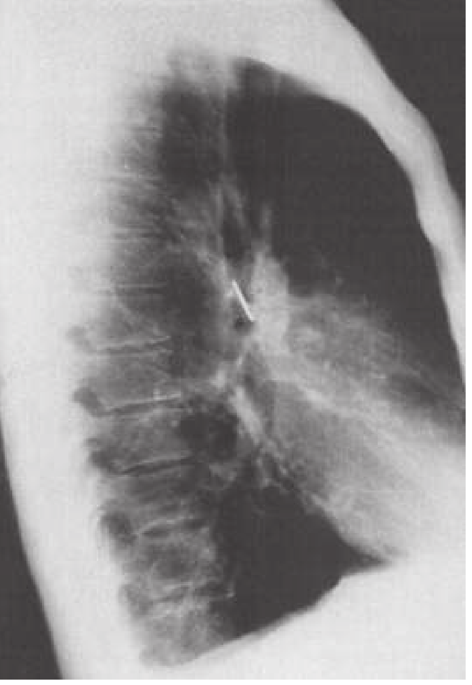
CT Scan (if stable and clinically indicated)
- More sensitive than plain X-ray
- CT virtual bronchoscopy has ~98% positive predictive value for tracheobronchial foreign bodies
- Risk of sending an unstable child to the CT scanner - only if the child is sufficiently stable
- MRI useful for radiolucent objects with high fat content (nuts)
Bronchoscopy - Gold Standard
- Rigid bronchoscopy under general anesthesia is both diagnostic AND therapeutic
- A strong history alone may prompt bronchoscopy even if X-ray is normal
- Flexible bronchoscopy may be used diagnostically first, then rigid bronchoscopy for removal
- K.J. Lee's Essential Otolaryngology; Rosen's Emergency Medicine
Treatment
Immediate/Emergency Management (Based on Severity)
If the Child is in Respiratory Arrest or Critical Obstruction:
For infants (up to 1 year):
- 5 back blows - held head-down, firm blows between shoulder blades
- 5 chest thrusts - two fingers on the lower sternum
- Alternate back blows and chest thrusts; do NOT use abdominal thrusts (Heimlich) in infants under 1 year
- If unconscious: begin chest compressions (CPR), visualize oropharynx between cycles - remove foreign body only if visible
- Direct/video laryngoscopy + Magill forceps removal if object visualized
- Never perform blind finger sweeps - this pushes the object deeper
If basic maneuvers fail:
- Emergency intubation - can push a tracheal foreign body into right mainstem bronchus (then withdraw tube above carina to ventilate left lung)
- Needle cricothyrotomy if intubation fails due to fixed proximal obstruction
If the Child is Stable (Partial Obstruction, Maintaining Oxygen):
- Keep child calm and sitting upright (position of comfort)
- Supplemental oxygen via face mask
- Do NOT attempt any oropharyngeal examination that might upset the child and precipitate complete obstruction
- Establish IV access
- Alert ENT (Otolaryngology), Anesthesia, and prepare for the operating room
- Urgent rigid bronchoscopy under general anesthesia with spontaneous ventilation
- Sharp objects are retrieved into the bronchoscope to minimize mucosal trauma
- Rarely, tracheotomy is required for very large or impacted foreign bodies
Post-Procedure Care
- Monitor for post-obstructive pulmonary edema, mucosal trauma, pneumothorax
- If object was present >2 weeks: treat for secondary infection/bronchiectasis
- Parental counseling and safety education to prevent recurrence
Summary Algorithm for a 1-Year-Old with Sudden Stridor
Sudden stridor in 1-year-old
         |
Is the child cyanosed/in arrest?
         |
    YES --> Back blows + chest thrusts --> Laryngoscopy + Magill forceps
            If fails: Intubate / Needle cricothyrotomy
            Emergency bronchoscopy
         |
    NO  --> Keep calm, O2, upright position
            CXR (AP + Lateral + expiratory/decubitus)
            Consider CT if stable and CXR inconclusive
            Urgent ENT consult
            Rigid bronchoscopy (diagnostic + therapeutic) under GA
Key Points to Remember
- Stridor = noisy breathing from partial airway obstruction - classify as inspiratory, expiratory, or biphasic to localize the level
- Sudden-onset stridor in a 1-year-old = foreign body aspiration until proven otherwise
- Most foreign bodies are radiolucent - a normal CXR does not rule out FBA; a strong history mandates bronchoscopy
- Bronchoscopy is the gold standard for both diagnosis and treatment
- In infants: back blows + chest thrusts (NOT Heimlich); never perform blind finger sweeps
- Always be prepared for complete airway obstruction - have resuscitation equipment at hand before examining
Sources: Bailey and Love's Short Practice of Surgery (28th Ed.) | K.J. Lee's Essential Otolaryngology | Rosen's Emergency Medicine | Tintinalli's Emergency Medicine
This is a shared conversation. Sign in to Orris to start your own chat.