
Endocrine gland
Endocrine Glands
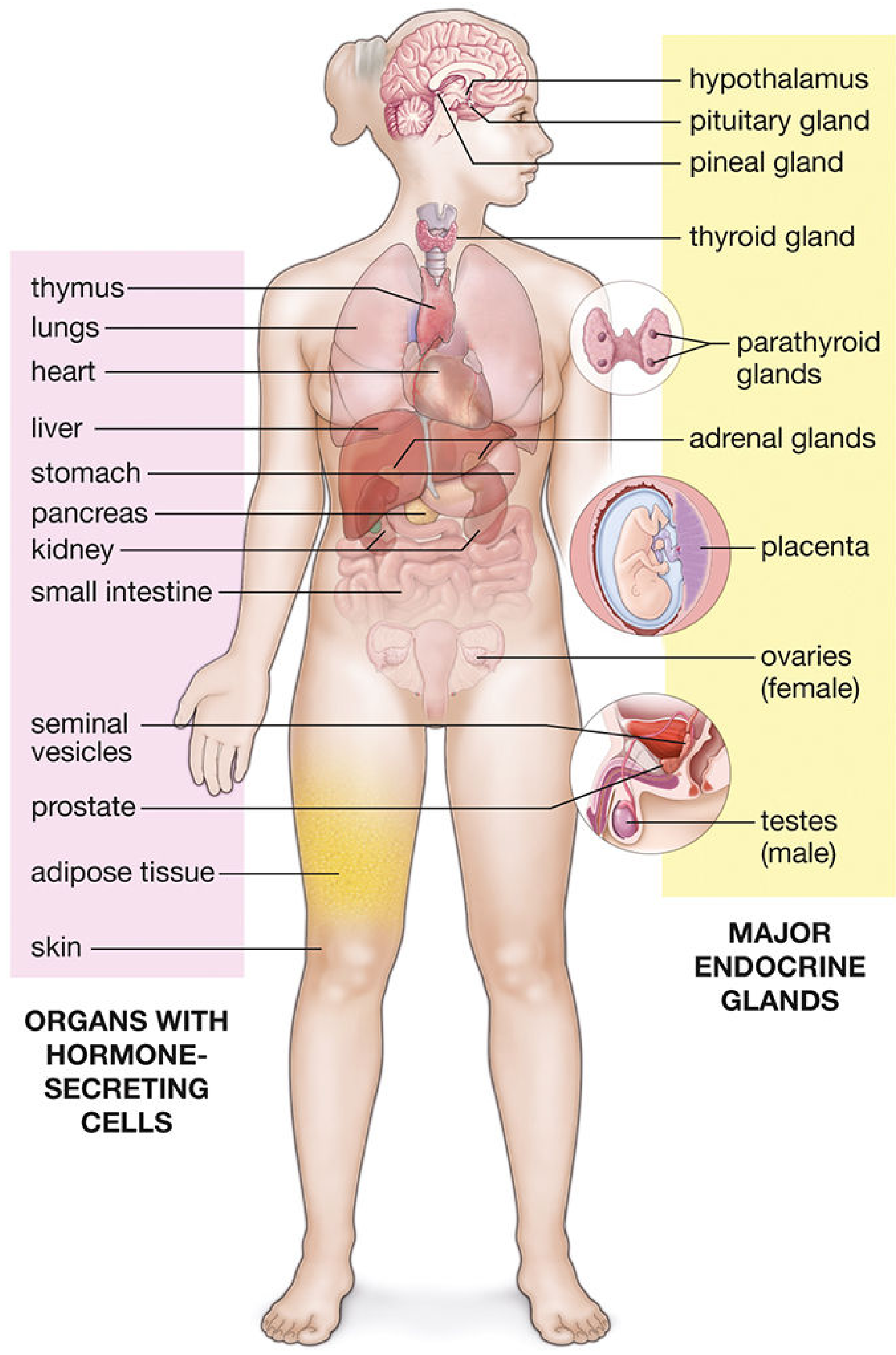
Definition
Endocrine vs. Exocrine Glands
| Feature | Endocrine | Exocrine |
|---|---|---|
| Ducts | Absent | Present |
| Secretion route | Bloodstream / lymphatics | Duct to epithelial surface |
| Examples | Thyroid, adrenal | Salivary, sweat, pancreas (exocrine portion) |
| Vascularization | Rich | Moderate |
The pancreas is unique - it is both an exocrine gland (secretes digestive enzymes into the duodenum via the pancreatic duct) and an endocrine gland (islets of Langerhans secrete insulin and glucagon into the blood). - Histology: A Text and Atlas
Types of Hormonal Secretion
| Mode | Mechanism |
|---|---|
| Endocrine | Hormone enters the bloodstream; acts on distant target cells |
| Paracrine | Hormone diffuses to adjacent cells; does not enter blood |
| Autocrine | Hormone acts on receptors of the same cell that produced it |
Chemical Classification of Hormones
| Class | Biosynthesis | Examples |
|---|---|---|
| Peptides/Proteins | Synthesized from amino acids via mRNA | Insulin, GH, PTH, ADH, oxytocin, ACTH, TSH, FSH, LH |
| Steroids | Derived from cholesterol | Cortisol, aldosterone, testosterone, estradiol, progesterone |
| Amines | Derived from tyrosine | Thyroid hormones (T3, T4), epinephrine, norepinephrine |
- Costanzo Physiology, 7th Ed., p. 399
Classic Endocrine Glands and Their Hormones
| Gland | Key Hormones | Major Actions |
|---|---|---|
| Hypothalamus | TRH, CRH, GnRH, GHRH, somatostatin, dopamine | Regulates anterior pituitary secretion |
| Anterior Pituitary | TSH, ACTH, FSH, LH, GH, Prolactin | Regulates other endocrine glands and tissues |
| Posterior Pituitary | ADH (vasopressin), Oxytocin | Water reabsorption; milk ejection, uterine contractions |
| Thyroid | T3, T4 (amines) | Metabolism, growth, CNS maturation, thermogenesis |
| Parathyroid | PTH, Calcitonin | Calcium homeostasis (PTH raises Ca²+; calcitonin lowers Ca²+) |
| Adrenal Cortex | Cortisol (glucocorticoid), Aldosterone (mineralocorticoid), DHEA | Gluconeogenesis, Na+ retention, anti-inflammatory |
| Adrenal Medulla | Epinephrine, Norepinephrine | Sympathetic "fight-or-flight" response |
| Pancreas (islets) | Insulin (β cells), Glucagon (α cells) | Lower / raise blood glucose |
| Testes | Testosterone | Spermatogenesis, male secondary sex characteristics |
| Ovaries | Estradiol, Progesterone | Female reproductive cycle, pregnancy maintenance |
| Placenta | hCG, hPL, Estradiol, Progesterone, Estriol | Maintains pregnancy; growth-hormone-like effects |
| Kidney | Renin, 1,25-(OH)2 Vitamin D (calcitriol) | Angiotensin conversion; intestinal Ca²+ absorption |
| Pineal gland | Melatonin | Circadian rhythm regulation |
- Costanzo Physiology, 7th Ed., Table 9.2
Embryological Origins of Endocrine Glands
- CNS: Posterior pituitary, pineal gland
- Neural crest: Adrenal medulla (chromaffin cells)
- Gut tube epithelium: Anterior pituitary, thyroid, parathyroid glands
- Histology: A Text and Atlas, p. 1972
Regulation of Hormone Secretion
- A target organ secretes a hormone (e.g., thyroid hormone T3/T4)
- Rising levels feed back to the hypothalamus and anterior pituitary, suppressing releasing hormones (TRH) and tropic hormones (TSH)
- This creates a "hypothalamic-pituitary-target organ axis" with exquisite sensitivity
-
Neural input to the hypothalamus (stress, circadian rhythm, emotions)
-
Serum ion/nutrient levels (e.g., Ca²+ regulates PTH; glucose regulates insulin)
-
Other hormones (positive or negative feedback from adjacent glands)
-
Histology: A Text and Atlas, p. 2002; Costanzo Physiology, p. 399
The Diffuse Neuroendocrine System (DNES)
- GI tract - secretin, CCK, gastrin, GIP
- Kidney - renin, erythropoietin
- Heart - ANP (atrial natriuretic peptide)
- Liver - IGF-1
- Adipose tissue - adipokines (leptin, adiponectin, resistin)
- Skin - Vitamin D precursor activation
Endocrine vs. Nervous System - Comparison
| Feature | Endocrine System | Nervous System |
|---|---|---|
| Communication medium | Hormones (via blood) | Neural impulses + neurotransmitters |
| Speed | Slower | Fast |
| Duration of response | Prolonged | Brief |
| Target range | Wide (body-wide) | Specific (neuromuscular junction, synapse) |
Clinical Principles of Endocrine Disease
- Hormone overproduction - most commonly from increased cell number (hyperplasia) or tumor. E.g., Graves disease (TSH-receptor antibodies mimic TSH → hyperthyroidism)
- Hormone underproduction - from gland destruction (tuberculosis of adrenals), autoimmunity (Hashimoto thyroiditis), surgical removal (accidental parathyroid removal during thyroidectomy), or genetic abnormalities
- Altered tissue response - hormone receptor mutations (e.g., insulin resistance in type 2 diabetes, largely driven by adipose tissue signaling)
- Endocrine tumors - many are hormonally active (cause overproduction); some are non-functional but cause local compression or metastasize (e.g., thyroid cancer without hyperthyroidism)
- Costanzo Physiology, 7th Edition, Ch. 9 - Endocrine Physiology (pp. 395-458)
- Histology: A Text and Atlas with Correlated Cell and Molecular Biology, Ch. 21 - Endocrine System (pp. 1970-2002)
Casts in urine
urinary casts urine microscopy RBC WBC granular hyaline
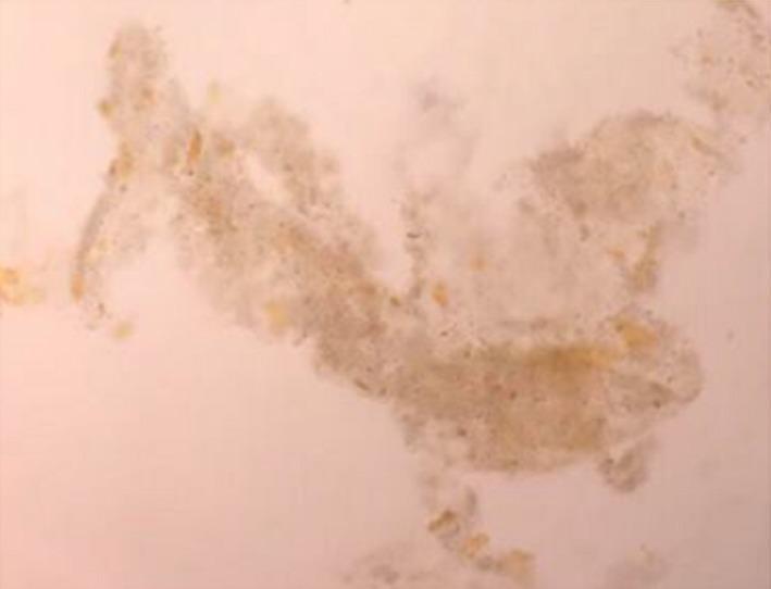
This diagnostic urine microscopy image at 400x magnification demonstrates multiple granular casts characteristic of acute tubular necrosis (ATN). The casts exhibit a classic 'muddy brown' coloration and granular texture, composed of degenerating tubular epithelial cells and debris. Notably, these casts also show distinct yellow to golden-brown pigmentation consistent with bile staining, a finding associated with severe hyperbilirubinemia or obstructive jaundice. The morphology includes elongated cylindrical structures alongside irregular and fragmented forms. The background of the slide is relatively clear and light, facilitating the high-contrast visualization of the darkly pigmented casts. This finding is clinically significant in the context of hepatorenal syndrome or bile acid nephropathy, where high levels of bilirubin can directly contribute to renal tubular injury and the formation of pigmented casts.
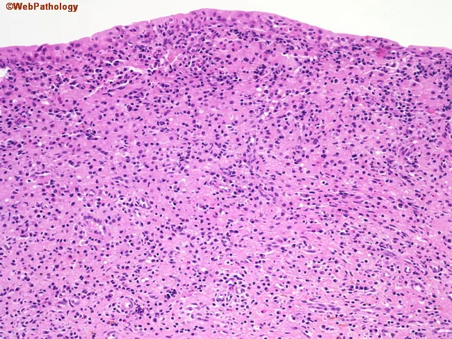
Histology of malakoplakia in the urinary bladder mucosa. Specimen: bladder mucosa biopsy from cystoscopy. Modality: light microscopy of H&E-stained sections. Morphology: sheets of large macrophages with abundant foamy, granular cytoplasm (von Hansemann cells) scattered among lymphocytes and plasma cells in the lamina propria; occasional multinucleated giant cells. Within macrophages, basophilic, calcium- and iron-containing inclusions (Michaelis–Gutmann bodies) may be seen, giving targets/granular calcific appearances. The background epithelium shows chronic cystitis with neutrophils and surface erosion. The histiocytes may be arranged in granulomatous nodules or diffuse infiltrates; plasma cells and fibroblastic reaction may be present. The pattern is an abnormal inflammatory response due to defective phagolysosome function in macrophages, resulting in undigested bacterial debris from organisms like Escherichia coli or Proteus mirabilis; common bladder involvement. Clinically, malakoplakia presents with frequency, dysuria, hematuria; cystoscopy reveals plaque-like or mass-like lesions mimicking neoplasia. Diagnostic significance: malakoplakia should be distinguished from bladder carcinoma; Michaelis–Gutmann bodies are pathognomonic but not always present. Potential clinical use cases: educational histopathology, differential diagnosis in elderly women with hematuria and chronic UTIs, guiding antibiotic therapy targeting intracellular bacteria. Correlation with urine culture and imaging aids in confirming infection-driven pathology. In research contexts, malakoplakia serves as a model of impaired intracellular digestion and macrophage dysfunction.
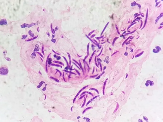
A light microscopy image obtained from a urine cytology slide stained with a fungal stain (e.g., lactophenol cotton blue) demonstrates numerous fusiform to sickle-shaped macroconidia with 1 to 4 septa, embedded in a refractile background. The macroconidia are slender, curved, and multi-septate, consistent with Fusarium species, most commonly F. solani, F. oxysporum, or F. moniliforme. Occasional hyphal elements may be seen, but the diagnostic hallmark in this preparation is the characteristic banana-shaped spindle-like conidia with multiple internal septa. This morphology supports a diagnosis of fusariosis involving the urinary tract in an adult male with chronic liver disease; the clinical history notes hematuria and negative urine cytology for malignant cells, highlighting an infectious etiology rather than neoplasia. Fusarium is a filamentous, hyaline fungus widely distributed in soil and plants in tropical and subtropical climates and is an important opportunistic pathogen in immunocompromised hosts. It is associated with keratitis, endophthalmitis, onychomycosis, burn wound infections, catheter-related fungemia, and, less commonly, urinary tract infections. Morphologic identification on cytology or histopathology is vital for rapid diagnosis. Clinically relevant terms include fungal keratitis, sinusitis, disseminated fusariosis, amphotericin B therapy, voriconazole susceptibility, and drug resistance. This image is valuable for educational reference, differential diagnosis of septate hyaline molds, and microbiology/pathology training.
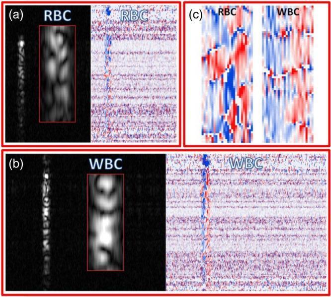
This diagnostic imaging panel displays Optical Coherence Tomography (OCT) intensity and phase-change signals for human blood cells, specifically erythrocytes (RBC) and leukocytes (WBC). Panel (a) illustrates the signal modulation of RBCs, where the intensity image shows a granular speckle pattern and the phase-change map displays a horizontal distribution of red and blue shifts. Panel (b) shows WBCs, characterized by a more defined, brighter central region in the intensity scan compared to the diffuse RBC speckle. Panel (c) provides a high-magnification comparison of the phase-change signals: RBCs exhibit larger, mosaic-like patches of red and blue interference signals, while WBCs demonstrate a finer, more vertically oriented and chaotic color distribution. These differences in statistical speckle properties and phase modulation are used to distinguish cell types and infer dynamic properties within coherent imaging modalities. The visual data highlights the application of OCT signal analysis in hematology and microstructural tissue characterization.
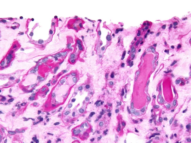
Imaging modality: Brightfield light microscopy of a renal cortical biopsy section stained with Periodic Acid-Schiff (PAS). The section demonstrates tubulointerstitial injury with intratubular casts. In myeloma cast nephropathy, casts formed from monoclonal light chains may be PAS-negative, appearing pale pink (left side of the reference image), in contrast to hyaline casts, which are PAS-positive and stain dark pink (right side). Myelomatous (light-chain) casts obstruct tubule lumens, often with relative sparing of glomeruli in the presented field, and are composed of monoclonal immunoglobulin light chains that precipitate within distal tubules and collecting ducts. The PAS counterstain highlights basement membranes and surrounding stroma, facilitating assessment of cast distribution and tubular injury. Clinically, this pattern supports a diagnosis of cast nephropathy secondary to multiple myeloma or Waldenström-like disorders with excessive free light chains. The differential includes simple hyaline casts and other tubular casts seen in dehydration, nephrotoxicity, or ATN; immunostaining and light-chain restriction studies may be required for confirmation. The diagnostic significance lies in identifying myeloma cast nephropathy, which guides oncologic therapy and renal prognosis. Potential clinical uses include assessment of renal involvement in plasma cell dyscrasias, monitoring treatment response, and prognostication in multiple myeloma.
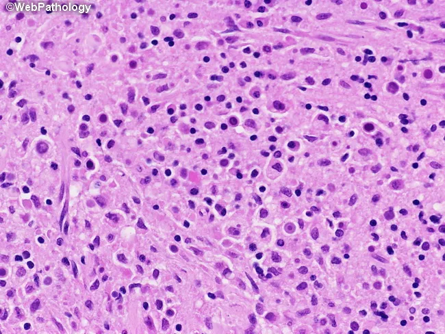
Light microscopy of a bladder tissue biopsy stained with hematoxylin and eosin (H&E) at high power (approximately 400x total magnification) demonstrates classic malakoplakia of the urinary bladder. The lamina propria and overlying urothelium contain numerous foamy, granular histiocytes (von Hansemann cells) characterized by abundant eosinophilic cytoplasm and eccentric, round to oval nuclei. Within the histiocytes and in the interstitial space, basophilic, targetoid inclusions are visible: Michaelis–Gutmann bodies, laminated calcium phosphate deposits surrounding bacterial remnants. These Michaelis–Gutmann bodies appear as centripetally layered, bull’s-eye–like structures and can be highlighted by special calcium stains (e.g., von Kossa) or demonstrated with periodic acid–Schiff when necessary; their presence is diagnostic for malakoplakia. The inflammatory background is mixed, with scattered lymphocytes and plasma cells, and occasional multinucleated giant cells forming a granulomatous milieu. Clinically, malakoplakia reflects impaired macrophage bactericidal function often in the setting of chronic bacterial infection, most commonly Escherichia coli. The overall pattern mimics neoplastic processes and may present as a bladder mass or mimicking urothelial carcinoma; thus, recognition of von Hansemann cells and Michaelis–Gutmann bodies is essential for accurate diagnosis. This image supports teaching, differential diagnosis, and research on macrophage lysosomal storage-like disorders and infectious granulomatous inflammation in the genitourinary tract for education.
red blood cell cast RBC cast glomerulonephritis urine microscopy
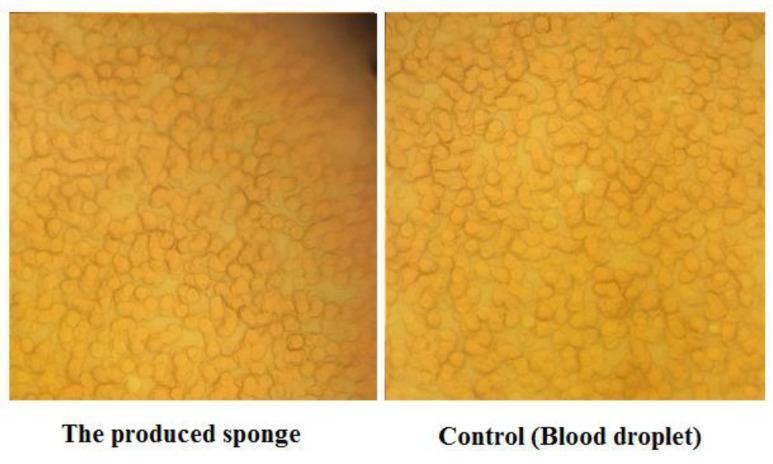
This diagnostic image provides a side-by-side comparison of red blood cell (RBC) morphology viewed under light microscopy at 100x magnification. The left panel shows RBCs within a produced gelatin sponge matrix, while the right panel serves as a control showing a standard blood droplet. Both images display a dense field of closely packed, circular to slightly oval erythrocytes characterized by a uniform yellowish-orange hue. In the gelatin sponge sample, the RBCs maintain their healthy, biconcave-typical morphology without signs of toxicity or structural deformation, though they exhibit a subtle increase in cellular clumping compared to the more even distribution in the control. A slightly darker background in the sponge sample suggests the presence of the cross-linked gelatin matrix. This comparison is used to evaluate the biocompatibility of hemostatic sponge materials and their interaction with cellular blood components during the coagulation process.
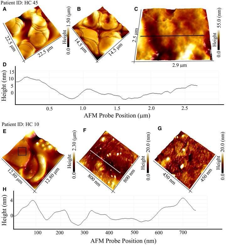
This composite diagnostic image presents Atomic Force Microscopy (AFM) analysis of red blood cell (RBC) membrane topography from two different subjects (HC-45 and HC-10). The figure comprises 3D-rendered topographical maps and corresponding sectional height profiles. Panels A-C display RBCs from subject HC-45, showing a relatively smooth, featureless membrane surface; the sectional profile in Panel D reveals broad, low-frequency height variations (up to 15 nm) attributed to natural cell corrugation and minor lipid bilayer damage. In contrast, Panels E-G display RBCs from subject HC-10, where high-resolution 3D AFM imaging reveals the presence of distinct protein aggregates, characterized as fibrillar and spherical particles. The sectional profile in Panel H quantifies these surface features, showing high-frequency height fluctuations (peaks ~4 nm) corresponding to the distribution of these aggregates. This comparison illustrates the use of nanoscale imaging to detect protein fibril accumulation on the RBC membrane, a marker relevant in neurodegenerative research such as Alzheimer's disease pathology.
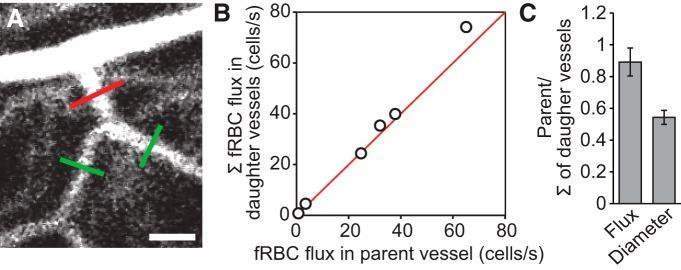
This composite educational graphic illustrates the analysis of red blood cell (RBC) flux at microvascular bifurcations in the retina. Panel A is a diagnostic confocal microscopy image showing a vascular bifurcation; a red line indicates a perpendicular line scan across the parent vessel, while green lines indicate scans across the daughter vessels (scale bar: 40 µm). Panel B is a scatter plot comparing fluorescent RBC (fRBC) flux in the parent vessel against the sum of fRBC fluxes in the daughter vessels (measured in cells/s). The data points align closely with a red unity line, demonstrating mass conservation of blood flow at branching points. Panel C is a bar graph showing the ratio of the parent vessel to the sum of daughter vessels for both RBC flux (approximately 0.9) and vessel diameter (approximately 0.54). These panels collectively demonstrate a validated methodology for measuring absolute hemodynamic parameters and provide physiological insights into the relationship between vascular geometry and blood flow distribution in the human-related biomedical research context.
Casts in Urine
What Are Urinary Casts?
"A cast is a protein coagulum that is formed in the renal tubule and traps any tubular luminal contents within the matrix." - Campbell-Walsh-Wein Urology
Conditions That Favor Cast Formation
- Concentrated, acidic urine (low pH promotes Tamm-Horsfall gelation)
- Low urine flow rate / oliguria (increases contact time)
- Proteinuria (provides matrix material)
- Tubular injury (releases cellular debris into the lumen)
Classification of Urinary Casts
1. Hyaline Casts
- Composition: Pure Tamm-Horsfall mucoprotein, no cellular inclusions
- Appearance: Colorless, nearly transparent; low refractive index; easily missed on bright-field microscopy; best seen with phase-contrast
- Significance: Can be normal (a few per low-power field is acceptable). Seen transiently after vigorous exercise, fever, or heat exposure. Increased numbers indicate kidney disease (e.g., pyelonephritis, chronic renal disease) or dehydration
2. Granular Casts
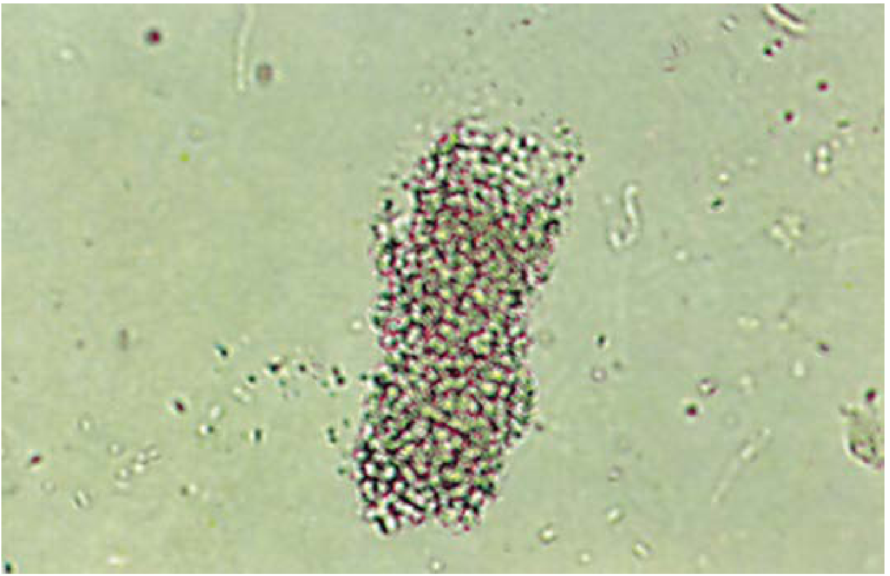
- Composition: Granules derived from plasma protein aggregates (fibrinogen, immune complexes, globulins), or cellular remnants of lysed leukocytes, erythrocytes, or renal tubular cells. Fine salt precipitates and lysosomes may also contribute
- Appearance: Fine or coarse granules; vary from pale to dark brown
- Significance: Seen in glomerular and tubular diseases, tubulointerstitial disease, pyelonephritis, viral infections, chronic lead poisoning, renal allograft rejection, and post-exercise. The classic "muddy brown" coarsely granular cast in the context of AKI is highly associated with acute tubular necrosis (ATN). With prolonged stasis, large granules progressively dissolve into fine granules, then transition to waxy casts
3. Waxy Casts
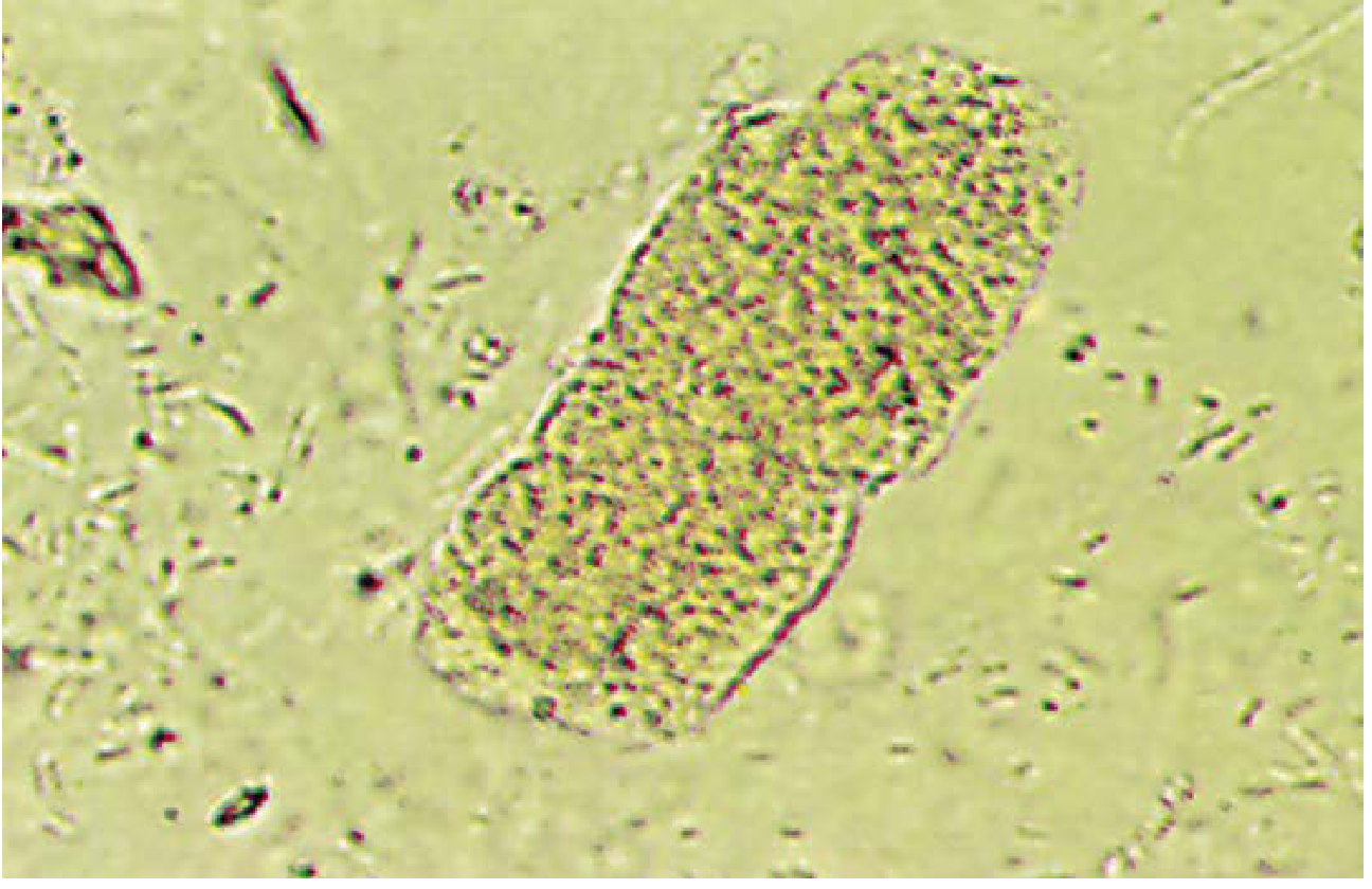
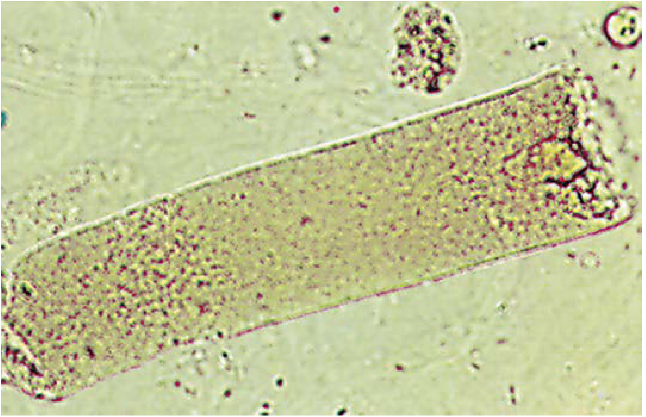
- Composition: Represent the end-stage degeneration of cellular or granular casts following prolonged stasis
- Appearance: Highly refractile (high refractive index); homogeneously smooth with sharp margins, blunted ends, and characteristic cracks or fissures along lateral margins (indicating brittleness). Easily visible on bright-field
- Significance: Imply localized nephron obstruction and oliguria. Associated with chronic renal failure, tubular atrophy and degeneration, and renal allograft rejection. When unusually broad (wider than normal tubules), called "renal failure casts" - indicate end-stage renal disease and extreme urine stasis
4. RBC (Erythrocyte) Casts - Pathognomonic Significance
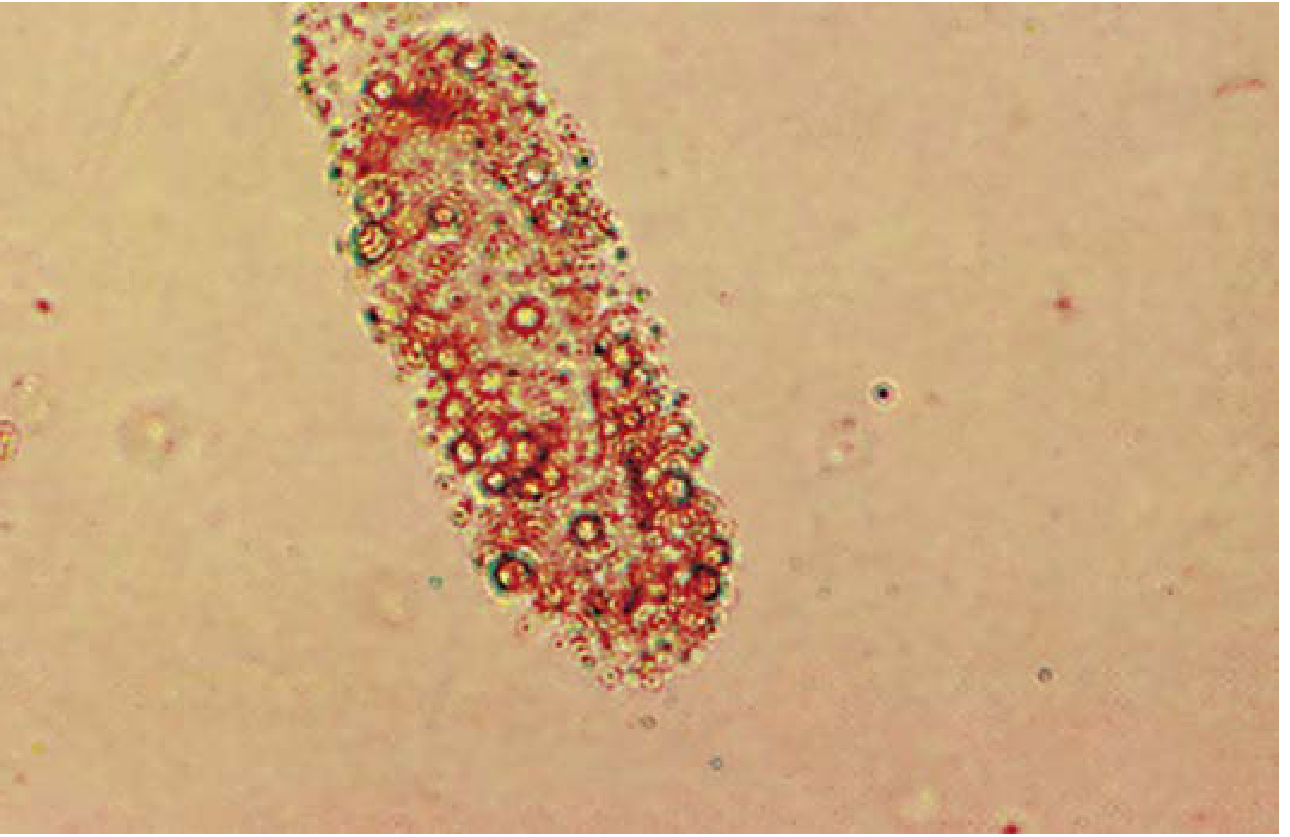
- Formation: Glomerular damage allows RBCs to escape into the tubule. If concomitant proteinuria is present and conditions are right, RBCs become trapped in the Tamm-Horsfall matrix in the distal nephron
- Appearance: Yellow under low power; RBC outlines sharply defined in at least part of the cast. With prolonged stasis, RBCs lyse and the cast becomes a reddish-brown hemoglobin (blood) cast
- Significance: Pathognomonic for glomerular bleeding - a definitive sign of glomerulonephritis or vasculitis affecting the glomerulus
- Diseases: Acute proliferative GN, IgA nephropathy, lupus nephritis (SLE), subacute bacterial endocarditis, ANCA-associated vasculitis, renal infarction, post-infectious GN. Rarely, severe pyelonephritis (tubulointerstitial disease allows transtubular RBC entry)
5. WBC (Leukocyte) Casts
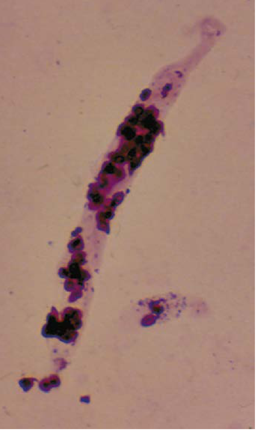
- Formation: Leukocytes (primarily neutrophils) enter tubular lumina from the interstitium during inflammation
- Appearance: Refractive; granulated; multilobated nuclei may be visible. Phase-contrast microscopy helps delineate nuclear segmentation
- Significance: Reflect tubulointerstitial disease with neutrophilic exudate and interstitial inflammation
- Diseases: Acute pyelonephritis (most common), acute interstitial nephritis (AIN), lupus nephritis, active proliferative GN (via complement-mediated chemotaxis), nephrotic syndrome
6. Renal Tubular Epithelial (RTE) Cell Casts
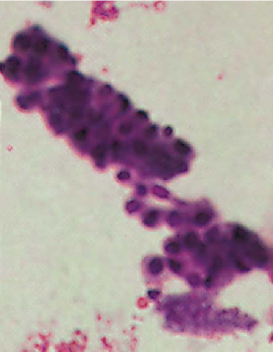
- Appearance: Cells with singular, round nuclei (distinguishing feature from WBCs, which have multilobed nuclei). Difficult to distinguish from leukocyte casts on bright-field alone; phase-contrast or Papanicolaou staining required
- Significance: Indicate renal tubular epithelial damage
- Diseases: Acute tubular necrosis (ATN) - hallmark finding, viral disease (CMV, other viruses), drug toxicity (heavy metals, ethylene glycol, salicylate poisoning), renal transplant rejection (an early and reliable indicator), AIN, glomerular disease with heavy proteinuria
7. Fatty Casts
- Composition: Lipid-laden renal tubular cells incorporated into the cast matrix; lipid droplets appear as oval fat bodies
- Appearance: Refractile fat droplets; show "Maltese cross" birefringence under polarized light (cholesterol esters)
- Significance: Seen with heavy proteinuria and are a hallmark of nephrotic syndrome. Also in hypothyroidism and lipiduria
8. Pigmented Casts
| Pigment Cast | Appearance | Clinical Association |
|---|---|---|
| Hemoglobin cast | Yellow to red-brown color; smooth surface | Intravascular hemolysis (hemoglobinuria), degenerated RBC casts |
| Myoglobin cast | Pigmented, morphologically similar to hemoglobin cast | Rhabdomyolysis - AKI (myoglobin precipitates in acidic urine) |
| Bilirubin cast | Yellow-brown pigmented cast | Jaundice with elevated direct (conjugated) bilirubin |
| Hemosiderin cast | Hemosiderin granules from pigment-laden tubular cells | Chronic intravascular hemolysis |
9. Crystal Casts
- Contain urates, calcium oxalate, or sulfonamide crystals trapped in a visible matrix
- Polarizable crystals within cast matrix
- Indicate tubular/collecting duct crystal deposition (crystalline nephropathy)
- Hematuria (tubular damage) usually accompanies them
10. Myeloma (Light Chain) Casts
- Composition: Monoclonal immunoglobulin light chains (Bence Jones protein) precipitating in distal tubules and collecting ducts
- On biopsy: Light-chain casts are PAS-negative (pale pink), unlike hyaline casts which are PAS-positive (dark pink)
- Significance: Cast nephropathy in multiple myeloma - a major cause of AKI in myeloma. Casts obstruct tubules, trigger giant cell reaction, and cause tubular atrophy
11. Other Cast Types
| Cast | Association |
|---|---|
| Bacterial | Bacterial kidney infections (pyelonephritis) |
| Fungal | Fungal kidney infections |
| Mixed | Combined findings (e.g., RBC + WBC in lupus nephritis) |
Summary Table - Cast Types and Clinical Significance
| Cast Type | Key Finding | Diseases |
|---|---|---|
| Hyaline | Normal, few acceptable | Normal, dehydration, exercise, CKD |
| Granular (muddy brown) | Degenerated cells/protein | ATN, GN, tubulointerstitial disease |
| Waxy | Prolonged oliguria/stasis | CKD, ESRD, transplant rejection |
| RBC cast | Glomerular bleeding | GN (IgA, lupus, post-infectious, ANCA) |
| WBC cast | Tubular/interstitial inflammation | Pyelonephritis, AIN, GN |
| RTE cell cast | Tubular necrosis | ATN, drugs, viral, transplant rejection |
| Fatty cast | Heavy proteinuria | Nephrotic syndrome |
| Hemoglobin cast | Hemolysis or RBC lysis | Intravascular hemolysis, GN |
| Myoglobin cast | Rhabdomyolysis | Crush injury, myopathies |
| Bilirubin cast | Conjugated hyperbilirubinemia | Obstructive jaundice, hepatic failure |
| Broad/Renal failure cast | End-stage nephron | ESRD |
| Myeloma cast | Light chains | Multiple myeloma |
Clinical Memory Aid
- RBC cast = Glomerulonephritis (nephritic syndrome) - NEVER normal
- WBC cast = Pyelonephritis or AIN - infection/inflammation in the kidney
- Muddy brown granular cast = ATN (ischemic or nephrotoxic AKI)
- Fatty cast / oval fat bodies = Nephrotic syndrome
- Waxy/broad cast = Chronic kidney disease / ESRD (very slow flow)
- Hyaline cast = Often normal; increases in kidney disease
- Campbell-Walsh-Wein Urology - Cast formation and Tamm-Horsfall protein
- Henry's Clinical Diagnosis and Management by Laboratory Methods - Detailed cast morphology (all microscopy images)
- Comprehensive Clinical Nephrology, 7th Edition - Table of casts and clinical associations
- Rheumatology, 2-Volume Set (Elsevier 2022) - Casts in rheumatologic renal disease