
Describe the normal histology of skin and relevant soft tissues (epidermis, dermis, subcutis, adnexal structures). Use Bailey and Love Short Practice of Surgery 28th Edition, Sabiston Textbook of Surgery 21st Edition, and Shwartz's Principles of Surgery 11th Edition, and any other reliable and latest version of anatomy textbooks, and for each part state what reference the information was extracted out of.
Reading File
Now let me search the library for the relevant books and content on skin histology.
Finding Sources
Finding Sources
Finding Sources
Finding Sources
All three requested books are available. Now let me search for skin histology content across them simultaneously.
Finding Sources
Finding Sources
I have key leads. Now let me read the specific skin histology sections from Junqueira's, the surgical textbooks, and Fitzpatrick's in parallel.
Finding Sources
Reading File
Excellent content from Junqueira's. Let me continue reading more of this section and simultaneously search for skin content in Bailey & Love, Sabiston, and Schwartz.
Reading File
Finding Sources
I have exact file paths and line numbers. Now let me read from Bailey & Love, Schwartz, Sabiston, and continue Junqueira's in parallel.
Reading File
Reading File
Reading File
Reading File
Excellent content from all sources. Let me continue reading the Bailey & Love section and Junqueira dermis/subcutis sections.
Reading File
Reading File
Reading File
Excellent content from all sources. Let me read more from Schwartz and the Junqueira subcutis/adnexal sections to complete the picture.
Reading File
Reading File
I now have comprehensive content from all sources. Let me read the Schwartz dermal components and subcutis section plus the Bailey & Love dermis continuation.
Reading File
Reading File
I have extensive content from all three surgical textbooks and Junqueira's histology. Let me now fetch the key images and also read the Schwartz subcutis section and Junqueira's sweat gland section in parallel.
Reading File
Reading File
Loading Image
I now have comprehensive data from all sources. Let me fetch one more key diagram and read the Schwartz subcutis section.
Reading File
Loading Image
I now have all the content I need. Let me compile the comprehensive response.
Normal Histology of Skin and Relevant Soft Tissues
Overview
The skin is the largest organ in the body, accounting for approximately 15-20% of total body weight and presenting 1.5-2 m² of surface area in adults. It is composed of three distinct layers: the epidermis (outermost), the dermis, and the subcutaneous tissue (hypodermis/subcutis). The epidermis is of ectodermal origin; the dermis and subcutis are of mesodermal origin.
Sources: Junqueira's Basic Histology - Text and Atlas, 17th Edition, p. 900; Schwartz's Principles of Surgery, 11th Edition, Chapter 16
Figure 1 - Layers and Strata of the Epidermis (H&E micrograph + diagram)
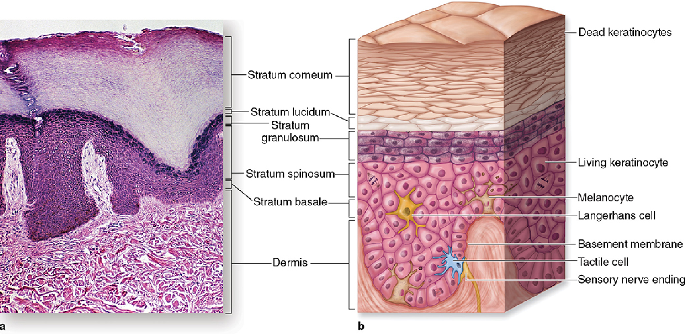
Figure 18-2 from Junqueira's Basic Histology, 17th Edition - Layers or strata of the epidermis in thick skin. (a) H&E micrograph (x100); (b) diagram illustrating the three important non-keratinocyte epidermal cells: melanocytes, Langerhans cells, and Merkel cells.
Figure 2 - Summary Table: Skin and Subcutaneous Layers
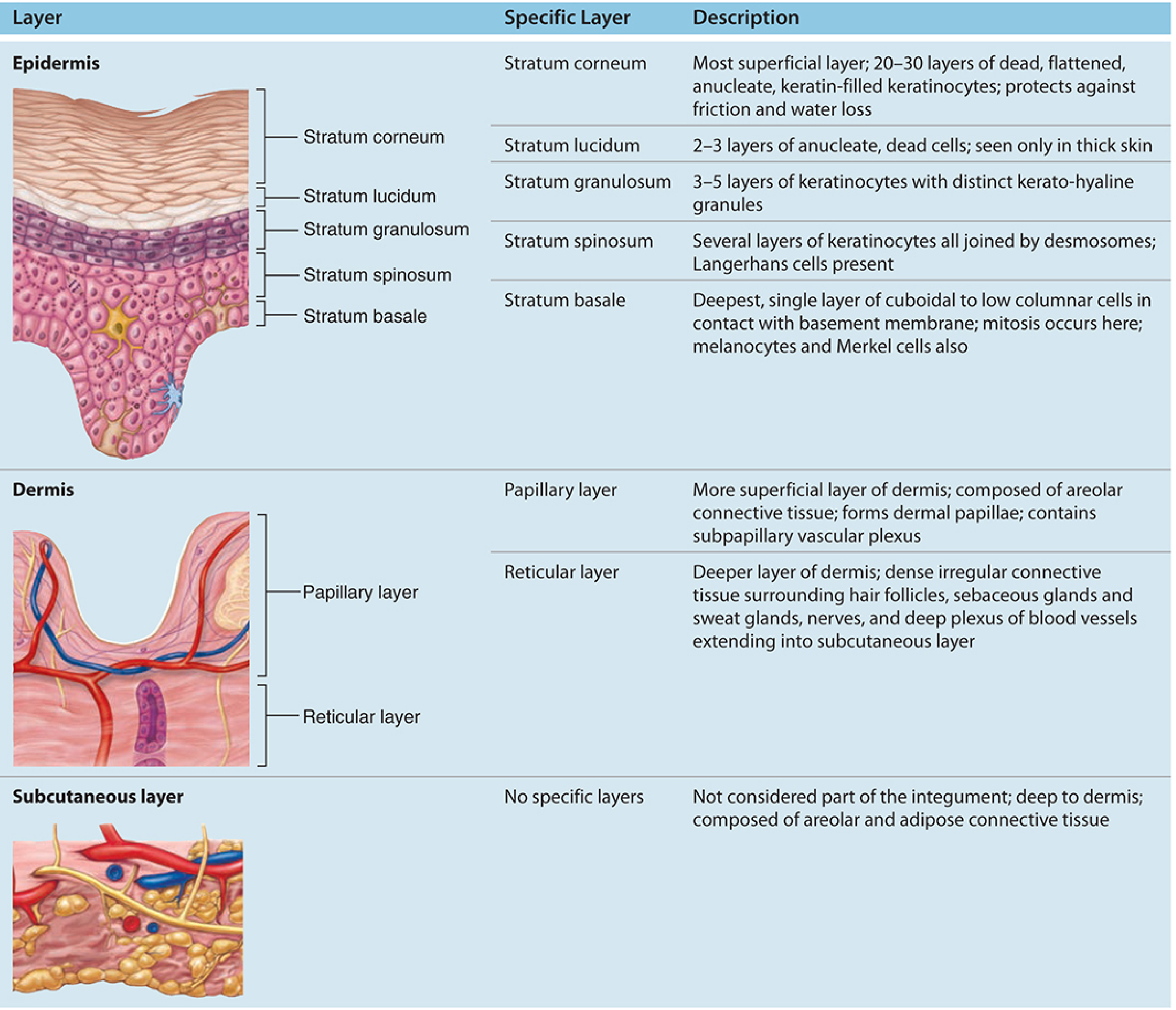
From Junqueira's Basic Histology, 17th Edition - Table 18-1: Summary of skin and subcutaneous layers, p. 909
1. Epidermis
General Structure
The epidermis constitutes approximately 5% of the skin and is composed of keratinized, stratified squamous epithelium organized into five strata (layers). The epidermis is avascular; it receives nutrients by diffusion from the dermal vasculature through the basement membrane. Its thickness is highly variable: from 75-150 µm in thin skin (eyelids) up to 0.4-1.5 mm in thick skin (palms and soles).
Bailey and Love's Short Practice of Surgery, 28th Edition, Chapter 45; Schwartz's Principles of Surgery, 11th Edition, p. 541
The epidermis consists primarily of continually regenerating keratinocytes (approximately 90% of cells). The remaining cells include melanocytes, Langerhans cells, and Merkel cells. All epidermal appendages (sweat glands, pilosebaceous follicles) are derived from this tissue.
Schwartz's Principles of Surgery, 11th Edition, p. 541
The Five Strata (Deep to Superficial)
a) Stratum Basale (Germinative Layer)
- The deepest, single layer of cuboidal to low-columnar cells resting directly on the basement membrane.
- Cells contain abundant intermediate filaments (keratin tonofibrils) attached to hemidesmosomes at the basal surface and desmosomes laterally.
- This is the mitotically active layer - continuous cell division replenishes cells lost from the surface. The entire epidermal life cycle takes approximately 30 days.
- The stratum basale also contains melanocytes and Merkel cells (described below).
Junqueira's Basic Histology, 17th Edition, p. 902-903; Schwartz's Principles of Surgery, 11th Edition, p. 541; Bailey and Love, 28th Edition, Chapter 45
b) Stratum Spinosum (Prickle Cell Layer)
- A thick layer of polyhedral cells, especially prominent in the epidermal ridges.
- Cells have central nuclei and nucleoli; cytoplasm actively synthesizes keratins.
- Keratin filaments assemble into microscopically visible bundles called tonofibrils, converging and terminating at numerous desmosomes that hold the layered cells together.
- The cells extend slightly around tonofibrils on both sides of each desmosome, producing the characteristic appearance of short "spines" or prickles at the cell surfaces (hence the name).
- Langerhans cells are found in this layer (described below).
- In thick skin subjected to continuous friction and pressure, this layer is thicker with more abundant tonofibrils and desmosomes.
Junqueira's Basic Histology, 17th Edition, p. 904-905
c) Stratum Granulosum
- Consists of 3-5 layers of flattened cells undergoing terminal differentiation (keratinization).
- Cytoplasm is filled with intensely basophilic, non-membrane-bound masses called keratohyaline granules, which contain filaggrin and other proteins that cross-link keratin filaments into large cytoplasmic structures.
- Smaller organelles called lamellar granules (membrane-coating granules) undergo exocytosis in this layer, secreting a lipid-rich layer around the cells that makes the epidermis impermeable to water.
- Together, the lipid envelope and the keratin-filled cells determine most of the physical barrier properties of the epidermis.
Junqueira's Basic Histology, 17th Edition, p. 905-907
d) Stratum Lucidum
- A thin, translucent band seen only in thick skin (palms and soles).
- Consists of 2-3 layers of anucleate, dead cells.
- Here proteins are dispersed through the tonofibril bundles, giving this layer a regular, "clear" (lucid) appearance.
- Essentially absent in eyelid skin.
Bailey and Love's Short Practice of Surgery, 28th Edition, Chapter 45; Junqueira's Basic Histology, 17th Edition, p. 907
e) Stratum Corneum
- The most superficial layer, comprising 15-20 layers of squamous, fully keratinized cells (squames) filled with filamentous keratins.
- Cells have lost their nuclei and cytoplasm and consist only of flattened, keratinized structures bound by hydrophobic, lipid-rich intercellular cement.
- Keratin filaments contain at least six different polypeptides (molecular masses 40-70 kDa), synthesized during differentiation in immature layers.
- Squames undergo continuous disintegration or desquamation (shedding) at the surface as desmosomes and lipid-rich cell envelopes break down.
Junqueira's Basic Histology, 17th Edition, p. 907-908; Bailey and Love's Short Practice of Surgery, 28th Edition, Chapter 45
Non-Keratinocyte Cells of the Epidermis
Melanocytes
- Dendritic cells of neural crest origin, usually located in the stratum basale (approximately 1 per 10 basal cells).
- Synthesize melanin (a brown-black pigment) within specialized organelles called melanosomes.
- Melanosomes are transferred via dendritic processes to keratinocytes in the strata granulosum and spinosum, providing protection against ultraviolet radiation (UVR).
- Ethnic differences in skin colour are determined by variations in the amount, combination, and distribution of melanin within keratinocytes - not by differences in the number of melanocytes.
- UV light stimulates melanin synthesis, leading to tanning.
Bailey and Love's Short Practice of Surgery, 28th Edition, Chapter 45; Junqueira's Basic Histology, 17th Edition, p. 910-912
Langerhans Cells
- Bone marrow-derived antigen-presenting dendritic cells primarily found in the stratum spinosum.
- They lack desmosomes and tonofibrils (visible as clear cells in H&E), but are identified by CD1a surface marker and characteristic cytoplasmic Birbeck granules (tennis racket-shaped on electron microscopy).
- Function as sentinels of the immune system, capturing and processing antigens to present to T lymphocytes.
Junqueira's Basic Histology, 17th Edition, p. 913-914
Merkel Cells
- Found in the stratum basale, most abundant in skin of the fingertips and external genitalia.
- Function as mechanoreceptors (slow-adapting type I receptors for discriminative touch).
- Contain characteristic dense-core cytoplasmic granules near the basolateral membrane, in direct synaptic contact with expanded disc-like endings of unmyelinated sensory nerve fibers.
- Granules contain classic neurotransmitters (serotonin) and neuropeptides.
- Clinically important: Merkel cell carcinomas are rare but highly aggressive, with twice the mortality of melanoma.
Junqueira's Basic Histology, 17th Edition, p. 913-915
Dermal-Epidermal Junction (Basement Membrane Zone)
The basement membrane lies between the stratum basale and the dermis. It consists of:
- Lamina lucida (electron-clear): contains laminin and heparan sulfate
- Lamina densa (electron-dense): contains type IV collagen
- Anchoring fibrils: composed of type VII collagen, inserting into the basal lamina and binding the dermis to the epidermis
The basal layer attaches to the basement membrane zone via hemidesmosomes. Dermal papillae interdigitate with epidermal ridges (rete pegs) to strengthen adhesion between layers, especially in skin subject to friction.
Sabiston Textbook of Surgery, 21st Edition, p. 3516; Schwartz's Principles of Surgery, 11th Edition, p. 2039; Junqueira's Basic Histology, 17th Edition, p. 918
2. Dermis
General Structure
The dermis constitutes approximately 95% of the skin. It is a mesoderm-derived connective tissue layer that supports the epidermis, anchors it to the subcutaneous tissue, and houses the neurovascular supply, epidermal appendages (adnexal structures), and supportive cells. Its thickness varies by body region from 0.6 mm (eyelids) to a maximum of 4 mm on the back.
Bailey and Love's Short Practice of Surgery, 28th Edition, Chapter 45; Schwartz's Principles of Surgery, 11th Edition, p. 2067
Layers of the Dermis
The dermis is divided into two sublayers with indistinct boundaries:
a) Papillary Layer
- The superficial, thinner layer, including the dermal papillae that interdigitate with epidermal pegs.
- Composed of loose (areolar) connective tissue with:
- Types I and III collagen fibers (arranged loosely)
- Fibroblasts, scattered mast cells, dendritic cells, and leukocytes
- A capillary and lymphatic network (subpapillary vascular plexus)
- From this layer, anchoring fibrils of type VII collagen insert into the basal lamina, helping bind the dermis to the epidermis.
- The interdigitation increases surface area, enhancing resistance to shear forces and facilitating nutrient diffusion across the dermal-epidermal junction.
Bailey and Love's Short Practice of Surgery, 28th Edition, Chapter 45; Junqueira's Basic Histology, 17th Edition, p. 918; Schwartz's Principles of Surgery, 11th Edition, p. 2067
b) Reticular Layer
- The deeper, much thicker layer, consisting of dense irregular connective tissue.
- Predominantly type I collagen bundles arranged parallel to the skin surface, interwoven with elastic fibers - providing mechanical strength and elasticity.
- Contains fewer cells but more fibers than the papillary layer.
- Proteoglycans rich in dermatan sulfate occur abundantly among the collagen and elastic fibers.
- Houses hair follicles, sebaceous glands, sweat glands, nerves, and deep blood vessel plexuses.
Bailey and Love's Short Practice of Surgery, 28th Edition, Chapter 45; Junqueira's Basic Histology, 17th Edition, p. 918-919; Schwartz's Principles of Surgery, 11th Edition, p. 2067
Fibers and Ground Substance of the Dermis
- 98% of the dry weight of the dermis is collagen.
- Typically 80-90% type I collagen and 8-12% type III collagen (ratio approximately 4:1).
- Types IV and VII collagen are present in smaller quantities at the dermo-epidermal junction.
- Elastic fibers are thinner and more perpendicularly oriented in the papillary dermis, becoming thicker and parallel in the reticular dermis. They can stretch to twice their original length and return to original configuration.
- The fibrous network lies within a hydrated amorphous ground substance of proteoglycans and glycosaminoglycans, which can contain up to 1000 times their weight in water.
- Both collagen and elastic fibers are in a constant state of turnover regulated by matrix metalloproteinases (MMPs) and fibroblasts. UV radiation induces MMP activity, accelerating degradation and leading to wrinkling.
Schwartz's Principles of Surgery, 11th Edition, p. 2069-2072
Cells of the Dermis
- Fibroblasts: The fundamental cells of the dermis. Spindle- or stellate-shaped with well-developed rough endoplasmic reticulum, responsible for producing all dermal fibers and ground substance. Can differentiate into myofibroblasts (expressing smooth muscle actin), which mediate wound contraction.
- Mast cells: Present in the papillary dermis, rich in histamine and heparin granules.
- Dermal dendrocytes and macrophages: Antigen-presenting and phagocytic cells.
- Vascular pericytes, smooth muscle cells: Associated with the dermal vasculature.
Schwartz's Principles of Surgery, 11th Edition, p. 2083
Vascular Supply of the Dermis
- Terminal vessels and capillaries fill the dermis and penetrate the dermal papillae to perfuse the cellular elements of the dermis and epidermis.
- A network of vessels just below the dermis - the subdermal plexus - supplies the overlying skin and is important in thermoregulation.
- Bailey and Love describes the body in terms of angiosomes: three-dimensional segments of tissue each with an arterial supply and venous drainage; blood equilibrates between neighbouring angiosomes via "choke" vessels.
Schwartz's Principles of Surgery, 11th Edition, p. 4039; Bailey and Love's Short Practice of Surgery, 28th Edition, Chapter 45
3. Subcutis (Hypodermis)
The subcutaneous tissue (hypodermis) lies beneath the dermis and corresponds to the superficial fascia of gross anatomy.
- Composed of richly vascularized loose connective tissue containing pockets of lipid-laden adipocytes (fat lobules) separated by fibrous septae.
- The septae contain cellular components similar to the dermis, as well as neurovascular structures supplying the cutaneous tissue and the deepest portions of sweat glands.
- Functions: insulation (thermoregulation), energy storage, cushioning and protection from mechanical forces, and allowing the skin to glide over underlying tissues.
- Binds the skin loosely to the underlying muscle and fascia.
- Contains any remnants of the panniculus carnosus (vestigial muscle layer).
Junqueira's Basic Histology, 17th Edition, p. 900-901; Schwartz's Principles of Surgery, 11th Edition, p. 2104; Bailey and Love's Short Practice of Surgery, 28th Edition, Chapter 45
4. Adnexal Structures (Skin Appendages)
Adnexal structures are epidermal derivatives that span both epidermal and dermal layers. They are of critical clinical importance because in partial-thickness injuries they serve as sources of re-epithelialization.
Bailey and Love's Short Practice of Surgery, 28th Edition, Chapter 45; Schwartz's Principles of Surgery, 11th Edition, p. 1988
a) Hair Follicles
- Tubular invaginations of the epidermis from which hair shafts grow. The face has approximately 600 hairs/cm² and the remainder of the body approximately 60/cm².
- The growing follicle has a terminal dilation called the hair bulb, into which a single dermal papilla inserts, bringing a capillary network.
- The hair root consists of three concentric layers from outermost to innermost:
- Cuticle: thin, heavily keratinized squamous cells
- Cortex: heavily keratinized, densely packed cells
- Medulla (in thick hairs): large, vacuolated, moderately keratinized cells
- Keratinocytes of the hair bulb matrix proliferate rapidly, take up melanosomes, and undergo keratinization to form the hair.
- Hair keratin is harder and more compact than epidermal keratin.
- An arrector pili muscle (strips of smooth muscle) is inserted into the wall of the hair follicle; contraction elevates the hair in response to cold and emotional stress.
- Hair growth is discontinuous, with alternating phases of growth (anagen) and rest (telogen).
- All skin bears hair except the glabrous skin of palms, soles, lips, glans penis, clitoris, and labia minora.
Junqueira's Basic Histology, 17th Edition, p. 920-921; Bailey and Love's Short Practice of Surgery, 28th Edition, Chapter 45
b) Sebaceous Glands
- Hair follicle appendages situated between the follicle and arrector pili muscle.
- Contraction of the arrector pili muscle compresses the gland, releasing sebum (holocrine secretion) along the elevated hair.
- Holocrine secretion: the entire sebocyte cell dies and contributes to the secretory product. Basal cuboidal cells on the gland capsule proliferate and differentiate centrally into large sebocytes filled with small lipid droplets; their nuclei shrink and undergo autophagy; near the duct the cells disintegrate, releasing sebum.
- Sebum is a complex mixture of wax esters, squalene, cholesterol, and triglycerides with weak antibacterial and antifungal properties. It helps maintain the stratum corneum and hair shafts.
- Sebum production increases greatly at puberty, stimulated primarily by testosterone in men and ovarian/adrenal androgens in women.
- Present in greatest numbers on the face and scalp.
Junqueira's Basic Histology, 17th Edition, p. 921-923; Schwartz's Principles of Surgery, 11th Edition, p. 2060; Bailey and Love's Short Practice of Surgery, 28th Edition, Chapter 45
c) Sweat Glands
Eccrine (Merocrine) Sweat Glands
- The most common type; distributed throughout the entire body surface except the lips.
- Tubular-shaped exocrine glands with a secretory coil and an excretory duct that opens onto the skin surface as a sweat pore.
- Present in greatest numbers on the palms, soles, axillae, and forehead.
- Secrete sweat (hypotonic NaCl solution) via exocytosis.
- Produce approximately 10 L/day in an adult; the most effective means of thermoregulation in humans via evaporative heat loss.
- Activated by the cholinergic system; respond to thermoregulatory stimuli and emotional stimuli.
- The bulk of the gland resides within the dermis; the coiled secretory portion may extend into the subcutis.
- Surrounded by a layer of myoepithelial cells that contract and assist in secretion.
Schwartz's Principles of Surgery, 11th Edition, p. 2047-2048; Bailey and Love's Short Practice of Surgery, 28th Edition, Chapter 45
Apocrine Sweat Glands
- Found in the axillae, groins (inguinoperineal region), areola, eyelids (Moll's glands), and external auditory canal.
- Larger glands; cells undergo decapitation secretion (a portion of apical cytoplasm pinches off with secretory products into the lumen).
- Activated by adrenergic system; become functionally active at puberty under the influence of sex hormones.
- Secretion is initially odorless; characteristic malodor develops after bacterial degradation.
- May have served a pheromone-producing function (now considered vestigial in humans).
- Also surrounded by myoepithelial cells.
Schwartz's Principles of Surgery, 11th Edition, p. 2048-2049; Bailey and Love's Short Practice of Surgery, 28th Edition, Chapter 45; Junqueira's Basic Histology, 17th Edition, p. 920
d) Nails
- Keratinaceous structures overlying the distal phalanges.
- Composed of three parts:
- Nail root: proximal portion, continuous with the germinal nail matrix (the source of nail growth)
- Nail plate: overlies the nail bed (shape determined by the underlying phalanx)
- Free edge: overlies a thickened portion of epidermis called the hyponychium
- Function: protect the distal digits and augment pulp function as a source of counter-pressure.
Schwartz's Principles of Surgery, 11th Edition, p. 2062
5. Sensory Structures
The dermis contains multiple sensory receptor types:
- Meissner's corpuscles: encapsulated mechanoreceptors in dermal papillae, especially in fingertips - mediate discriminative touch (fast-adapting).
- Pacinian corpuscles: deep in dermis/subcutis, layered onion-like capsule - respond to pressure and vibration.
- Ruffini endings: in the reticular dermis - respond to skin stretch.
- Free nerve endings: throughout the dermis and epidermis - mediate pain, temperature, and crude touch.
- Merkel cell-neurite complexes: in stratum basale - slow-adapting, discriminative touch (see above).
Junqueira's Basic Histology, 17th Edition, p. 913-715
Summary Table of Skin Layers and Histological Features
| Layer | Composition | Key Histological Features |
|---|---|---|
| Stratum basale | Single layer of cuboidal/columnar keratinocytes + melanocytes + Merkel cells | Mitosis; hemidesmosomes; tonofibrils |
| Stratum spinosum | Multiple layers of polyhedral keratinocytes + Langerhans cells | Desmosomes; tonofibrils; "prickle" appearance |
| Stratum granulosum | 3-5 layers of flattened keratinocytes | Basophilic keratohyaline granules; lamellar granules |
| Stratum lucidum | 2-3 anucleate cell layers (thick skin only) | Translucent/clear; only palms and soles |
| Stratum corneum | 15-20 layers of dead squames | Anucleate; filled with fibrillar keratin; continuous desquamation |
| Papillary dermis | Loose connective tissue | Type I & III collagen; capillary loops; anchoring fibrils |
| Reticular dermis | Dense irregular connective tissue | Coarse type I collagen bundles; elastic fibers; adnexal structures |
| Subcutis/Hypodermis | Loose connective tissue + adipocytes | Fat lobules in fibrous septae; superficial fascia level |
Reference Summary
| Information | Source |
|---|---|
| Overview, epidermis layers (5 strata), melanocytes, dermis layers, subcutis, hair, sebaceous glands, eccrine/apocrine glands, Langerhans cells, Merkel cells | Junqueira's Basic Histology - Text and Atlas, 17th Edition (Chapter 18) |
| Epidermis (5%), dermis (95%), keratinocytes, melanocytes, dermis papillary/reticular, adnexal structures, hair follicles, sebaceous/eccrine/apocrine glands, skin dimensions, blood supply, angiosomes | Bailey and Love's Short Practice of Surgery, 28th Edition (Chapter 45) |
| Epidermis (stratum basale to corneum), 4-5 layers, keratinocyte life cycle, pilosebaceous units, sweat glands, nails, dermis architecture (collagen types, elastic fibers, ground substance), fibroblasts, hypodermis | Schwartz's Principles of Surgery, 11th Edition (Chapter 16) |
| Basement membrane zone (lamina lucida, lamina densa, anchoring fibrils), keratinocyte immune function, skin barrier, reepithelialization | Sabiston Textbook of Surgery, 21st Edition (Chapter 23) |
| Skin grafting anatomy (dermal papillae, subdermal plexus, skin appendages in subcutis) | Schwartz's Principles of Surgery, 11th Edition (Chapter 131, Skin Grafts) |
This is a shared conversation. Sign in to Orris to start your own chat.