
Pineal gland anatomy
Reading File
The pineal gland is a small midline endocrine structure in the brain, best known for producing melatonin. It sits in the epithalamus, posterior to the third ventricle, between the two superior colliculi. It is about 5-8 mm in length and weighs roughly 100-180 mg in adults.
Searching sources...
Finding Sources
Searching the Web
pineal gland anatomy location structure blood supply
Reading File
Reading File
Loading Image
Reading File
Reading File
Loading Image
Reading Web Page
https://teachmeanatomy.info/neuroanatomy/structures/pineal-g…
Here is a thorough overview of pineal gland anatomy drawn from authoritative medical textbooks.
Pineal Gland Anatomy
Overview
The pineal gland (also called the epiphysis cerebri or pineal body) is a small, pine cone-shaped neuroendocrine organ. Rene Descartes famously called it the "seat of rational thought (the soul)" - but clinically it is significant mainly as the sole source of melatonin and as a site for rare tumors.
- Dimensions: ~5-8 mm long, 3-5 mm in diameter
- Weight: 100-180 mg in adults
- Shape: flattened, pine cone-shaped (hence "pineal," from Latin pinea = pine cone)
- Histology: A Text and Atlas, p. 2003; Robbins Pathologic Basis of Disease, p. 1041
Location and Relations
The pineal gland is a midline structure located in the epithalamus, tucked at the posterior wall of the third ventricle where the two thalamic halves join.
- It develops from neuroectoderm of the posterior roof of the diencephalon
- It remains attached to the brain by a short stalk (pineal stalk / habenular commissure region)
- Immediately inferior and anterior lie the superior colliculi of the midbrain (important for visual reflexes)
- The pineal recess of the third ventricle extends into the stalk
- Posteriorly it sits in the quadrigeminal cistern
- It is surrounded by connective tissue derived from the pia mater
Vascular Supply
The pineal gland has one of the most profuse blood supplies in the brain - second only to the kidney.
- Arterial supply: Posterior choroidal arteries (about 10 branches arising from the posterior cerebral artery)
- Venous drainage: Internal cerebral veins
The rich vascularity reflects its endocrine secretory function and explains why it sits outside the blood-brain barrier (it is a circumventricular organ).
- TeachMeAnatomy; Junqueira's Basic Histology, p. 1045
Nerve Supply
The pineal gland receives unmyelinated sympathetic nerve fibers that enter via the stalk. These fibers originate from the superior cervical ganglion and relay light/dark signals from the retina via:
- Retinohypothalamic tract (retina → hypothalamus)
- Suprachiasmatic nucleus (SCN) of the hypothalamus
- Sympathetic fibers → superior cervical ganglion → pineal
These sympathetic fibers end directly among pinealocytes, with some forming synapses. They stimulate melatonin release during darkness and inhibit it during daylight.
- Junqueira's Basic Histology, p. 1046
Histology
The pineal gland is covered by pia mater, which sends connective tissue septa into the parenchyma, dividing it into lobules. Two main cell types are present:
1. Pinealocytes (majority, ~95%)
- Large secretory cells with slightly basophilic cytoplasm and euchromatic (pale, irregular) nuclei
- Ultrastructurally: secretory vesicles, abundant mitochondria
- Have long cytoplasmic processes that terminate as dilated endings near capillaries (endocrine release pattern)
- Produce melatonin (a low-molecular-weight tryptophan derivative)
- Silver impregnation stains reveal slender processes resembling primitive neuronal precursors
2. Interstitial glial cells (~5%)
- Modified astrocytes that stain positively for GFAP (glial fibrillary acidic protein)
- Have darker, more elongated nuclei compared to pinealocytes
- Located mainly in perivascular areas and between groups of pinealocytes
- Junqueira's Basic Histology, p. 1045-1046; Robbins, p. 1041
Histology of the pineal gland (H&E):
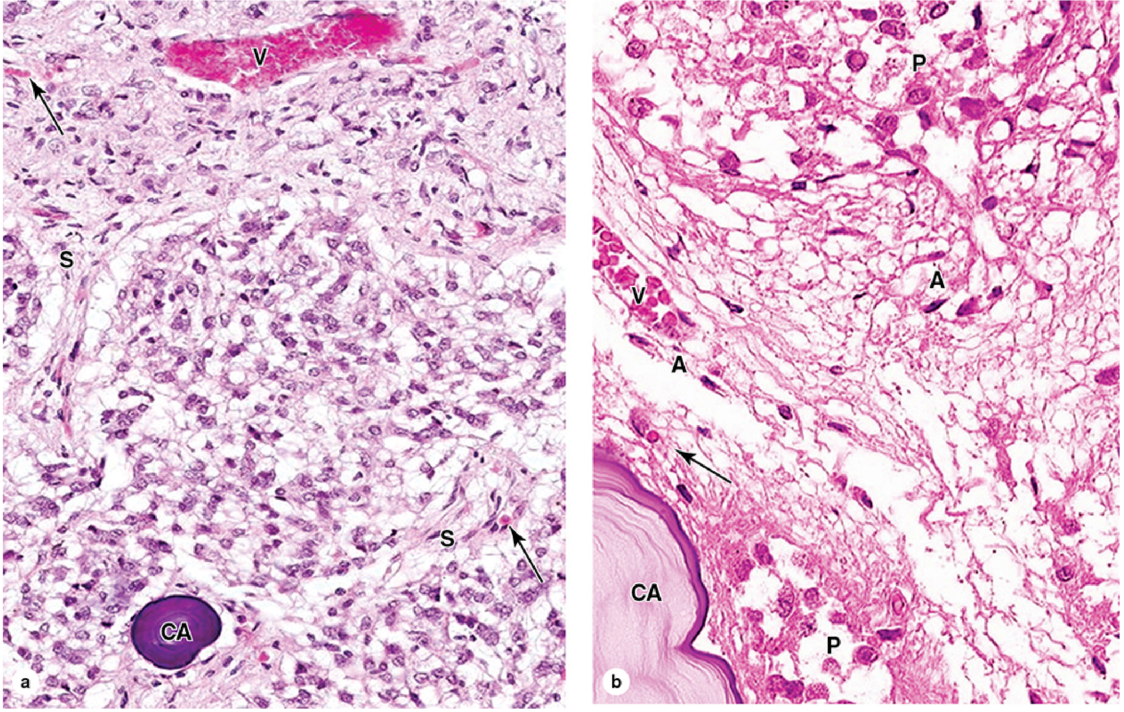
Corpus Arenacea ("Brain Sand")
A hallmark feature of the pineal gland is the presence of corpora arenacea - calcified concretions of calcium and magnesium salts formed by mineralization of extracellular protein deposits around pinealocyte processes.
- They increase in number with age (commonly visible on plain skull X-rays and CT in adults from the 2nd decade onward)
- Of unknown physiologic significance, but serve as a clinically useful anatomic landmark - midline calcification on skull X-rays
- Shift of pineal calcification off midline suggests an intracranial space-occupying lesion
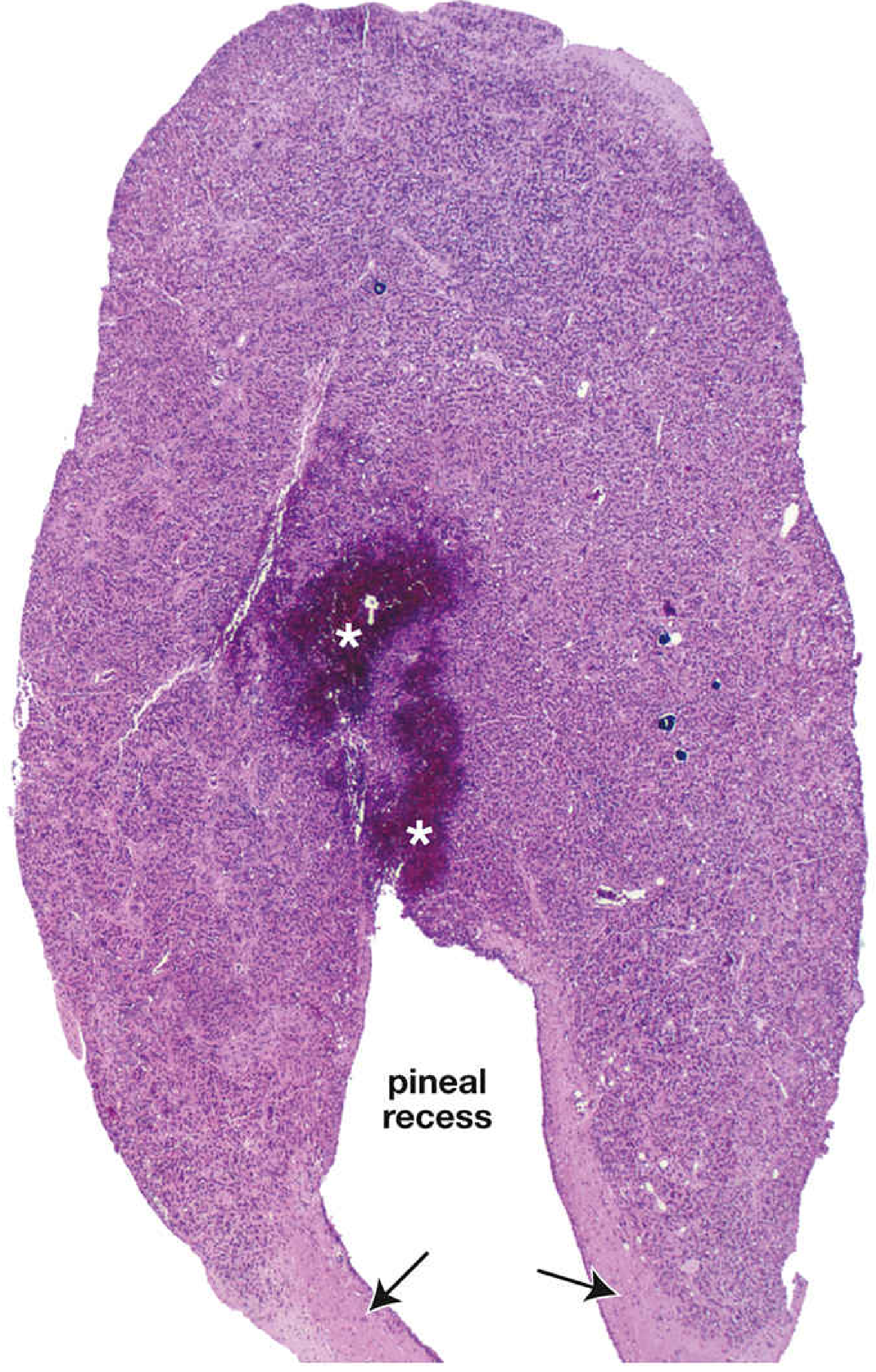
Infant Pineal Gland (gross histology)
Function Summary
| Function | Mechanism |
|---|---|
| Circadian rhythm regulation | Melatonin secretion driven by SCN light-dark signals |
| Sleep-wake cycle | High melatonin at night promotes sleep onset |
| Reproductive maturation | High melatonin → delayed puberty; low melatonin → precocious puberty |
| Endocrine modulation | Influences hypothalamus, pituitary, and adrenal rhythms |
Clinical Notes
- Pinealomas (pineal tumors) are rare; most are germ cell tumors from residual embryonic tissue. They are divided into pineoblastomas (poorly differentiated, aggressive) and pineocytomas (well differentiated, less aggressive).
- Parinaud syndrome (dorsal midbrain compression causing upgaze palsy) can result from pineal region tumors compressing the superior colliculi.
- Because it lies outside the blood-brain barrier, the pineal gland is accessible to blood-borne substances - relevant for drug delivery and pathogen entry.
- Pineal calcification is a useful radiologic marker; displacement suggests mass effect.
- Robbins Pathologic Basis of Disease, p. 1041-1042; Fischer's Mastery of Surgery
This is a shared conversation. Sign in to Orris to start your own chat.