
Muscle tension dysphonia in detail according to standard ENT textbook in simplified manner also give its grades and management
muscle tension dysphonia laryngoscopy vocal folds
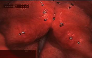
This diagnostic endoscopic image provides a superior laryngoscopic view of the larynx in a patient with Muscle Tension Dysphonia (MTD). The image demonstrates significant supraglottic hyperfunction characterized by both anteroposterior and lateral compression of the glottic aperture. The false vocal folds (ventricular folds) exhibit medial bulging, while the epiglottis and arytenoid tissues show signs of anterior-posterior narrowing, collectively obscuring the true vocal folds. The mucosa appears erythematous, consistent with laryngeal strain. Several white light reflections (artifacts) are visible across the mucosal surface due to the endoscopic light source. This clinical photograph is used to illustrate the functional laryngeal changes associated with primary MTD, where excessive muscular tension leads to abnormal glottal configuration despite the absence of structural lesions like polyps or nodules. The image is relevant for otorhinolaryngology and speech-language pathology education regarding voice disorders and laryngeal assessment.
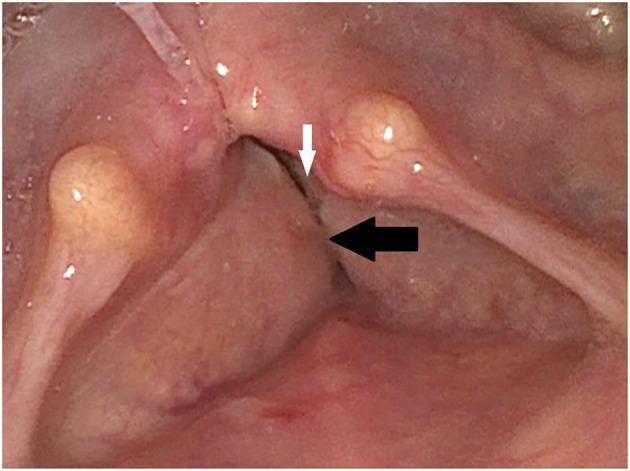
This endoscopic clinical photograph displays the glottal and supraglottal regions of the larynx during laryngoscopy. The image demonstrates bilateral vocal cord palsy, with the true vocal cords (indicated by the white arrow) appearing in a fixed, medial position. The true vocal cords exhibit a narrow glottal gap, suggesting impaired abduction. Positioned superior to the true cords are hyperplastic vestibular folds (false vocal cords, indicated by the black arrow), which appear significantly enlarged and medially displaced. This hyperplasia results in partial obscuration of the underlying true vocal folds. Anatomical landmarks such as the arytenoid eminences and the interarytenoid area are visible but show signs of crowding due to the soft tissue enlargement. This finding is clinically relevant to the diagnosis of bilateral vocal fold immobility and compensatory muscle tension or secondary hyperplasia of the supraglottic structures, which typically presents with symptoms of dyspnea, inspiratory stridor, and dysphonia.
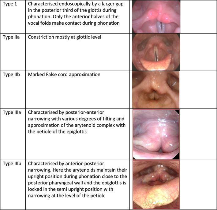
A clinical comparison chart displaying five endoscopic views of the human larynx, illustrating different types of Muscle Tension Dysphonia (MTD) patterns (Types I–III). Each entry consists of a diagnostic photograph and a brief descriptive text of the glottic and supraglottic configurations during phonation. Type 1 shows a posterior glottic gap where only the anterior halves of the vocal folds contact. Type IIa involves constriction primarily at the glottic level, while Type IIb demonstrates marked approximation of the false vocal cords (ventricular folds). Type IIIa is characterized by posterior-anterior narrowing with tilting and approximation of the arytenoid complex toward the epiglottic petiole. Type IIIb shows anterior-posterior narrowing with the arytenoids positioned near the posterior pharyngeal wall and the epiglottis locked in a semi-upright position. These laryngeal hyperfunction patterns are used in otorhinolaryngology and speech-language pathology to classify primary MTD, typically associated with voice misuse or abuse rather than organic pathology.
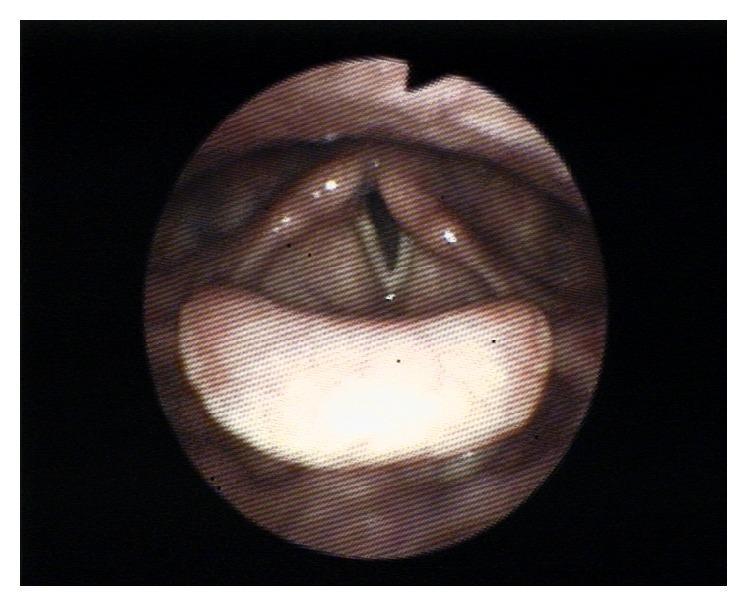
This diagnostic fiberoptic laryngoscopy image provides a superior view of the larynx during attempted phonation. The primary finding is unilateral left vocal cord paralysis. While the right vocal cord appropriately adducts toward the midline, the left vocal cord remains fixed in a more lateral, paramedian position. This asymmetry results in an incomplete glottic closure, manifesting as a visible V-shaped gap (glottal insufficiency) between the true vocal folds. Anatomical landmarks include the epiglottis in the foreground, the false vocal folds, and the arytenoid cartilages posteriorly. The left vocal fold appears relatively flaccid and lacks the tension seen on the contralateral side. This clinical finding is characteristic of recurrent laryngeal nerve neuropraxia or injury, typically presenting with symptoms of dysphonia or hoarseness due to the air leak during phonatory efforts.
Muscle Tension Dysphonia (MTD) - Complete Overview
What is MTD?
- Vocal fold position and tensioning relative to each other
- Position of the larynx relative to the rest of the vocal tract
"Muscle tension dysphonia is defined as a hyperfunctional posturing of the vocal folds. This hyperfunction causes tightness and dysphonia with harshness, breathiness, choppy vocal production, or tremor."
- Cummings Otolaryngology Head and Neck Surgery
Primary vs. Secondary MTD
| Type | Description |
|---|---|
| Primary MTD | The excessive muscle tension IS the cause of the voice disorder - no underlying structural or neurological cause |
| Secondary MTD | Excessive muscle tension develops as a COMPENSATION for an underlying disorder (e.g. vocal fold paresis, nodules, reflux, respiratory problem). The laryngeal muscles try to "make up" for a deficiency |
Causes / Predisposing Factors
- Vocal misuse/abuse - shouting, straining, talking over noise
- Poor vocal technique - especially in professional voice users (teachers, singers, lawyers)
- Stress, anxiety, and depression - emotional arousal directly increases laryngeal muscle tension
- Conversion disorders (psychogenic dysphonia)
- Poor posture and breathing patterns
- Chronic upper respiratory infections - initial muscle compensations that persist
- Extraoesophageal/laryngopharyngeal reflux - mucosal irritation triggering hyperfunction
- Working in dusty, smoky, or fume-laden environments
- Prolonged speaking in poor acoustic environments
Symptoms
- Hoarseness - may be harsh, breathy, or strained in quality
- Pitch abnormalities - voice too high or too low, reduced pitch range
- Globus sensation - feeling of tightness, constriction, or a "lump in the throat"
- Effortful voice production
- Discomfort or pain on speaking or singing
- Vocal fatigue - especially toward end of the day or after prolonged use
- Voice variability - may be noticed during the consultation itself
Diagnosis
Clinical Examination
- Laryngoscopy is the key investigation - reveals the characteristic patterns (see grades below)
- Stroboscopy may show reduced mucosal wave, incomplete closure, supraglottic constriction
- Probe voice therapy during the consultation - voice variability that responds to simple techniques suggests MTD
On Laryngoscopy - Key Signs
- Sustained supraglottal constriction during phonation (anteroposterior or lateromedial)
- Posterior glottic gap
- False vocal cord approximation
- Larynx held high in the neck (laryngeal elevation)
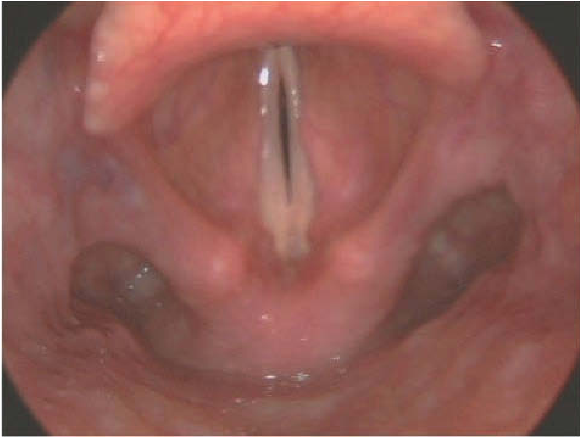
Classification / Grades of MTD
Morrison & Rammage Classification (based on endoscopic appearance)
| Type | Endoscopic Finding | Mechanism |
|---|---|---|
| Type I | Posterior glottic gap - only anterior halves of vocal folds contact during phonation; a triangular gap remains posteriorly | Reduced thyroarytenoid (TA) contraction with increased posterior laryngeal tension |
| Type IIa | Glottic-level constriction - constriction mostly at the glottic level | Overall increase in intrinsic laryngeal muscle tension |
| Type IIb | False cord (ventricular fold) approximation - marked medial bulging and approximation of false cords, partially obscuring true folds | Ventricular fold hyperfunction - the most dramatic pattern visually |
| Type IIIa | Posterior-anterior narrowing - tilting and approximation of the arytenoid complex toward the petiole of the epiglottis | Anteroposterior supraglottic compression dominant |
| Type IIIb | Anterior-posterior narrowing - arytenoids remain upright close to posterior pharyngeal wall; epiglottis locked in semi-upright position with narrowing at its petiole | Complete supraglottic constriction in the AP plane |
| Type IV (Isometric) | Normal-appearing larynx with no visible tension patterns, but voice is abnormal - tension cannot be seen endoscopically | Entirely intrinsic/muscular; also called "isometric" MTD |
Note: These patterns may also be seen to a mild degree in the normal population - clinical correlation is always required.
Management
1. Treat the Underlying Cause (Secondary MTD)
- Treat reflux (LPR/GORD) with PPI therapy, dietary advice
- Treat structural lesions (nodules, polyps, etc.) if present
- Treat nasal/sinus conditions affecting resonance
2. Voice Therapy (Primary Treatment - Direct & Indirect)
| Technique | How it helps |
|---|---|
| Yawn-sigh method | Opens and relaxes the larynx; reduces supraglottic constriction |
| Chewing technique | Relaxes jaw, tongue, and laryngeal muscles during phonation |
| Vocal function exercises | Strengthening and balance of intrinsic laryngeal muscles |
| Airflow/Flow phonation | Encourages breathy, easy onset; reduces hard glottal attacks |
| Resonance therapy | Optimizes forward resonance (humming, nasal consonants) |
| Deconstruction therapy | Reduces constriction by targeting specific muscle groups |
| Easy vocal onset | Replaces hard glottal attacks with gentle onset |
| Confidential voice therapy | Soft, low-intensity voicing to reduce laryngeal tension |
| Accent Method | Rhythmic breath-voice coordination to improve tension balance |
| Laryngeal manipulation | Manual therapy to lower laryngeal position and reduce extrinsic tension |
| CBT-enhanced voice therapy | Addresses psychological factors alongside voice technique |
- Vocal hygiene program - adequate hydration, avoid irritants (caffeine, alcohol, smoking), steam inhalation
- Patient education and counselling - understanding the condition
- Vocal rest - appropriate periods of silence, especially after acute episodes
- Elimination of voice abuse - identifying and stopping harmful vocal behaviors
- Avoidance of laryngeal irritants
- General relaxation - progressive muscle relaxation, mindfulness
- Vocal diary - self-monitoring of voice use patterns
- Hierarchy analysis - graded exposure to voice-demanding situations
3. Laryngeal Manual Therapy
- Applying gentle downward pressure and massage to the thyrohyoid space
- Releasing hyoid bone and thyroid cartilage tension
- Most effective in patients with palpable suprathyoid/thyrohyoid tenderness
- Evidence shows significant improvement when combined with voice therapy
4. Psychological/Behavioural Intervention
- Cognitive Behavioural Therapy (CBT) - particularly important when anxiety, depression, or psychogenic factors are prominent
- Helps address perpetuating factors to prevent relapse
- Recent studies show CBT-enhanced voice therapy produces broader and more lasting improvement than voice therapy alone
5. Medical Treatment
- LPR/GORD management - PPIs, alginate preparations, dietary modification
- Botulinum toxin injection - occasionally used in severe or refractory cases (into thyroarytenoid muscle), though evidence is limited
Key Points Summary
| Feature | Detail |
|---|---|
| Most common functional voice disorder | Yes - one of the biggest causes of voice problems |
| Diagnosis of exclusion | Often, but can coexist with organic pathology |
| Primary cause | Vocal misuse, poor technique, stress |
| Gold standard investigation | Laryngoscopy ± stroboscopy |
| First-line treatment | Voice therapy (direct + indirect) |
| Adjunct treatment | Laryngeal manual therapy, CBT, treat underlying cause |
| Prognosis | Generally good with voice therapy; relapse if predisposing factors not addressed |