
okayy see i have an exam in 12 hours so i will ask you diffferent questions regarding anatomy, physio and histo for everything i want you to give me simple and right answers pleasee i want the answer to be very simple and probabbly some trick to remember
in which part of the stomach are goblet and parietal cells present
- Parietal cells - found in the body/fundus of the stomach (oxyntic glands)
- Goblet cells - NOT normally present in the stomach at all. Their presence in the stomach = intestinal metaplasia (a pre-cancerous change)
Goblet Cells & Parietal Cells in the Stomach
Parietal Cells
- Found in the fundus and body of the stomach (oxyntic glands)
- They secrete HCl and intrinsic factor
Goblet Cells
- NOT normally present in the stomach
- If goblet cells appear in the stomach, it means intestinal metaplasia has occurred - this is a pathological finding (seen in chronic atrophic gastritis, H. pylori infection, and is a pre-cancerous change for gastric cancer)
Memory Tricks
"Parietal cells Pump acid in the Proper stomach" (fundus/body = the proper/main stomach)
Goblet cells belong in the gut (intestine), not the stomach. If they show up in the stomach, the stomach is trying to become intestine - that's intestinal metaplasia. Think: "Goblets don't belong on the stomach table"
then how is mucus produceded in stomach
- Surface mucous cells - line the entire stomach surface
- Mucous neck cells - sit in the necks of gastric glands
How Mucus is Produced in the Stomach
| Cell | Location | What it does |
|---|---|---|
| Surface mucous cells | Lines the entire stomach surface + gastric pits | Secretes thick, viscous mucus + bicarbonate |
| Mucous neck cells | Neck of gastric glands | Secretes thinner mucus |
Key Point for Exam
Goblet cells use goblets (cups) to secrete mucus in the intestine. The stomach uses surface mucous cells - same job, different cell, different location.
cells of crypts of Lieberkuhn?
Cells of Crypts of Lieberkühn
| Cell | Location in Crypt | Function |
|---|---|---|
| Stem cells | Lower crypt | Give rise to all other cells |
| Enterocytes | Migrate up to villi | Absorption |
| Goblet cells | Throughout crypt | Mucus secretion |
| Enteroendocrine cells | Throughout crypt | Hormones (GIP, secretin, CCK etc.) |
| Paneth cells | BASE only (never migrate up) | Antimicrobials - defensins, lysozyme |
Key Exam Points
- Paneth cells = bottom of crypt - they are the only cells that stay at the base and do NOT migrate up to villi
- Paneth cells have bright pink (eosinophilic) granules - easy to spot in histology
- All other cells are born in the crypt, migrate UP to the villi, live ~3-5 days, then shed
Memory Trick
Paneth, Enterocytes, Goblet, Stem, Enteroendocrine
what is this crypts of lieberkuhn
Crypts of Lieberkühn - What Are They?
- The hills = villi (absorb nutrients)
- The valleys/pits between the hills = Crypts of Lieberkühn
Simple Summary
| Feature | Detail |
|---|---|
| Other name | Intestinal glands |
| Location | Small intestine (between villi) AND large intestine (no villi there, only crypts) |
| Shape | Test-tube shaped pits going DOWN into the mucosa |
| Main job | Cell production - constantly making new cells to replace the ones shed off villi |
The Crypt-Villus Axis (super important!)
VILLUS (tip) ← cells migrate UP and die here
    ↑
    ↑  cells travel upward
    ↑
CRYPT (base) ← cells are BORN here (stem cells live here)
Memory Trick
"Crypts = Cradle" - where new intestinal cells are born and raised before moving up.
so not in stomach?
GI Tract - What's Where
| Structure | Location |
|---|---|
| Gastric glands (oxyntic glands, pyloric glands) | Stomach |
| Crypts of Lieberkühn | Small intestine + Large intestine |
| Villi | Small intestine only |
| Rugae | Stomach only |
The Stomach Has Its OWN Glands:
- Cardiac glands - in cardia
- Oxyntic (fundic) glands - in fundus & body (have parietal + chief cells)
- Pyloric glands - in antrum/pylorus (mostly mucus)
Memory Trick
"Lieberkühn = Intestinal only" The stomach is too acidic and tough - it has its own VIP glands (gastric glands), not crypts!
- Stomach → gastric glands + gastric pits
- Intestine → crypts of Lieberkühn
Question N17 Mucous cells are most often organized as acini rather than cylindrical tubules. Answer: FALSE
The Answer is FALSE - and here's why:
The Correct Fact (from Junqueira's Histology):
Mucous cells are organized as short tubules (NOT acini) Serous cells are organized as acini
Simple Rule to Remember:
| Cell Type | Shape of Secretory Unit |
|---|---|
| Serous cells | Acini (round, berry-like clusters) |
| Mucous cells | Tubules (short tube-shaped) |
Example from Salivary Glands:
- Sublingual gland = mostly mucous tubules (mostly mucous)
- Parotid gland = purely serous acini
- Submandibular = mixed (both)
Memory Trick:
"Serous = Spherical (acini)" - both start with S "Mucous = Tubes" - mucus flows through TUBES (like a pipe)
Question N16 Which section of the digestive tract has the most goblet cells? Answer: Large intestine i was thinking smalll intestine
Why Large Intestine has the MOST Goblet Cells
The Logic:
| Section | Goblet Cells | Why? |
|---|---|---|
| Small intestine | Present but fewer | Has villi for absorption - focus is on absorbing nutrients |
| Large intestine | MOST abundant | No digestion here - main job is lubrication to move dry, hard stool along |
Why you thought small intestine - understandable!
The Logic to Remember:
The further down the gut, the drier and harder the contents → more mucus needed → more goblet cells
Stomach → 0 goblet cells
Small intestine → some goblet cells
Large intestine → MOST goblet cells ✓
Memory Trick:
"Large intestine = Large number of goblet cells" The colon needs to LUBRICATE hard stool - it needs all the mucus it can get!
large intestine does not have villi?
Villi - Where and Where NOT
| Structure | Villi? | Crypts? |
|---|---|---|
| Stomach | ❌ No | ❌ No (has gastric pits/glands) |
| Small intestine | ✅ YES | ✅ Yes (crypts of Lieberkühn) |
| Large intestine | ❌ No | ✅ Yes (crypts of Lieberkühn only) |
Why no villi in large intestine?
What does large intestine mucosa look like then?
- Flat surface (no villi)
- Deep crypts packed with goblet cells
- That's why it looks smooth compared to the velvety small intestine
Memory Trick - The 3 V's:
"Villi = Small intestine only" Villi are Very important for Vitamin & nutrient absorption = Small intestine's job
"Small = has villi, Large = no villi" - the SMALL intestine is the one doing the big absorption work!
what is the function of goblet cells and also how is water and electrolyte absorbed in LI and also does it not have microvilli as well?
All Three Answers:
1. Function of Goblet Cells
- Mucus lubricates the gut lining so stool can pass easily
- Mucus protects the epithelium from digestive enzymes and bacteria
- In the large intestine especially - keeps the dry, compacting stool moving smoothly
"Goblet = Cup of mucus" - shaped like a goblet/wine cup, filled with mucus granules
2. How Water & Electrolytes are Absorbed in Large Intestine
- Na⁺ is actively pumped out (via Na⁺/K⁺ ATPase on basolateral side)
- Water follows passively by osmosis
- Cl⁻ follows Na⁺ (electrochemical gradient)
- About 1.5L of water is absorbed daily in the colon
3. Microvilli - YES! Large intestine DOES have microvilli!
| Structure | Large Intestine? |
|---|---|
| Villi (finger-like mucosal folds) | ❌ NO |
| Microvilli (tiny projections on individual cells = brush border) | ✅ YES |
The Key Distinction:
Villi = visible folds of the mucosa (macro structure, seen with naked eye/low power) Microvilli = tiny projections on each individual cell surface (micro structure, only seen with electron microscope) = form the "brush border"
Question N9 A parotid gland biopsy of a 48-year-old woman shows inflammatory infiltrates in the interlobular connective tissue with damage to acinar cells and striated ducts. In this case, resorption of which of the following will be most altered by destruction of the striated ducts? Answer:
Answer: Sodium (Na⁺)
What do Striated Ducts do?
| Action | What happens |
|---|---|
| Reabsorb Na⁺ (main job) | Pumps sodium OUT of saliva back into blood |
| Secrete K⁺ | Puts potassium INTO saliva |
| Reabsorb Cl⁻ | Takes chloride back |
| Secrete HCO₃⁻ | Adds bicarbonate into saliva |
Why striated ducts? (The histology hint)
Memory Trick:
"Striated ducts = Sodium stealers" They steal Na⁺ back from saliva, just like the kidney tubules steal Na⁺ back from urine That's why they're called the "kidney of the salivary gland"!

Question N10 The branching structures shown in the photomicrograph below (a scanning electron micrograph taken from the region between two hepatocytes) are involved in which of the following?
Answer: Bile secretion / Bile transport
What are Bile Canaliculi?
- They are tiny grooves/channels formed between two adjacent hepatocytes
- NOT separate tubes - they are formed by the plasma membranes of two neighboring hepatocytes coming together
- Lined with microvilli on the inside (to increase surface area for bile secretion)
- Sealed by tight junctions on the edges (to prevent bile leaking into blood)
Bile Flow Direction:
Hepatocyte secretes bile
        ↓
Bile canaliculi (between hepatocytes)
        ↓
Canal of Hering
        ↓
Bile ductules → Bile ducts → Common bile duct → Duodenum
Key Exam Points:
- Bile canaliculi have no wall of their own - made by the hepatocyte membranes themselves
- The microvilli inside = seen in SEM as the "branching/finger-like" structures in the image
- Function = initial collection point for bile
Memory Trick:
"Canaliculi = little canals between liver cells" Think of two houses sharing a gutter between them - that gutter = bile canaliculus!
Question N9 Which of the following locations in the mucosa provides a niche for multipotent gastric stem cells? Answer: Isthmus of glandular epithelium can u explain this i dont understand anything
Let me explain from scratch!
First - understand the structure of the stomach wall:
STOMACH LUMEN (inside)
        |
   GASTRIC PIT  ← opening at surface
        |
    ISTHMUS     ← narrow middle zone ← STEM CELLS LIVE HERE
        |
    NECK        ← mucous neck cells here
        |
    BASE        ← parietal, chief cells here
What is the ISTHMUS?
- Isthmus = where stem cells sit
- From here, stem cells can go UP → become surface mucous cells
- From here, stem cells can go DOWN → become parietal cells, chief cells, neck cells
Why is this important?
Compare with intestine:
| Location | Stem cell niche |
|---|---|
| Stomach | Isthmus of gastric gland |
| Small/Large intestine | Base of crypt of Lieberkühn |
Memory Trick:
"Isthmus = Waist of the gland = where stem cells WAIT before deciding which direction to go"
can u please show me an easy diagram so i can remeber better
Simple labeled diagram of a gastric gland and gastric pit in the stomach wall for medical students. Show a cross-section of the stomach mucosa with these clearly labeled parts from top to bottom: "Stomach Lumen" at the top, then "Gastric Pit (opening)", then "Isthmus - STEM CELLS HERE" with a highlight or star, then "Neck - mucous neck cells", then "Base - parietal cells (HCl) and chief cells (pepsin)". Show arrows pointing upward from the isthmus labeled "migrate UP → surface mucous cells" and arrows pointing downward labeled "migrate DOWN → parietal, chief cells". Clean, colorful, easy to read, educational medical diagram style.
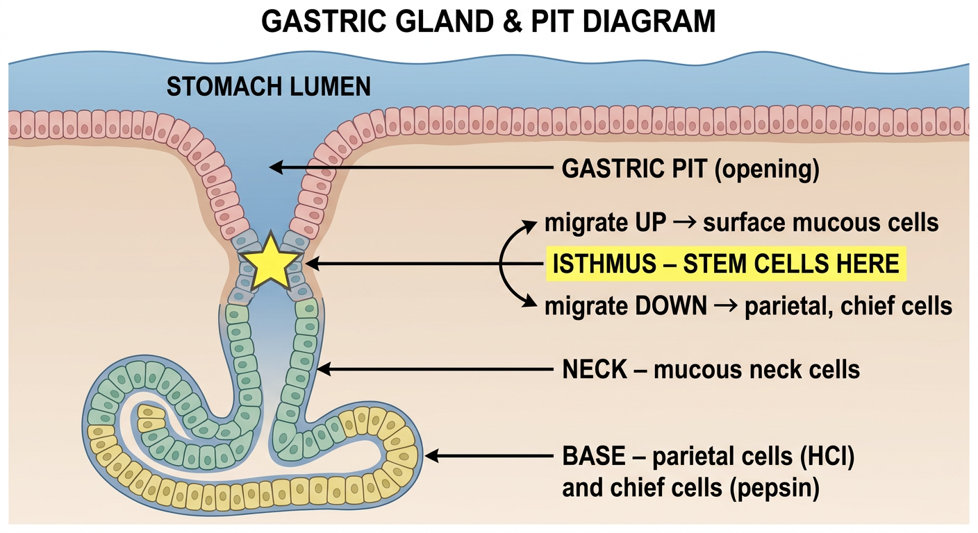
Quick recap from the diagram:
- Gastric pit = the opening at the top (surface)
- Isthmus = the narrow "waist" = where stem cells live ⭐
- Stem cells migrate UP → become surface mucous cells
- Stem cells migrate Down → become parietal cells (HCl) and chief cells (pepsin)
- Base = deepest part = working cells (parietal + chief)
One liner to remember:
"Stem cells sit at the WAIST (isthmus) of the gastric gland - they send kids UP and DOWN"
what is antrum
Stomach Regions - Simple Map
ESOPHAGUS
    ↓
CARDIA  ← where esophagus meets stomach
    ↓
FUNDUS  ← dome-shaped top part
    ↓
BODY    ← main middle part (biggest region)
    ↓
ANTRUM  ← lower part, funnel-shaped
    ↓
PYLORUS ← exit valve → leads to duodenum
    ↓
DUODENUM
What's special about the Antrum?
| Feature | Detail |
|---|---|
| Other name | Pyloric antrum |
| Main cells | G cells (secrete gastrin) |
| Glands | Pyloric glands - mostly mucus secreting |
| Function | Grinds food, mixes it, releases gastrin |
| No parietal cells | ✅ correct - parietal cells are in fundus/body only |
Key Exam Fact:
Antrum = home of G cells → G cells make Gastrin → Gastrin stimulates parietal cells (in body/fundus) to make HCl
Memory Trick:
"Antrum = Announcer" - it announces to the rest of the stomach "hey, food is here, make acid!" via gastrin.
"G cells in G-astric antrum make G-astrin" - all G's!
What's the difference between fundus and antrum glands?2 absorption?
Fundus/Body Glands vs Antrum Glands
Part 1 - The Difference:
| Feature | Fundic/Oxyntic Glands (Fundus + Body) | Pyloric Glands (Antrum) |
|---|---|---|
| Other name | Oxyntic glands | Pyloric glands |
| Main cells | Parietal cells + Chief cells | G cells + mucous cells |
| Parietal cells secrete | HCl + Intrinsic factor | ❌ absent |
| Chief cells secrete | Pepsinogen | ❌ absent |
| G cells secrete | ❌ absent | Gastrin |
| Gland shape | Long, straight, deep | Shorter, more coiled/branched |
| Main function | Digestion (acid + enzymes) | Hormonal control + mucus |
Simple way to remember:
Fundus/Body = Factory - makes the actual acid and enzymes Antrum = Manager - sends hormones (gastrin) telling the factory what to do
Part 2 - Absorption in the Stomach?
| Substance | Absorbed in stomach? |
|---|---|
| Water | tiny amount |
| Alcohol | ✅ YES - absorbed here (that's why it hits fast!) |
| Aspirin (weak acid) | ✅ small amount |
| Nutrients (glucose, protein, fat) | ❌ NO - absorbed in small intestine |
Memory Trick:
"Only ASS gets absorbed in the stomach" Alcohol, Salicylates (aspirin), Some water - everything else waits for the small intestine!
g cells in atnrum helps in vitamin b12 absorption?
Vitamin B12 Absorption - The Correct Story
The key player is: PARIETAL CELLS (not G cells)
Parietal cells (fundus/body)
        ↓ secrete
INTRINSIC FACTOR (IF)
        ↓ binds to
Vitamin B12 in stomach
        ↓ IF-B12 complex travels to
Terminal ILEUM
        ↓ absorbed here
Into bloodstream
G cells vs Parietal cells - Don't confuse them!
| Cell | Location | Secretes | Role in B12? |
|---|---|---|---|
| G cells | Antrum | Gastrin | ❌ NO role in B12 |
| Parietal cells | Fundus/Body | HCl + Intrinsic Factor | ✅ YES - make intrinsic factor |
What does Gastrin (from G cells) actually do?
Why B12 deficiency happens:
Memory Trick:
"Intrinsic Factor from Parietal cells = B12's VIP escort" Without the escort (IF), B12 can't get into the terminal ileum party! G cells = gastrin only, nothing to do with B12!

what is this?
This is a Hepatic Lobule histology slide!
What the Letters Mean:
| Letter | Structure | How to identify |
|---|---|---|
| C | Central Vein | Large vessel in the CENTER of the lobule |
| V | Portal Vein (branch) | Largest vessel in the portal triad - thin wall, big lumen |
| A | Hepatic Artery (branch) | Small vessel, THICK wall, small lumen |
| D | Bile Duct | Lined by cuboidal cells, carries bile AWAY from liver |
The Big Picture - Classic Hepatic Lobule:
         Portal triad          Portal triad
         (V + A + D)           (V + A + D)
              \                    /
               \                  /
        Blood flows INWARD → toward center
                        ↓
                  CENTRAL VEIN (C)
                        ↓
              Blood drains OUT to hepatic vein → IVC
Memory Trick for Portal Triad:
"VAD is at the Periphery" Vein, Artery, Duct = always found together at the corners Central vein = alone in the Center
How to tell V, A, D apart in histology:
- V (portal vein) = BIGGEST lumen, thinnest wall
- A (artery) = small lumen, THICK muscular wall
- D (bile duct) = lined by cuboidal epithelium (not flat endothelium like vessels)
B) Oligohydramnios explain in simple terms
Oligohydramnios - Super Simple Explanation
What is it?
- Normal amniotic fluid = ~800mL at term
- Oligohydramnios = significantly LESS than normal
Where does amniotic fluid come from?
Causes (remember with RUOK):
| Cause | Example |
|---|---|
| Rupture of membranes | Most common cause - fluid leaks out |
| Urinary tract problem | Renal agenesis (no kidneys), urethral obstruction |
| Outflow obstruction | Posterior urethral valves (boys) |
| Kidneys absent/abnormal | Potter sequence |
Consequences - Potter Sequence:
No kidneys/urine → No amniotic fluid
        ↓
Baby gets squished in womb
        ↓
1. Pulmonary hypoplasia (lungs can't develop - most lethal!)
2. Limb deformities (clubfoot)
3. Flat face / Potter facies
4. Intrauterine growth restriction
Pulmonary hypoplasia is the most dangerous consequence - babies die because lungs never developed properly
vs Polyhydramnios (opposite):
| Oligohydramnios | Polyhydramnios | |
|---|---|---|
| Fluid | TOO LITTLE | TOO MUCH |
| Baby can't... | PEE (kidney/urinary problem) | SWALLOW (GI obstruction, e.g. esophageal atresia, anencephaly) |
Memory Trick:
"Oligo = no PEE → kidney problem" "Poly = can't SWALLOW → GI/neuro problem"
"Oligo = Output problem (urine)" "Poly = swallowing Problem"
Question N13 stimulation of the sympathetic nerve fibres causes segmental contraction Answer: 0
Answer: FALSE (0)
The correct fact:
From Guyton & Hall: "Stimulation of the sympathetic nervous system INHIBITS activity of the gastrointestinal tract"
Sympathetic vs Parasympathetic on the GUT:
| Sympathetic ("fight or flight") | Parasympathetic ("rest and digest") | |
|---|---|---|
| Effect on motility | INHIBITS (slows/stops) | STIMULATES (increases) |
| Effect on secretions | Decreases | Increases |
| Sphincters | Contracts (closes) | Relaxes (opens) |
| Blood flow to gut | Decreases | Normal/increases |
So what DOES cause segmental contraction?
Memory Trick:
"Sympathetic = Scared = STOPS digesting" When you're scared/running from danger, your body shuts down the gut "Para = PROMOTES digestion" (rest and digest)
Sphincters are the EXCEPTION: sympathetic CONTRACTS sphincters (to hold everything in while you run!)
Question N28 Atrial natriuretic peptide increases NaCl reabsorption in distal tubule/collecting tubule and duct Answer: 0
Answer: FALSE (0)
The correct fact:
ANP acts on the distal tubule / collecting tubule and duct to cause ↓ NaCl reabsorption (NOT increase!)
What ANP actually does:
When is ANP released?
ANP's goal = get rid of Na⁺ and water = lower blood pressure
| Action | Effect |
|---|---|
| ↓ NaCl reabsorption in distal tubule/collecting duct | More Na⁺ lost in urine |
| Water follows Na⁺ out | More urine output |
| ↓ Renin release | Less angiotensin II → less aldosterone |
| Vasodilation | Lower blood pressure |
| ↓ Aldosterone directly | Less Na⁺ reabsorption |
Compare the hormones:
| Hormone | Site | Effect on NaCl |
|---|---|---|
| Aldosterone | Distal tubule/collecting duct | ↑ reabsorption (keeps Na⁺) |
| ANP | Distal tubule/collecting duct | ↓ reabsorption (loses Na⁺) |
| ADH | Collecting duct | ↑ water reabsorption only |
Memory Trick:
"ANP = A-Natriuretic Peptide = makes you pee Na⁺" "Natri-URETIC" = puts sodium in the URINE The name literally tells you it LOSES sodium - NOT reabsorbs it!
"ANP is the heart saying: TOO MUCH FLUID - DUMP IT!"
Question N29 Prolonged treatment with diuretic drugs that block Na channels in the late distal tubules and cortical collecting tubules, can lead to hypokalemia. Answer: 0
Answer: FALSE (0)
The correct fact:
Why? You need to understand the Na-K exchange:
Na⁺ comes IN (through ENaC channels)
        ↓
Creates negative charge inside cell
        ↓
K⁺ is SECRETED OUT into tubule lumen (to balance charge)
        ↓
K⁺ lost in urine
Now BLOCK the Na⁺ channel:
Na⁺ can't come in
        ↓
No negative charge created
        ↓
No driving force for K⁺ to leave
        ↓
K⁺ stays in blood → HYPERKALEMIA ✓
These drugs = Potassium-SPARING diuretics
| Drug | Mechanism | K⁺ effect |
|---|---|---|
| Amiloride | Blocks ENaC (Na channel) directly | ↑ K⁺ (hyperkalemia) |
| Triamterene | Blocks ENaC (Na channel) directly | ↑ K⁺ (hyperkalemia) |
| Spironolactone | Blocks aldosterone receptor | ↑ K⁺ (hyperkalemia) |
Contrast with other diuretics:
| Diuretic | Example | K⁺ effect |
|---|---|---|
| Loop diuretics | Furosemide | ↓ K⁺ (HYPOkalemia) |
| Thiazides | Hydrochlorothiazide | ↓ K⁺ (HYPOkalemia) |
| K⁺-sparing | Amiloride, Spironolactone | ↑ K⁺ (HYPERkalemia) |
Memory Trick:
"Block Na channel → K stays → HYPERkalemia" "K-sparing diuretics SPARE K (keep it) = HIGH K in blood" The name "potassium-SPARING" tells you K is saved = HIGH = hyperkalemia!
Question N32 The majority of progesterone is produced in Graafian follicle Answer: 0
Answer: FALSE (0)
The correct fact:
Progesterone is secreted by the corpus luteum - mainly in the second half (luteal phase) of the menstrual cycle.
What produces what - the full picture:
The life story of a follicle:
Primary follicle
        ↓ grows
Graafian follicle (mature follicle)
        ↓ OVULATION (LH surge)
Egg released → Corpus luteum forms from the leftover follicle
        ↓
Corpus luteum secretes PROGESTERONE (mainly) + some estrogen
        ↓ (if no pregnancy)
Corpus luteum degenerates → Corpus albicans (scar)
What does each structure make?
| Structure | Main hormone |
|---|---|
| Graafian follicle (theca + granulosa cells) | Estrogen (mainly) |
| Corpus luteum | Progesterone (mainly) + some estrogen |
| Placenta (during pregnancy) | Takes over progesterone after 8-10 weeks |
Memory Trick:
"Corpus Luteum = Progesterone Palace" After ovulation, the leftover follicle becomes the corpus luteum = the main progesterone factory
"Graafian = Estrogen, Corpus Luteum = Progesterone" Think: "G comes before C in alphabet" = Graafian (1st half = estrogen) → Corpus luteum (2nd half = progesterone)
Why does this matter clinically?
- No ovulation → no corpus luteum → no progesterone (seen in PCOS)
- Progesterone maintains pregnancy lining - without it → menstruation
- If pregnancy occurs, hCG keeps corpus luteum alive to keep making progesterone until placenta takes over
Question N3 Which of the following statements is NOT true? Answer: The adrenal medulla and the adrenal cortex function independently from each other why is this not true
Why They Do NOT Function Independently
The KEY connection - confirmed by textbook:
The adrenal medulla NEEDS cortisol from the cortex to function properly!
The Cortisol-Epinephrine Link:
Adrenal CORTEX
        ↓ produces cortisol
Cortisol drains into adrenal medulla via local blood flow
        ↓
Cortisol INDUCES the enzyme PNMT
(Phenylethanolamine-N-methyltransferase)
        ↓
PNMT converts: Norepinephrine → EPINEPHRINE
Summary of their interdependence:
| Interaction | Detail |
|---|---|
| Cortex → Medulla | Cortisol activates PNMT enzyme in medulla |
| PNMT | Converts norepinephrine → epinephrine |
| Result | 80% of medullary output is epinephrine (because of cortisol!) |
| Blood supply | Cortical blood flows THROUGH medulla (portal-like system) |
Also - shared blood supply:
Memory Trick:
"Cortex feeds the Medulla" "No Cortisol = No Epinephrine" (only norepinephrine would be made) "PNMT = Please Need My Cortisol" to make epinephrine!
Question N11 Cholecystokinin inhibits secretion from the exocrine pancreas Answer: 0
Answer: FALSE (0)
The correct fact:
CCK STIMULATES enzyme secretion from pancreatic acinar cells - it's one of CCK's PRIMARY jobs!
CCK - What it does (full picture):
Where is CCK made?
CCK's 3 main jobs:
| Action | Target | Effect |
|---|---|---|
| Stimulates enzyme secretion | Pancreatic acinar cells | Releases lipase, protease, amylase |
| Contracts gallbladder | Gallbladder | Squeezes bile into duodenum |
| Relaxes sphincter of Oddi | Bile duct sphincter | Opens it to let bile/enzymes flow |
CCK vs Secretin - don't confuse them!
| Hormone | Source | Stimulates |
|---|---|---|
| CCK | I cells (duodenum) | Pancreatic ENZYMES (acinar cells) + gallbladder contraction |
| Secretin | S cells (duodenum) | Pancreatic HCO₃⁻ + water (duct cells) - to neutralize acid |
Memory Trick:
"CCK = Calls the Cooking Kit" Fat/protein arrives in duodenum → CCK calls for digestive enzymes from pancreas + bile from gallbladder to cook (digest) the food!
"CCK = Contract the gallbladder, Call the enzymes"
Secretin = "Secret-in bicarbonate" - neutralizes the acid secretly arriving from stomach
Question N2 Major artery of the tongue is lingual artery which originates from
Answer: External Carotid Artery (ECA)
Branches of External Carotid Artery - The Full List:
| Letter | Branch | Supplies |
|---|---|---|
| S | Superior thyroid a. | Thyroid gland (1st branch) |
| A | Ascending pharyngeal a. | Pharynx |
| L | Lingual a. | Tongue ← your answer |
| F | Facial a. | Face |
| O | Occipital a. | Back of scalp |
| P | Posterior auricular a. | Behind ear |
| M | Maxillary a. | Deep face, teeth, nose |
| S | Superficial temporal a. | Scalp/temple |
The Lingual Artery specifically:
- Runs deep to the hyoglossus muscle
- Divides into branches to supply the whole tongue
- Important landmark: hypoglossal nerve (CN XII) runs nearby
Memory Trick:
"Some Angry Ladies Fight Over PMS" = 8 branches of ECA in order Lingual = L = 3rd branch = Licks the tongue (lingual = tongue)
Lingual artery → External Carotid Artery ✓
Question N3 General sensation from the pharyngeal part of the tongue are carried by:
Answer: Glossopharyngeal Nerve (CN IX)
Tongue Innervation - The Full Map:
- Anterior 2/3 = oral part
- Posterior 1/3 = pharyngeal part
Sensory Innervation:
| Part of tongue | General sensation | Taste |
|---|---|---|
| Anterior 2/3 | Lingual nerve (CN V3) - branch of trigeminal | Chorda tympani (CN VII) - facial nerve |
| Posterior 1/3 | CN IX (Glossopharyngeal) | CN IX (Glossopharyngeal) |
| Epiglottis/base | CN X (Vagus) | CN X (Vagus) |
Motor (movement of tongue):
Simple Summary Table:
| Nerve | Job on tongue |
|---|---|
| CN V3 (lingual) | General sensation - anterior 2/3 |
| CN VII (chorda tympani) | Taste - anterior 2/3 |
| CN IX | General sensation + taste - posterior 1/3 |
| CN X | Sensation + taste - epiglottis |
| CN XII | Movement (motor) |
Memory Trick:
"CN IX does it ALL in the posterior 1/3" - both sensation AND taste "Anterior 2/3 = needs TWO nerves (V3 + VII)" "Posterior 1/3 = just ONE nerve does both (IX)"
"Anterior = 5 & 7, Posterior = 9" (numbers go up as you go back in the mouth!)
Question N6 The anterior superior alveolar artery originates from: Answer:
Answer: Infraorbital Artery
The Full Chain:
External Carotid Artery (ECA)
        ↓
Maxillary Artery (3rd part)
        ↓
Infraorbital Artery
        ↓
Anterior Superior Alveolar Artery
(supplies upper front teeth + gums)
All Superior Alveolar Arteries:
| Artery | Origin | Supplies |
|---|---|---|
| Anterior superior alveolar | Infraorbital artery | Upper front teeth (incisors, canines) |
| Middle superior alveolar | Infraorbital artery | Upper premolars |
| Posterior superior alveolar | Maxillary artery directly (3rd part) | Upper molars |
Memory Trick:
"Anterior and Middle = Infraorbital (they're brothers from the same parent)" "Posterior = goes directly from Maxillary (it's independent)"
ECA → Maxillary → Infraorbital → Anterior Superior Alveolar
ECA (neck) → Maxillary (deep face) → Infraorbital (eye floor) → teeth
Question N7 Infection originating in the teeth can track into the cranial cavity through: Answer: The superior mesenteric vein
The Correct Answer: Pterygoid venous plexus → Cavernous sinus
How dental infection reaches the cranial cavity:
Tooth infection (abscess)
        ↓
Alveolar veins (drain teeth)
        ↓
Pterygoid venous plexus (in infratemporal fossa)
        ↓ (via emissary veins - NO VALVES!)
Cavernous sinus (inside skull)
        ↓
INTRACRANIAL SPREAD → cavernous sinus thrombosis!
Why this pathway is dangerous:
| Feature | Why dangerous |
|---|---|
| Valveless veins in face/head | Infection can spread in ANY direction |
| Pterygoid plexus connects to cavernous sinus | Direct route from teeth to brain |
| Facial vein also connects to cavernous sinus | Via ophthalmic veins |
The "Danger Triangle of the Face":
Memory Trick:
"Valveless veins = infection highway - no toll booths, no stopping!" "Teeth → Pterygoid plexus → Cavernous sinus = the dental-brain highway"
"Superior mesenteric vein" is in the BELLY - nothing to do with teeth!
Question N12 Which Mucosal fold relates to the pharyngotympanic tube? Answer: The palatoglossal fold
The Correct Answer: Salpingopharyngeal fold
What is the Pharyngotympanic Tube?
The Mucosal Folds Around the Tube Opening:
| Structure | What it is |
|---|---|
| Torus tubarius | Ridge of cartilage around the tube opening |
| Salpingopharyngeal fold | Mucosal fold running DOWNWARD from torus tubarius (covers salpingopharyngeus muscle) |
| Salpingopalatine fold | Smaller fold running toward soft palate |
| Fossa of Rosenmüller | Recess BEHIND the torus tubarius |
The Key Answer:
The mucosal fold that RELATES to the pharyngotympanic tube = Salpingopharyngeal fold "Salpingo" = Greek for tube (same root as fallopian tube = uterine tube)
The Pharyngeal Folds - Don't Confuse:
| Fold | Location | Related to |
|---|---|---|
| Palatoglossal fold | Mouth - between soft palate & tongue | Forms anterior pillar of tonsil |
| Palatopharyngeal fold | Between soft palate & pharynx | Forms posterior pillar of tonsil |
| Salpingopharyngeal fold | Nasopharynx | Pharyngotympanic tube ✓ |
Memory Trick:
"Salpingo = tube" (think salpingectomy = removal of fallopian TUBE) "Salpingopharyngeal = the TUBE fold in the pharynx" Wherever you see "salpingo" = it's related to a TUBE!
Question N13 The greater omentum turning posteriorly, ascends to become adherent to Answer:
Answer: Superior surface of the transverse colon + anterior layer of the transverse mesocolon
The Greater Omentum - Its Journey:
STOMACH (greater curvature)
        ↓ hangs down as 2 layers
Descends in front of intestines (like an apron)
        ↓ turns posteriorly at the bottom
Ascends back up BEHIND itself
        ↓ becomes adherent to
TRANSVERSE COLON (superior surface)
+ anterior layer of TRANSVERSE MESOCOLON
        ↓ continues up to
Posterior abdominal wall
Simple 4-layer structure:
- 2 layers going DOWN from stomach
- 2 layers coming back UP (fused to transverse colon)
Why is this clinically important?
- It migrates to sites of infection/inflammation
- Walls off perforations (e.g. perforated appendix)
- Prevents infection spreading throughout peritoneum
Memory Trick:
"Greater omentum = apron that folds up and sticks to the transverse colon" Down from stomach → fold → back up → STICKS to transverse colon
"Omentum = O for apron shape = hangs down from stomach, folds, comes back up to transverse colon"
cealic disease affects the pliace circullaris right??
Celiac Disease and Plicae Circulares - The Precise Answer
The PRIMARY target = Villi (villous atrophy)
The 3 levels of small intestine surface area - all affected:
Level 1: PLICAE CIRCULARES (circular folds of mucosa + submucosa)
         ↑ carry villi on their surface
Level 2: VILLI (finger-like projections) ← PRIMARY TARGET of celiac
         ↑ flattened/destroyed in celiac
Level 3: MICROVILLI (brush border on each cell)
         ↑ also damaged
What happens in celiac disease:
| Normal | Celiac disease |
|---|---|
| Tall villi | Flat villi (villous atrophy) |
| Normal crypts | Deep crypts (crypt hyperplasia) |
| Few intraepithelial lymphocytes | Many intraepithelial lymphocytes |
| Normal absorption | Malabsorption |
So to directly answer your question:
✅ Yes - plicae circulares ARE affected (they appear flattened/lost on imaging) But the direct/primary damage is to the villi - plicae circulares are affected secondarily because they carry the villi
Memory Trick:
"Celiac = Gluten kills the Villi" "No villi = no plicae appearance = no absorption = malabsorption" Celiac = proximal small intestine (duodenum/jejunum) - where plicae circulares are most prominent!
Question N28 The countercurrent multiplier mechanism makes renal medullary interstitial fluid hypoosmotic relative to the tubular fluid. Answer: false
Answer: FALSE
The correct fact:
The countercurrent multiplier makes the renal medullary interstitium HYPEROSMOTIC (very concentrated) - the OPPOSITE of what the statement says!
The Countercurrent Multiplier - Super Simple:
The goal: Create a concentration gradient in the medulla so the kidney can make concentrated urine
CORTEX          ~300 mOsm (normal)
    ↓
OUTER MEDULLA   ~600 mOsm
    ↓
INNER MEDULLA   ~1200 mOsm (very concentrated!)
How it works (simple version):
| Structure | What it does |
|---|---|
| Descending limb of loop of Henle | Permeable to water → water leaves → tubular fluid gets concentrated |
| Ascending limb of loop of Henle | Pumps Na⁺/K⁺/Cl⁻ OUT (impermeable to water) → makes medulla salty/hyperosmotic |
| Collecting duct | Passes through hyperosmotic medulla → water drawn out (with ADH) → concentrated urine |
Why "countercurrent"?
- Descending limb flows DOWN
- Ascending limb flows UP
- They run alongside each other in opposite directions = countercurrent
- This amplifies (multiplies) the osmotic gradient = multiplier
The key osmolality numbers:
| Location | Osmolality |
|---|---|
| Plasma/cortex | 300 mOsm |
| Outer medulla | 600 mOsm |
| Tip of loop (inner medulla) | 1200 mOsm |
| Maximum concentrated urine | 1200 mOsm |
Memory Trick:
"Countercurrent = Creates a CONE of concentration" Medulla gets MORE concentrated as you go deeper "Medulla = HYPER-osmotic" - the opposite of the statement!
Think: "The deeper you go in the medulla, the saltier it gets" - like going deeper into the ocean gets colder
Question N8 Since hypochlorhydria becomes more prevalent with increasing age, the physician of a retirement community informs residents about the phasic regulation of gastric acid secretion. In this regard, which of the following is a correct statement? Answer: D. During the gastric phase, gastric distention enhances acid secretion. can u please explain this whok
no just explain the question and the answer in simple terms i have exam in an hour
The 3 Phases of Gastric Acid Secretion
| Phase | Trigger | How |
|---|---|---|
| Cephalic | See/smell/think about food | Brain → vagus nerve → acid |
| Gastric | Food ENTERS stomach | Stomach stretch + proteins → gastrin → acid |
| Intestinal | Food enters duodenum | Initially small stimulation, then INHIBITION |
The Answer - Why D is correct:
"During the gastric phase, gastric distention enhances acid secretion"
What is hypochlorhydria?
Memory Trick:
"3 phases = CEG = Cephalic, gEastric, intestInal" Gastric phase = stretch = gastrin = acid ✓