
DLE based on iadvl rooks bolognia and fitzpatricks
discoid lupus erythematosus DLE skin lesions face scalp scarring alopecia
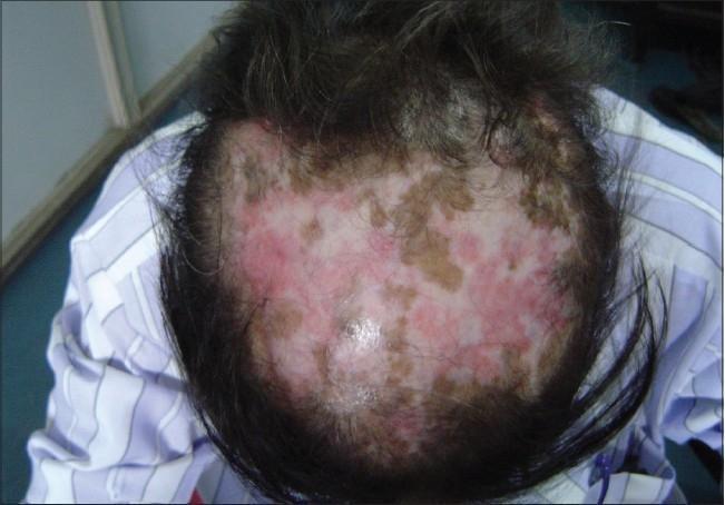
This clinical photograph displays a superior view of the scalp demonstrating classic features of Discoid Lupus Erythematosus (DLE). The image reveals extensive, patchy scarring alopecia (cicatricial alopecia) characterized by a significant reduction in hair density and visible destruction of hair follicles. The skin lesions exhibit a tri-colored, variegated appearance: erythematous (pink-red) areas indicating active inflammation, hyperpigmented (brown-tan) macules and patches, and focal regions of porcelain-white hypopigmentation suggestive of established dermal fibrosis and scarring. The distribution is irregular across the vertex and crown, showing the progressive nature of the follicular damage. This visual serves as a primary educational example of the cutaneous manifestations of chronic cutaneous lupus erythematosus on the scalp, emphasizing the transition from active inflammatory plaques to permanent, irreversible hair loss.
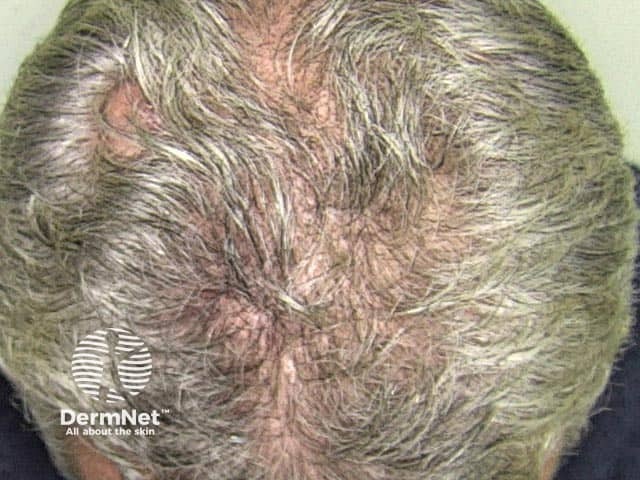
Clinical photography of the scalp in a patient with discoid lupus erythematosus (DLE) demonstrating cicatricial (scarring) alopecia in the occipital/posterior scalp. The image shows localized, erythematous to hypopigmented plaques with adherent scales and follicular plugging, surrounding areas of atrophic scarring and reduced hair density. The hair is finely distributed around the lesion with thinning indicative of irreversible follicular destruction. There is a central koebnerized-like scar with cribriform appearance and irregular, raised borders consistent with chronic inflammatory scarring alopecia. Vascularity appears mild, with pale, scarred skin in the center and erythematous periphery. The clinical presentation aligns with discoid lupus activity: chronic, scarring lesions that may progress to permanent hair loss if untreated. The image underscores typical features of DLE on the scalp: well-demarcated plaques, follicular plugging, hyperkeratosis, and central alopecic patches. Color balance and lighting emphasize subtle redness, scale, and atrophic skin. This photograph is useful for educational purposes, differential diagnosis against other causes of cicatricial alopecia (lichen planopilaris, frontal fibrosing alopecia, traction alopecia), and for teaching clinicians about the appearance of DLE lesions in acral and scalp regions. Potential clinical applications include diagnosis confirmation, treatment planning, dermatopathology correlation, and monitoring disease progression or response to antimalarial therapy.
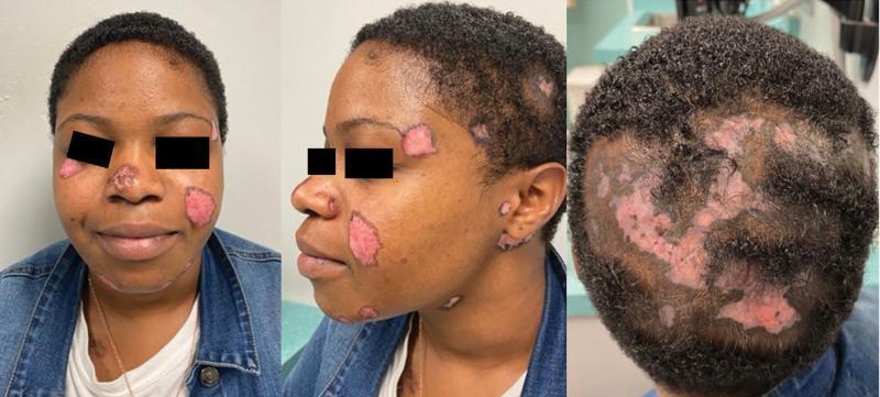
A composite of three clinical photographs showing the face and scalp of a patient with chronic cutaneous lupus erythematosus (CCLE), specifically discoid lupus erythematosus (DLE). The frontal and lateral views of the face demonstrate multiple well-demarcated, erythematous, atrophic patches of varying sizes. These lesions are located on the nasal bridge, cheeks, temples, jawline, and ears. Key morphological features include central atrophy and scarring with peripheral hyperpigmentation and active erythema. The posterior view of the scalp reveals significant multifocal cicatricial (scarring) alopecia. The hair loss areas directly correspond to the presence of large, irregular, confluent erythematous and depigmented patches. The scalp lesions show a complete loss of follicular ostia within the atrophic centers, characteristic of permanent hair loss in DLE. This visual series illustrates the classic distribution and morphology of discoid rash affecting both glabrous skin and hair-bearing areas in an adult patient.
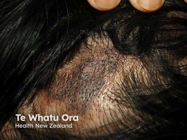
This dermatologic clinical photograph depicts a discoid lupus erythematosus (DLE) lesion on the scalp with resulting cicatricial (scarring) alopecia. The image shows an irregular, hypopigmented-to-erythematous plaque at the scalp margin with a central area of hair loss and complete disruption of hair follicles, producing permanent scarring. The patch is bordered by a slightly erythematous, scaly margin and perifollicular hyperpigmentation. Follicular openings are diminished or obliterated in the affected zone, consistent with cicatricial alopecia. The surrounding skin may appear atrophic, with subtle pigmentary changes typical of chronic discoid lupus. Clinically, the pattern is localized and plaque-like, most commonly affecting sun-exposed scalp regions; DLE lesions may coalesce or evolve over weeks to months. This entity is part of the lupus erythematosus spectrum and carries diagnostic significance for autoimmune dermatology; histopathology would reveal interface dermatitis with basal cell vacuolization, periadnexal lymphocytic infiltrate, follicular plugging, and dermal sclerosis. Differential diagnosis includes lichen planopilaris, frontal fibrosing alopecia, and other scarring alopecias. Management implications include antimalarial therapy, topical steroids, calcineurin inhibitors, photoprotection, and rheumatology assessment for systemic involvement. This image serves educational purposes for dermatology, rheumatology, and medical training in recognizing cicatricial alopecia patterns associated with discoid lupus.
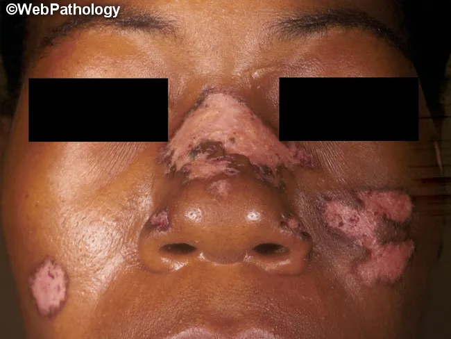
Clinical photography of facial skin illustrating discoid lupus erythematosus (DLE) with sun-exposed-area involvement. Frontal/anterior view of the face shows multiple erythromatous plaques on the cheeks, nasal bridge, perioral region, and ears, with overlying adherent scales and peripheral erythema. Lesions are firm and may exceed several centimeters; keratin plugs are often evident beneath scales when removed. Chronic plaques exhibit atrophic scarring and pigmentary alteration, including hypopigmentation in darker skin tones; depigmentation can be disfiguring and scalp involvement suggests long-standing, severe disease. Early lesions may be erythematous and edematous before plaque formation. The distribution pattern is predominantly head and neck in localized DLE but can extend to non-exposed sites in generalized disease; photosensitivity with seasonal exacerbations in spring/summer is typical. Clinically, the appearance—well-demarcated, scale-covered plaques on sun-exposed skin—helps distinguish DLE from seborrheic dermatitis, rosacea, and actinic damage; differential includes lupus erythematosus profundus, psoriasis, and granulomatous diseases. The image underscores the importance of sun protection, topical corticosteroids or calcineurin inhibitors, and potential antimalarial therapy in management. This photographic reference is valuable for dermatology education, augmenting recognition of cutaneous lupus manifestations, scarring alopecia risk, and the need for biopsy confirmation when systemic involvement is uncertain. Images like this support diagnostic training and patient counseling in lupus care.
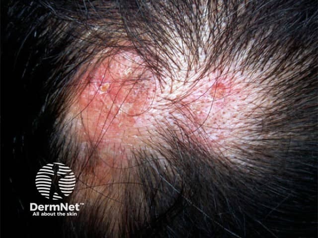
Clinical photography of a scalp lesion demonstrates classic discoid lupus erythematosus (DLE) features. The image shows well-demarcated, erythematous to violaceous plaques with adherent scale, follicular plugging, and central atrophy within the hair-bearing scalp. Hair loss is patchy and the affected areas may progress to irreversible scarring alopecia. The margins are often raised and inflammatory, with surrounding pigmentary changes that may include hyperpigmentation or hypopigmentation depending on skin phototype. These cutaneous plaques are typically sun-exposed site–predilection lesions and may appear in chronic, fluctuating courses. Clinically, DLE must be distinguished from seborrheic dermatitis, tinea capitis, lichen planopilaris, and psoriasis; biopsy or dermoscopy can aid in confirmation. If biopsy is performed, histology reveals interface dermatitis with basal layer vacuolization, thickened basement membrane, perivascular and periadnexal lymphocytic infiltrate, and follicular plugging; mucin deposition may be present. Management involves photoprotection, topical or intralesional corticosteroids for active plaques, and systemic agents such as antimalarials in extending disease, with caution for systemic lupus erythematosus. This image is valuable for dermatology education, lesion recognition, differential diagnosis training, and AI-enabled image retrieval for cutaneous lupus and scarring alopecia research. Keywords: discoid lupus erythematosus, DLE, scalp lesions, scarring alopecia, autoimmune skin disease, hair loss. Clinical relevance includes guiding biopsy decisions and treatment planning.
discoid lupus erythematosus histopathology interface dermatitis follicular plugging
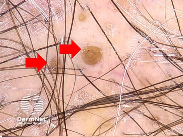
Modality and view: In vivo dermoscopy (dermoscopy) of a discoid lupus erythematosus plaque on scalp skin, captured with noninvasive, magnified close‑up imaging. The primary lesion shows characteristic follicular plugging: round to oval keratin-filled openings that appear as yellow‑brown to tan plaques at the follicular ostia. Surrounding scale is minimal to moderate, with a subtle erythematous halo and fine perifollicular hyperkeratosis evident around plugged follicles. Hair shafts traverse the surface; there is no gross necrosis or ulceration in this field. The plugs are well circumscribed, sometimes surrounded by faint brown pigmentation, which may reflect chronic sun exposure and pigmentary alteration. These dermoscopic findings correlate with discoid lupus erythematosus histopathology, where follicular plugging and interface dermatitis are common. The presence of follicular plugs in a lupus-type plaque is highly suggestive and helps distinguish DLE from inflammatory dermatoses such as seborrheic dermatitis or eczema, where oiliness and scaling predominate without discrete keratotic ostial plugs. Diagnostic significance: dermoscopic follicular plugging supports clinical suspicion of DLE and guides biopsy targeting if confirmation is needed. Potential clinical uses include monitoring activity, treatment response, and differentiation from other scarring alopecias. In clinical practice, this image aids education of residents and researchers in recognizing DLE dermoscopic signatures on scalp skin.
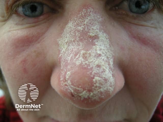
A high resolution clinical photograph documents localized cutaneous discoid lupus erythematosus of sun exposed facial skin, notably the nasal dorsum and surrounding perinasal cheeks. The frontal, close up view shows well demarcated erythematous plaques with adherent whitish scale and crusting, surface desquamation, and subtle follicular plugging. Lesions appear on the nasal bridge with partial peripheral involvement, displaying an atrophic center and peripheral erythema; mild telangiectasia may be evident in chronically sun exposed areas. This morphologic pattern—oval or polygonal plaques with silvery scale and scarring tendency—aligns with classic discoid lupus as a chronic inflammatory dermatosis within the spectrum of cutaneous lupus erythematosus. Although biopsy is not provided here, typical histopathology would reveal epidermal atrophy, interface dermatitis, follicular plugging, dermal lymphocytic infiltrates, and increased dermal mucin if sampled. Clinically, the image underscores the risk of pigmentary change and scarring on the nose with ongoing disease activity. Relevance to practice includes differentiating discoid lupus from seborrheic dermatitis, rosacea, psoriasis, or actinic keratosis in sun exposed facial zones. Management implications involve sun avoidance, topical anti inflammatory therapies, calcineurin inhibitors, and consideration of systemic antimalarials for disease control, with regular monitoring for evolution or systemic involvement. This image is educational for clinicians, students, and researchers.
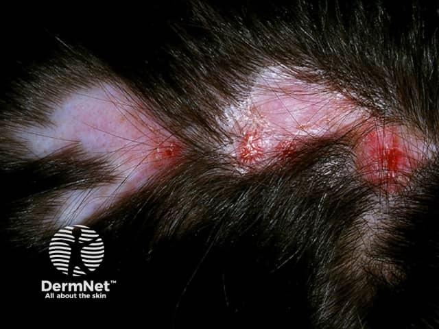
Clinical photography of a discoid lupus erythematosus lesion localized to the scalp/behind the ear demonstrates a solitary, well-demarcated erythematous plaque with adherent scale and thinning hair in the involved region. The plaque exhibits a raised red to violaceous border, a potentially atrophic center, and variable crusting, consistent with the classic morphology of DLE. Follicular plugging and surrounding erythema are common features, contributing to patchy hair loss (scarring alopecia) if chronic. The presentation is sun-exposed surface-limited, suggesting photosensitive cutaneous involvement. Imaging modality is non-invasive, relying on standard white-light clinical photography to document surface texture, scale, and border definition. Differential diagnoses include psoriasis of the scalp, tinea capitis, seborrheic dermatitis, cutaneous lupus erythematosus, lichen planus, and less likely basal cell carcinoma with ulceration on the scalp. Confirmation typically requires correlation with histopathology showing interface dermatitis with lymphocytic infiltrate and follicular involvement, and possibly direct immunofluorescence. Clinically, discoid lupus requires monitoring for progression to systemic lupus erythematosus, especially in patients with widespread disease or mucocutaneous involvement. Management emphasizes sun protection, avoidance of triggers, topical corticosteroids, calcineurin inhibitors, photoprotection, and, in selected cases, antimalarial therapy. This image is valuable for education on recognition, differentiation, and early diagnosis of discoid lupus erythematosus in dermatology training.
Note on library availability: The medical textbook library contains Fitzpatrick's Dermatology (9th ed.) and Rook's Dermatology (Dermatology 2-Volume Set 5e). IADVL Textbook and Bologna (Dermatology) were not found in the indexed collection. The content below is sourced from Fitzpatrick's and Rook's with supplementary clinical knowledge aligned with IADVL and Bologna perspectives where applicable.
Discoid Lupus Erythematosus (DLE)
Overview & Classification
- Acute cutaneous LE (ACLE) - malar rash, photosensitive eruption
- Subacute cutaneous LE (SCLE) - annular/papulosquamous
- Chronic cutaneous LE (CCLE) - includes classic DLE, hypertrophic DLE, mucosal DLE, lupus profundus/panniculitis, chilblain LE, and DLE-lichen planus overlap
Epidemiology
- Female predominance: the vast majority of patients are women
- Age: peak incidence in the 3rd-4th decade, but any age including children
- Race: more prevalent in African Americans and other pigmented skin types; Asian Indians can present with isolated macular hyperpigmentation
- Localized DLE: lesions confined to head and neck - most common form; only ~5% progress to SLE
- Generalized (disseminated) DLE: lesions both above and below the neck - higher risk of SLE, more recalcitrant to therapy
- Roughly 25% of SLE patients develop DLE at some point; conversely, 10-20% of DLE patients eventually meet SLE classification criteria, the majority doing so within 5 years - Rook's Dermatology 5e, p. 812; Fitzpatrick's, p. 1075
Clinical Features
Classic DLE
- Central atrophic scarring and hypopigmentation
- Peripheral hyperpigmentation and active erythema
- Follicular plugging (dilated, plugged follicular ostia) - Rook's Dermatology 5e, Ch. 69
Distribution
| Site | Notes |
|---|---|
| Face (malar, nose, ears) | Most common; conchal bowl + external auditory canal classically involved |
| Scalp | Involved in ~60%; scarring alopecia in ~1/3 |
| Nasolabial folds | Usually spared (like ACLE and SCLE) |
| Neck, V-area, extensor arms | Common sites |
| Palms/soles | Painful, disabling erosive lesions (uncommon) |
| Mucosae (lips, nasal, genital) | Occasional |
| Nails | Nail fold erythema, telangiectasia, pitting, paronychia |
- Localized DLE: head/neck only
- Generalized DLE: above AND below the neck; more recalcitrant, higher SLE risk - Fitzpatrick's, p. 1075
Scalp DLE
Photosensitivity
- DLE lesions can be potentiated by sunlight, though to a lesser extent than ACLE and SCLE
- Many patients have DLE on sun-protected skin - no absolute photodistribution
- The Koebner (isomorphic) phenomenon occurs - trauma can precipitate new lesions
Variants of CCLE
| Variant | Key Features |
|---|---|
| Classic DLE | Most common; discoid plaques, follicular plugging, scarring |
| Hypertrophic (verrucous) DLE | Thick hyperkeratotic scale; extensor arms, face, trunk; can mimic SCC, hypertrophic actinic keratosis |
| Mucosal DLE | Lips, nasal mucosa, conjunctivae, genital mucosa |
| Lupus profundus/panniculitis | Firm nodules, lower dermis/subcutis; 70% co-exist with DLE; can mimic breast carcinoma |
| Chilblain LE | Cold-induced acral lesions; associated with anti-Ro/SS-A; ~20% progress to SLE |
| LE tumidus (LET) | Most photosensitive; no scarring; some consider it intermittent CLE |
Pathogenesis
- Type I interferon upregulation plays a central role in CLE pathogenesis
- UV radiation induces keratinocyte apoptosis → release of nuclear antigens → autoantibody formation and leukocyte recruitment
- Inflammatory infiltrate: predominantly CD4+ T lymphocytes and macrophages in periappendageal and perivascular distribution
- The deeper extension of infiltrate into reticular dermis/subcutis distinguishes DLE from ACLE/SCLE - Fitzpatrick's, p. 1081
Histopathology
- Hyperkeratosis with follicular plugging
- Epidermal atrophy
- Vacuolar interface dermatitis at DEJ and follicular epithelium
- Thickened basement membrane (markedly so in DLE)
- Dense mononuclear infiltrate - CD4 T cells and macrophages; periappendageal and perivascular; extends into deep reticular dermis (deeper than ACLE/SCLE - key distinguishing feature)
- Dermal mucin deposition
- Melanophages in upper dermis
- Reduced/absent sebaceous glands
- In chronic lesions: dermal fibroplasia replaces the inflammatory infiltrate
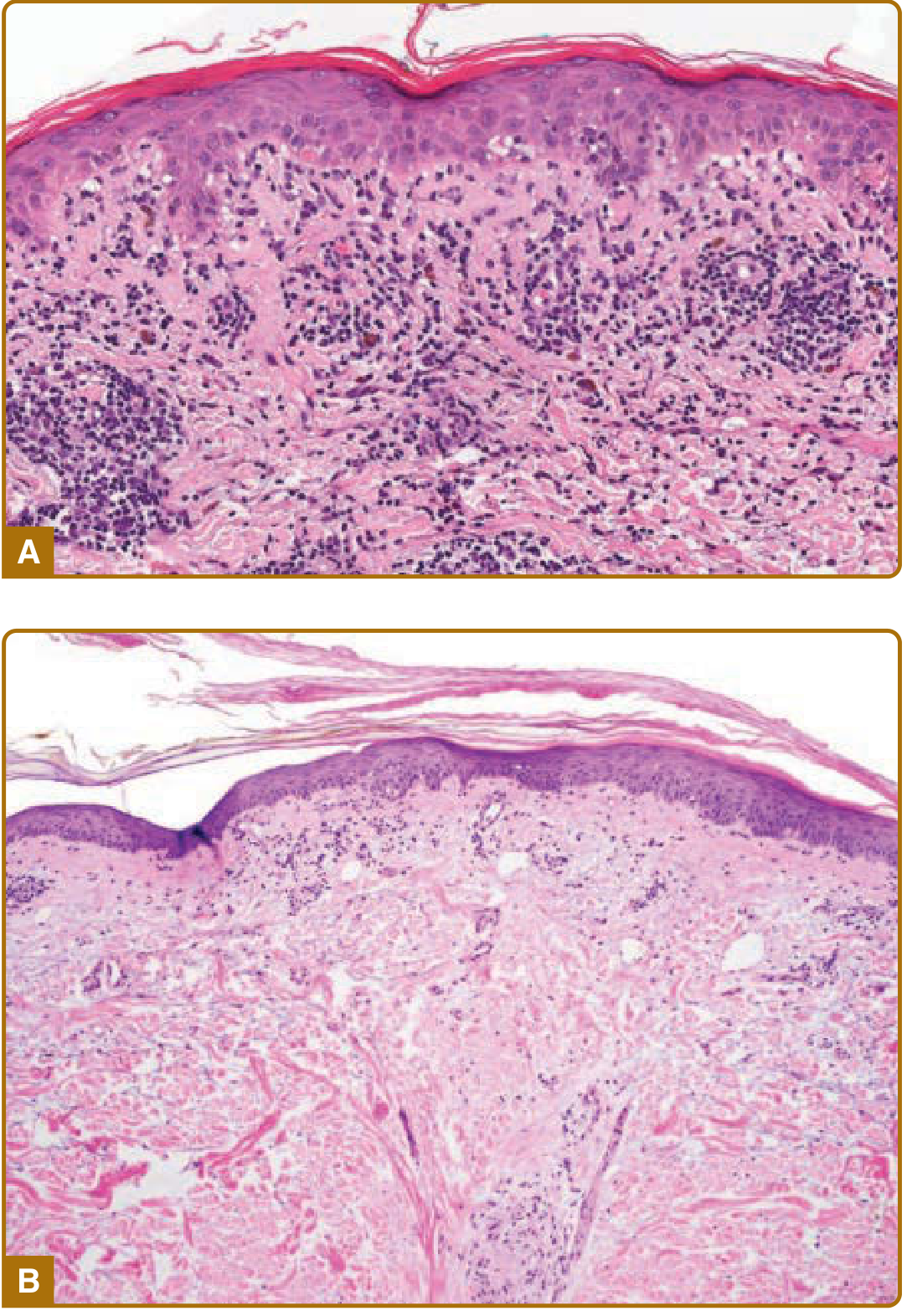
- Vacuolar interface alteration at epidermis AND follicular epithelium
- Interface change in follicles: usually vacuolar (not lichenoid)
- Perineural lymphoid infiltrate may be seen
- Fragments of "naked" hair shafts in giant cells
- Rook's Dermatology 5e, p. 3176-3192
Special staining: SCC risk
Immunofluorescence
Direct Immunofluorescence (DIF) - "Lupus Band Test"
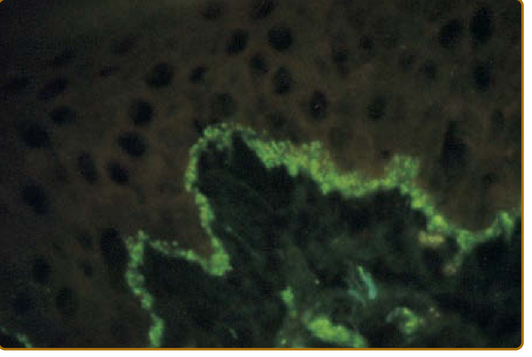
- Lesional DIF: Granular/linear band of immunoglobulin (IgG, IgA, IgM) and complement (C3, C4, C1q) deposits at the dermal-epidermal junction and/or follicular epithelium-dermis junction
- DIF of DLE lesional skin: positive in ~90% of cases (most reliable in CLE)
- Nonlesional DIF ("true lupus band test"): positive in SLE (especially sun-exposed nonlesional skin), typically negative in isolated DLE
- This helps distinguish DLE from SLE: in isolated DLE, nonlesional skin DIF is typically negative
Investigations / Laboratory Tests
| Investigation | Finding in DLE |
|---|---|
| ANA | Positive in only ~30-40% of isolated DLE (vs ~95% in SLE) |
| Anti-dsDNA | Distinctly uncommon in isolated DLE |
| Anti-Ro/SS-A | Rare in typical DLE; low levels sometimes by ELISA |
| Anti-ssDNA | Occasionally positive |
| Anti-U1 RNP | Sometimes, with mild SLE or MCTD overlap |
| Complement (C3/C4) | Modest depression occasionally |
| Hematology | Mild leukopenia, anemia (small percentage) |
| VDRL (BFP) | Small percentage |
Diagnosis
- Clinical evaluation (characteristic morphology and distribution)
- Trichoscopy (for scalp DLE)
- Histopathology (confirmatory - see above)
- DIF of lesional skin when differentiation from lichen planopilaris, mucous membrane pemphigoid is needed
- Suspend topical corticosteroids for several days before biopsy
Differential Diagnosis
- Seborrheic dermatitis, rosacea, psoriasis
- Hypertrophic actinic keratosis, keratoacanthoma, SCC (for hypertrophic DLE)
- Lichen planopilaris (scalp), frontal fibrosing alopecia
- Tinea capitis (in children)
- Granulomatous diseases
CLASI
Management
Principles
- Rule out underlying SLE at time of diagnosis
- Photoprotection - cornerstone of all CLE management
- Maximize local measures before adding systemic therapy
- Prefer non-immunosuppressive agents for CCLE (unlike ACLE which often needs systemic immunosuppression)
- Cigarette smoking impairs response to antimalarial therapy - advise cessation
Photoprotection
- Broad-spectrum, water-resistant sunscreen SPF ≥ 30 with efficient UVA blockade (avobenzone, micronized titanium dioxide, zinc oxide, ecamsule)
- Tightly woven clothing, broad-brimmed hats
- UV-blocking window films; acrylic shields over fluorescent lighting
- Corrective camouflage cosmetics (Dermablend, Covermark) - physical screen + cosmetic benefit
- Vitamin D supplementation (sun avoidance can cause deficiency) - Fitzpatrick's, p. 1083-1084
Local (Topical) Therapy
| Agent | Details |
|---|---|
| Topical corticosteroids | Super-potent (Class I): clobetasol propionate 0.05% or betamethasone dipropionate 0.05%. Twice daily x 2 weeks, then 2-week rest. Ointments for hyperkeratotic lesions. Scalp: Class I-II solutions/gels |
| Intralesional corticosteroids | Triamcinolone acetonide 2.5-5 mg/mL (face); up to 10 mg/mL for body lesions; every 4-6 weeks |
| Topical calcineurin inhibitors | Pimecrolimus 1% cream (= betamethasone valerate 0.1% for facial DLE - double-blind study); tacrolimus 0.1% ointment; use during steroid "rest" periods |
Systemic Therapy
- Hydroxychloroquine (HCQ) - aminoquinoline antimalarial; mainstay of CLE/DLE treatment; requires baseline ophthalmologic screening; smokers respond less well
- Chloroquine - alternative antimalarial
- Systemic retinoids (acitretin, isotretinoin) - particularly useful for hypertrophic DLE; isotretinoin effective for plantar DLE in one report
- Methotrexate
- Dapsone
- Mycophenolate mofetil (MMF)
- Thalidomide / Lenalidomide
- IVIg (intravenous immunoglobulin)
- Monoclonal antibodies: anifrolumab (anti-IFNAR1) - emerging therapy targeting the type I interferon pathway
From Rook's Dermatology 5e, p. 1407: "Active, inflammatory discoid lesions usually respond to oral antimalarial drugs (e.g. hydroxychloroquine) and corticosteroids (topical, intralesional). Second-line therapies for antimalarial-resistant cutaneous disease, such as systemic retinoids, methotrexate, dapsone, mycophenolate mofetil, thalidomide, lenalidomide, and IVIg..."
- Topical minoxidil (for scalp DLE with alopecia)
- Low-dose oral minoxidil
- Vitamin D supplementation
Prognosis & Complications
- Without treatment: indolent progression to large areas of cutaneous dystrophy and scarring alopecia - psychosocially and occupationally devastating
- With treatment: skin disease can be largely controlled
- Spontaneous remission occurs occasionally; rebound on stopping therapy is typical - slow taper recommended
- Squamous cell carcinoma (SCC): can develop in chronic smoldering DLE lesions - surveillance important
- Localized DLE: 5% risk of SLE progression
- Generalized/disseminated DLE: higher risk; most progression within 5 years
- Irreversible scarring alopecia in ~1/3 of patients with scalp DLE
- Death from SLE is distinctly uncommon in patients presenting initially with localized DLE
DLE vs SLE - Key Differentiating Points
| Feature | Isolated DLE | DLE in SLE |
|---|---|---|
| Lesion distribution | Head/neck (localized) or generalized | Any |
| ANA | ~30-40% positive | ~95% positive |
| Anti-dsDNA | Uncommon | Common (high titer) |
| Nonlesional DIF | Negative | Often positive |
| SLE progression | 5% (localized), higher (generalized) | Already systemic |
| Severity of systemic disease | Mild (those with DLE-dominant SLE tend to have less severe SLE) | Variable |
IADVL / Bolivian Context
- In darker skin phototypes (Fitzpatrick IV-VI), DLE presents with more prominent dyspigmentation: central hypopigmentation and marked peripheral hyperpigmentation; sometimes vitiligo-like depigmentation
- Asian Indians can present with isolated macular hyperpigmentation without classic erythema
- Post-inflammatory hyperpigmentation is the dominant cosmetic concern in pigmented skin
- The classic "carpet tack" sign and follicular plugging remain important diagnostic clues across all phototypes
- DLE in pigmented skin requires careful distinction from pigmentary disorders and tinea capitis
- Fitzpatrick's Dermatology (9th ed.), Chapter 61: Cutaneous Lupus Erythematosus, pp. 1074-1088
- Rook's Dermatology (5th ed.), Chapter 41 (Cutaneous LE) and Chapter 69 (Scarring alopecias), pp. 811-813, 1407> Library note: The indexed medical library contains Fitzpatrick's Dermatology (9th ed.) and Rook's Dermatology (Dermatology 2-Volume Set 5e). IADVL and Bolognias (Dermatology) were not found in the library database. The content below is drawn directly from Fitzpatrick's and Rook's with cross-referenced clinical knowledge. Where IADVL/Bolognias perspectives diverge or add, this is noted from general dermatology knowledge.
Discoid Lupus Erythematosus (DLE)
Classification
- ACLE (acute) - malar rash, photosensitive eruption
- SCLE (subacute) - annular / papulosquamous
- CCLE (chronic) - includes classic DLE, hypertrophic DLE, mucosal DLE, lupus profundus, chilblain LE, LE tumidus
Epidemiology
- Female predominance - vast majority are women
- Peak incidence: 3rd-4th decade; all ages including children
- Higher prevalence in African Americans and darker phototypes; also prevalent in Asia
- Localized DLE (head/neck only): ~5% risk of progression to SLE
- Generalized/disseminated DLE (above AND below neck): higher SLE risk, higher recalcitrance
- ~10-20% of DLE patients eventually meet SLE criteria; most progression within 5 years
- ~25% of SLE patients develop DLE at some point; these SLE patients tend to have milder systemic disease
- Fitzpatrick's, p. 1075; Rook's, p. 812
Clinical Features
Primary Lesion Evolution
- Early: Red-purple macule/papule → develops hyperkeratotic surface
- Evolved: Sharply demarcated, coin-shaped (discoid) erythematous plaque with prominent adherent scale extending into dilated follicular orifices
- Late/Scarred: Atrophic central scarring + hypopigmentation; hyperpigmentation + active erythema at periphery; telangiectasia; follicles devoid of hair
Distribution
| Site | Notes |
|---|---|
| Face | Any area including eyebrows, eyelids, nose, lips |
| Ears | Conchal bowl, outer external auditory meatus - classic site |
| Scalp | 60% involved; scarring alopecia in ~1/3; irreversible |
| Nasolabial folds | Usually spared |
| V-neck, extensor arms | Common in generalized DLE |
| Palms/soles | Painful erosive lesions; disabling |
| Mucosae | Lips, nasal mucosa, conjunctivae, genitalia |
| Nails | Nail fold erythema, telangiectasia, pitting, paronychia |
Localized vs Generalized
- Localized: head/neck only - most common; 5% SLE risk
- Generalized (disseminated): lesions below the neck in addition to head/neck; higher SLE risk; more recalcitrant
Photosensitivity
Clinical Images
CCLE Variants
| Variant | Key Features |
|---|---|
| Classic DLE | Most common; coin-shaped scarring plaques |
| Hypertrophic (verrucous) DLE | Thick hyperkeratotic scale; extensor arms, face, trunk; mimics SCC, keratoacanthoma, hypertrophic AK |
| Mucosal DLE | Lips, nasal mucosa, conjunctivae, genital mucosa |
| Lupus profundus/panniculitis | Firm nodules 1-3 cm; deep dermis/subcutis; saucerized depressions; 70% have overlying DLE; mimics breast carcinoma (lupus mastitis); dystrophic calcification |
| Chilblain LE | Cold-induced acral lesions; anti-Ro/SS-A associated; Raynaud's overlap; ~20% progress to SLE |
| LE tumidus (LET) | Most photosensitive subtype; no scarring/atrophy; best antimalarial response |
Pathogenesis
- Type I interferon pathway is central
- UV radiation → keratinocyte apoptosis → nuclear antigen exposure → autoantibody production, chemokine release, leukocyte recruitment
- Infiltrate: predominantly CD4+ T lymphocytes and macrophages in periappendageal and perivascular distribution
- The depth of infiltrate (into reticular dermis and subcutis) is the key histological distinction from ACLE/SCLE
- Cigarette smoking: independently associated with CLE and impairs antimalarial response (mechanism unrelated to drug metabolism)
Histopathology
| Feature | Detail |
|---|---|
| Hyperkeratosis | With follicular plugging |
| Epidermal atrophy | Variable |
| Interface dermatitis | Vacuolar basal cell degeneration at DEJ AND follicular epithelium |
| BM thickening | Markedly thickened basement membrane (more than ACLE/SCLE) |
| Inflammatory infiltrate | Dense CD4 T cells + macrophages; periappendageal and perivascular; extends into deep reticular dermis - key distinguishing feature |
| Dermal mucin | Increased |
| Melanophages | Present in upper dermis |
| Sebaceous glands | Reduced or absent |
| Chronic stage | Dense infiltrate replaced by dermal fibroplasia |
| Special: follicular | Interface change in follicles usually vacuolar (not lichenoid); perineural lymphoid infiltrate may be seen |
Immunofluorescence - "Lupus Band Test"
| Test | DLE | SLE |
|---|---|---|
| Lesional DIF | Positive ~90%; granular/linear IgG, IgA, IgM, C3 band at DEJ and/or follicular epithelium | Positive |
| Nonlesional DIF (sun-exposed) | Usually negative in isolated DLE | Positive (important distinction) |
| Nonlesional DIF (sun-protected) | Negative | Positive in ~50% |
Laboratory Investigations
| Test | Finding in Isolated DLE |
|---|---|
| ANA | Positive in ~30-40% (vs ~95% in SLE); low titer |
| Anti-dsDNA | Distinctly uncommon - if positive, suspect SLE |
| Anti-Ro/SS-A, Anti-La/SS-B | Rare in typical DLE |
| Anti-ssDNA | Occasionally positive |
| Anti-U1 RNP | Sometimes; suggests MCTD overlap |
| Complement C3/C4 | Modest depression in small % |
| CBC | Mild leukopenia, mild anemia in small % |
| VDRL (BFP) | Small % |
| ESR, globulins | Modest elevations in some |
Diagnosis
- Characteristic clinical morphology (carpet tack sign, follicular plugging, central atrophy, peripheral hyperpigmentation)
- Trichoscopy for scalp disease: large yellow dots, arborizing vessels, follicular plugs, late-stage follicular obliteration
- Histopathology (biopsy from active edge of lesion)
- DIF when DD from lichen planopilaris, mucous membrane pemphigoid is needed
- Suspend topical steroids before biopsy for several days
Differential Diagnosis
- Seborrheic dermatitis, rosacea, psoriasis
- Hypertrophic actinic keratosis, keratoacanthoma, squamous cell carcinoma (especially hypertrophic DLE)
- Lichen planopilaris - most important DD on scalp
- Frontal fibrosing alopecia
- Tinea capitis (pediatric)
- Sarcoidosis, granulomatous rosaces
Management
Principles
- Exclude underlying SLE at diagnosis
- Photoprotection - cornerstone for all CLE
- Maximize local measures before systemic agents
- For CCLE: prefer non-immunosuppressive modalities (unlike ACLE which requires systemic immunosuppression)
- Stop smoking - impairs antimalarial efficacy; smoking also independently associated with CLE
- Vitamin D supplementation (risk of deficiency from sun avoidance)
Photoprotection
- SPF ≥ 30 broad-spectrum, water-resistant sunscreen with UVA cover (avobenzone, micronized TiO2, zinc oxide, ecamsule)
- Tightly woven clothing, broad-brimmed hats
- UV-blocking films on home/car windows; acrylic shields over fluorescent lights
- Avoid photosensitizing drugs: hydrochlorothiazide, tetracycline, griseofulvin, piroxicam
Local Therapy
| Agent | Regimen |
|---|---|
| Superpotent topical CS (clobetasol 0.05%, betamethasone dipropionate 0.05%) | Twice daily × 2 weeks, then 2-week rest; ointments for hyperkeratotic DLE; solutions/gels for scalp |
| Intralesional triamcinolone acetonide | 2.5-5 mg/mL (face); up to 10 mg/mL (body); every 4-6 weeks |
| Pimecrolimus 1% cream | Equivalent to betamethasone valerate 0.1% for facial DLE (double-blind RCT); use during steroid rest periods |
| Tacrolimus 0.1% ointment | Effective for DLE; can compound with clobetasol for recalcitrant CLE |
Systemic Therapy
- Hydroxychloroquine - mainstay; antimalarial; steroid-sparing; monitor ophthalmologically
- Chloroquine - alternative
- Systemic retinoids (acitretin, isotretinoin)
- Methotrexate
- Dapsone
- Mycophenolate mofetil
- Thalidomide / Lenalidomide
- IVIg
- Anifrolumab (anti-IFNAR1 monoclonal antibody) - targets type I interferon receptor; approved for SLE, showing efficacy in refractory CLE
Prognosis & Complications
- Without treatment: indolent progression to scarring alopecia and cutaneous dystrophy
- Spontaneous remission occasional; rebound on stopping treatment is typical - slow taper recommended
- Squamous cell carcinoma in longstanding chronic DLE - rare but significant
- Irreversible scarring alopecia in ~1/3 with scalp involvement
- Localized DLE: low mortality, 5% SLE risk; generalized: higher risk
- SLE developing in DLE patients tends to have milder systemic manifestations
Notes on Darker Skin (IADVL-relevant context)
- In Fitzpatrick skin types IV-VI (common in South Asian/Indian patients), DLE presents with more prominent dyspigmentation: central hypopigmentation ± vitiligo-like depigmentation, marked peripheral hyperpigmentation
- Asian Indians may present with isolated macular hyperpigmentation as the dominant feature
- Post-inflammatory hyperpigmentation is a major concern and cosmetic burden
- Carpet tack sign and follicular plugging remain diagnostically important across all phototypes
- Trichoscopy shows scattered dark-brown to bluish discoloration in darker phototypes - Rook's, p. 1407
- Fitzpatrick's Dermatology, 9th ed., Chapter 61 (Sontheimer), pp. 1074-1088
- Rook's Dermatology, 5th ed., Chapter 41 (Cutaneous LE, pp. 810-813) and Chapter 69 (Scarring alopecias, pp. 1407-1408)