
Thyroid pathology explain
Reading File
Searching sources...
Thyroid pathology covers a broad set of conditions - hypothyroidism, hyperthyroidism, thyroiditis, and thyroid neoplasms (benign and malignant). Let me pull comprehensive coverage from the medical library.
Finding Sources
Finding Sources
Finding Sources
Finding Sources
Robbins Pathology is the best source for thyroid pathology. Let me read it along with related sections in parallel.
Reading File
Finding Sources
Finding Sources
Reading File
Finding Sources
Excellent - I have rich content from Robbins. Let me continue reading for hypothyroidism, Graves, Hashimoto, goiter, and tumors sections which are all in this same block.
Reading File
Reading File
I now have excellent, comprehensive content from Robbins covering all major thyroid pathologies. Let me fetch a few key images and compile the full answer.
Loading Image
Reading File
I now have comprehensive content. Let me compile the full detailed response.
Thyroid Pathology - Complete Overview
Source: Robbins, Cotran & Kumar Pathologic Basis of Disease
Anatomy & Normal Physiology
The thyroid gland consists of two lateral lobes connected by a thin isthmus, anterior to the larynx. It develops embryologically from pharyngeal epithelium descending from the foramen cecum at the base of the tongue - explaining ectopic thyroid tissue (lingual thyroid) seen occasionally. The gland is divided by fibrous septa into lobules, each containing 20-40 colloid-filled follicles lined by cuboidal to low columnar epithelium.
The diagram below shows the hypothalamic-pituitary-thyroid axis:
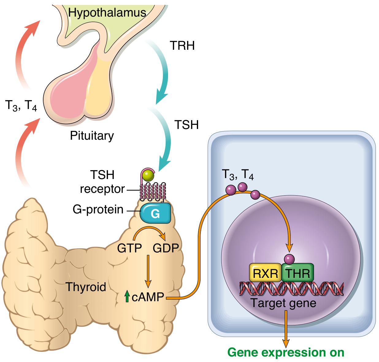
Hormone synthesis: TSH from the anterior pituitary binds the TSH receptor on follicular cells → activates Gs protein → raises cAMP → stimulates thyroid hormone synthesis. Follicular cells convert thyroglobulin into T4 (thyroxine) and T3 (triiodothyronine). In the periphery, T4 is deiodinated to T3, which binds thyroid hormone nuclear receptors with 10-fold greater affinity. Net effect: increased basal metabolic rate, carbohydrate/lipid catabolism, protein synthesis, and critical fetal brain development.
The gland also contains C cells (parafollicular cells) that secrete calcitonin, promoting calcium absorption into bone and inhibiting osteoclasts.
Classification of Thyroid Diseases
- Hyperthyroidism (thyrotoxicosis)
- Hypothyroidism
- Thyroiditis
- Goiter
- Tumors (adenoma, carcinoma)
1. HYPERTHYROIDISM (Thyrotoxicosis)
A hypermetabolic state caused by elevated free T3/T4.
Major causes (in order of frequency):
- Graves disease (~85% of cases)
- Hyperfunctional multinodular goiter
- Hyperfunctional thyroid adenoma
- Iodine-induced hyperthyroidism
- Subacute thyroiditis (transient release, not true hyperfunction)
- TSH-secreting pituitary tumor (rare)
Clinical features:
- Skin: warm, soft, flushed (vasodilation), heat intolerance, increased sweating
- Cardiovascular: tachycardia, palpitations, cardiomegaly, atrial fibrillation (especially in elderly), congestive heart failure
- Metabolic: weight loss despite increased appetite, muscle wasting
- Neuromuscular: anxiety, irritability, fine tremor, hyperreflexia
- GI: increased bowel motility, diarrhea
- Eyes: exophthalmos (specific to Graves disease - see below)
- Reproductive: oligomenorrhea, infertility
Graves Disease
The most common cause of hyperthyroidism. It is an autoimmune disorder caused by thyroid-stimulating immunoglobulins (TSI) - IgG antibodies that bind and activate the TSH receptor, mimicking TSH and causing unregulated thyroid hormone production.
Pathogenesis: Loss of T-regulatory cell tolerance → T helper cells sensitized to thyroid antigens → B cells produce anti-TSH receptor antibodies (TSI/TRAb).
Morphology:
- Diffusely enlarged gland (2-3x normal)
- Microscopically: tall, columnar follicular epithelium with papillary infoldings, reduced colloid with scalloped ("moth-eaten") edges
- Lymphocytic infiltrates with germinal centers
Unique features of Graves (not seen in other causes):
- Ophthalmopathy: exophthalmos due to inflammatory cell infiltration and glycosaminoglycan deposition in the retro-orbital space and extraocular muscles
- Dermopathy (pretibial myxedema): skin thickening due to glycosaminoglycan accumulation - despite the name, this is a feature of Graves hyperthyroidism, not hypothyroidism
2. HYPOTHYROIDISM
Defined as deficiency of thyroid hormone.
Primary causes:
| Category | Examples |
|---|---|
| Autoimmune | Hashimoto thyroiditis (most common in iodine-sufficient areas) |
| Iatrogenic | Surgery, radioiodine ablation, external radiation |
| Drug-induced | Lithium, amiodarone, methimazole, propylthiouracil |
| Iodine deficiency | Endemic (still common worldwide) |
| Congenital | PAX8, FOXE1 mutations; biosynthetic defects |
Secondary ("central") hypothyroidism:
- Pituitary failure (low TSH)
- Hypothalamic failure (low TRH) - rare
Clinical features (adults - myxedema):
- Generalized slowing of metabolic processes
- Cold intolerance, weight gain, fatigue, lethargy
- Constipation, dry coarse skin, hair loss
- Periorbital puffiness, macroglossia, hoarse voice
- Bradycardia, diastolic hypertension
- Hyponatremia, hyperlipidemia
- Myxedematous coma (life-threatening) in severe untreated cases
Congenital hypothyroidism (cretinism):
- Due to iodine deficiency in endemic areas or genetic defects
- Severe intellectual disability, short stature, coarse facial features, umbilical hernia
- Now rare due to iodine supplementation and neonatal screening
3. THYROIDITIS
Hashimoto Thyroiditis
The most common cause of hypothyroidism in iodine-sufficient areas. Primarily affects middle-aged women.
Pathogenesis: T cell-mediated autoimmune destruction of follicular epithelium. Both CD8+ cytotoxic T cells (direct killing) and CD4+ T helper cells (inducing apoptosis via Fas-FasL) contribute. Anti-TPO (antithyroid peroxidase) and anti-thyroglobulin antibodies are present but are markers rather than primary effectors.
Morphology:
- Symmetric thyroid enlargement
- Dense lymphocytic infiltration with germinal centers
- Oxyphilic (Hurthle/Askanazy) change - follicular cells acquire abundant granular eosinophilic cytoplasm
- Follicle destruction and atrophy
Clinical features:
- Painless thyroid enlargement - may mimic neoplasm
- Gradual hypothyroidism (TSH rises as T4/T3 fall)
- Initial transient thyrotoxicosis possible ("hashitoxicosis")
- Associated autoimmune diseases: type 1 DM, adrenal autoimmunity, SLE, Sjogren, myasthenia gravis
- Increased risk of extranodal marginal zone B-cell lymphoma (rare) and possibly papillary carcinoma
Subacute Granulomatous Thyroiditis (de Quervain Thyroiditis)
Triggered by viral infection (coxsackievirus, mumps, SARS-CoV-2, adenovirus). Peak age 40-50 years, F:M ~4:1.
Morphology: Disrupted follicles with foreign-body giant cells engulfing colloid → granulomas; replaced by fibrosis.
Clinical course (4 phases):
- Thyrotoxic phase: Release of stored hormone → mild hyperthyroidism, neck pain, fever, elevated ESR
- Euthyroid phase
- Hypothyroid phase: Transient, as follicles are depleted
- Recovery: Most return to normal thyroid function
Chronic Lymphocytic (Painless/Silent) Thyroiditis
Presumed autoimmune. Common in the postpartum period (up to 5% of women - "postpartum thyroiditis"). Anti-TPO antibodies present. Similar triphasic course to subacute, but without pain or granulomas. Up to 30% develop permanent hypothyroidism.
Riedel Thyroiditis
Rare; dense fibrous replacement of thyroid parenchyma, extending into surrounding structures. Associated with IgG4-related disease. Can mimic malignancy clinically.
4. GOITER
Enlargement of the thyroid, reflecting compensatory response to impaired hormone synthesis.
Diffuse Nontoxic (Simple) Goiter
Caused by iodine deficiency or goitrogens → low T4 → elevated TSH → gland hyperplasia.
Multinodular Goiter (MNG)
Results from repeated cycles of hyperplasia and involution → asymmetric growth → nodule formation. Microscopically: nodules of varying size, some colloid-rich, some with hemorrhage, cysts, or calcification.
- Nontoxic MNG: Most common; euthyroid or hypothyroid
- Toxic MNG (Plummer disease): One or more autonomous hyperfunctioning nodules (often due to activating TSH receptor mutations) → hyperthyroidism without ophthalmopathy
5. THYROID TUMORS
Thyroid Adenoma
- Benign follicular neoplasm, encapsulated
- Most are nonfunctioning ("cold" nodule on scan); rarely autonomous ("hot" / toxic)
- Morphology: uniform follicular cells, intact capsule - NO capsular or vascular invasion
- Fine-needle aspiration (FNA) cannot reliably distinguish follicular adenoma from follicular carcinoma
Malignant Tumors
| Feature | Papillary Ca | Follicular Ca | Medullary Ca | Anaplastic Ca |
|---|---|---|---|---|
| Frequency | ~85% | 5-15% | 5% | <5% |
| Origin | Follicular cells | Follicular cells | C cells | Follicular cells |
| Key molecular alteration | BRAF V600E, RET/PTC fusions | RAS mutations, PAX8-PPARG fusion | RET mutations (MEN2) | TP53, TERT promoter |
| Spread | Lymphatic (early) | Hematogenous | Lymphatic + hematogenous | Local invasion |
| Prognosis | Excellent (>95% 10-yr survival) | Good (minimally invasive) to poor (widely invasive) | Intermediate | Very poor (months) |
| Radioiodine responsive | Yes | Yes | No | No |
Papillary Thyroid Carcinoma (PTC)
Most common thyroid malignancy. Can occur at any age; strongly associated with prior radiation exposure (especially childhood).
Molecular: BRAF V600E mutation (~60%), RET/PTC gene rearrangements.
Morphology:
- Papillary fronds with fibrovascular cores
- Characteristic nuclear features (pathognomonic):
- Optically clear ("Orphan Annie eye") nuclei
- Nuclear grooves and intranuclear pseudoinclusions
- Psammoma bodies (laminated calcifications) - highly specific
- Sclerotic stroma
Behavior:
- Lymphatic spread to regional nodes is common but does not significantly worsen prognosis
- 5-20% local/regional recurrence; 10-15% distant metastases
- Prognosis worsened by: age >55, extrathyroidal extension, distant metastases
Follicular Thyroid Carcinoma (FTC)
More common in iodine-deficient areas. Peak age 40-60 years.
Key diagnostic criterion: Capsular and/or vascular invasion on histology - cannot be diagnosed on FNA alone.
Morphology:
- Uniform follicular cells forming follicles with colloid (resembles normal thyroid)
- Oncocytic (Hurthle cell) variant: cells with abundant eosinophilic granular cytoplasm
- No Orphan Annie nuclei, no psammoma bodies
Behavior: Spreads hematogenously (lung, bone, liver) rather than to lymph nodes.
Medullary Thyroid Carcinoma (MTC)
Arises from C cells (parafollicular cells) → secretes calcitonin (useful tumor marker).
- Sporadic (70%): Usually unilateral, presents in adults
- Familial (30%): Associated with MEN2A and MEN2B syndromes, RET proto-oncogene mutations (germline); often bilateral and multicentric
- Morphology: polygonal to spindle cells in nests/sheets, amyloid deposits in stroma (derived from procalcitonin)
- Lab: elevated serum calcitonin; CEA also elevated
- Genetic testing for RET mutations is mandatory in all patients
Anaplastic (Undifferentiated) Thyroid Carcinoma
Highly aggressive. Usually in elderly patients, often arising from a pre-existing differentiated thyroid carcinoma (with TP53 or TERT promoter mutations driving dedifferentiation).
- Rapid growth, infiltrates and compresses trachea/esophagus
- Morphology: bizarre pleomorphic giant cells, spindle cells, wide necrosis
- Mean survival: 6 months; does not take up radioiodine
Summary Table: Key Distinguishing Features
| Condition | Key Lab Findings | Key Histology | Unique Clinical Feature |
|---|---|---|---|
| Graves disease | Low TSH, high T3/T4, +TRAb/TSI | Tall columnar cells, scalloped colloid | Exophthalmos, pretibial myxedema |
| Hashimoto | High TSH, low T4, +anti-TPO | Oxyphilic change, germinal centers | Most common hypothyroidism |
| De Quervain | Low TSH (early), high ESR | Granulomas, giant cells | Painful, post-viral |
| Papillary Ca | Normal or low TSH | Orphan Annie nuclei, psammoma bodies | Best prognosis; radiation-linked |
| Follicular Ca | - | Capsular/vascular invasion (no FNA diagnosis) | Hematogenous spread |
| Medullary Ca | High calcitonin | Amyloid stroma | RET mutation; MEN2 |
| Anaplastic Ca | - | Bizarre giant cells, necrosis | Worst prognosis |
Source: Robbins, Cotran & Kumar Pathologic Basis of Disease, Chapter 24 - Endocrine Pathology
This is a shared conversation. Sign in to Orris to start your own chat.