
Floppy infant
Reading File
Now let me search the medical library for floppy infant content in parallel.
Finding Sources
Finding Sources
Searching PubMed
"floppy infant" hypotonia etiology diagnosis
Excellent results. Now let me read the key sections from Bradley & Daroff and Adams & Victor in parallel.
Reading File
Reading File
Let me get more content - the full differential from Bradley & Daroff and also search for clinical approach, investigations, and specific conditions.
Reading File
Finding Sources
I have rich content. Let me read a bit more of the localization/workup section from Bradley & Daroff.
Reading File
Let me get the workup/investigations and specific conditions section:
Reading File
Reading File
I now have enough content for a comprehensive answer. Let me also fetch the key clinical figure:
Loading Image
Finding Sources
I now have comprehensive content to deliver a thorough answer. Here is the complete response:
Floppy Infant (Neonatal/Infantile Hypotonia)
A "floppy infant" describes a neonate or young infant with abnormally reduced muscle tone (hypotonia). It is one of the most common neuromuscular presentations in pediatrics and represents a diagnostic challenge because causes span the entire neuraxis - from the cerebral cortex down to the muscle fiber itself.
Normal Posture vs. Hypotonic Infant
A normal infant lying supine demonstrates hip and knee flexion (lower extremities clear of the table), elbow flexion, and internal rotation at the shoulders.
A hypotonic infant lies with:
- Lower extremities in external rotation, lateral aspects of thighs/knees touching the table
- Upper extremities extended at the sides or abducted with slight elbow flexion, lying against the table ("frog-leg posture")
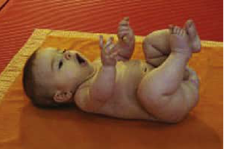
Normal infant posture: legs flexed, arms adducted. A hypotonic infant lies flat with frog-leg posturing.
Examination Maneuvers
| Maneuver | Normal Response | Hypotonic Response |
|---|---|---|
| Traction response (pull to sit) | Elbow/knee/ankle flexion; minimal head lag; head stays erect | Excessive head lag; no limb flexion |
| Vertical suspension (hands under axillae) | Infant stays suspended; head upright | Infant slips through examiner's hands; head falls forward |
| Horizontal suspension (prone over hand) | Head above horizontal; limbs flexed | Infant drapes over hand; head and limbs hanging limply |
- Bradley and Daroff's Neurology in Clinical Practice, p. 520
Epidemiology of Causes
Based on current literature, 60-80% of infant hypotonia is due to central (brain) causes, while 15-30% is due to peripheral causes (motor unit disorders). - Bradley and Daroff's Neurology in Clinical Practice, p. 521
Differential Diagnosis - Full Classification
I. Cerebral Hypotonia (~60-80%)
Chromosomal/Genetic Disorders
- Prader-Willi syndrome - profound neonatal hypotonia, poor feeding, failure to thrive; later hyperphagia, intellectual disability, hypogonadism; caused by loss of paternal 15q11-q13 (deletion or maternal uniparental disomy); diagnosed by methylation-specific PCR
- Trisomy 21 (Down syndrome)
- Other chromosomal anomalies
Chronic Nonprogressive Encephalopathy
- Hypoxic-ischemic encephalopathy (HIE) - low Apgar scores, lactic acidosis, multi-organ involvement; hypotonia typically gives way to spasticity
- Cerebral dysgenesis (in utero infection, toxic exposure, vascular insult, inborn error of metabolism)
- Key features: encephalopathy, seizures, irritability, poor feeding; reflexes and power relatively preserved
Chronic Progressive Encephalopathy
- Zellweger syndrome spectrum (ZSS) - peroxisome biogenesis defects (PEX gene mutations, most commonly PEX1); neonatal hypotonia + seizures + craniofacial dysmorphism + hepatic dysfunction; elevated plasma very-long-chain fatty acids (VLCFAs - C26:0 and C26:1); stippled patellae on X-ray
- Lysosomal storage disorders (GM1/GM2 gangliosidosis, Niemann-Pick)
- Mitochondrial encephalomyopathies
Benign Congenital Hypotonia
- Diagnosis of exclusion (retrospective); family history common; normal tone achieved eventually; intellectual disability may appear later
II. Combined Cerebral + Motor Unit Disorders
- Acid maltase deficiency (Pompe disease, GSD type II) - progressive muscular atrophy + cardiomegaly + macroglossia + hepatomegaly + hypotonia; rapidly fatal in infancy without treatment (enzyme replacement therapy with alglucosidase alfa available)
- Congenital myotonic dystrophy - maternal transmission; polyhydramnios, reduced fetal movements, neonatal respiratory failure; "tent-shaped" mouth
- Syndromic congenital muscular dystrophies
- Congenital disorders of glycosylation
- Infantile neuroaxonal dystrophy
III. Spinal Cord Disorders
- Acquired spinal cord lesions - birth trauma, especially after forceps/vacuum delivery
- Spinal Muscular Atrophy (SMA) type I - Werdnig-Hoffmann disease - most important LMN cause; autosomal recessive SMN1 gene mutation; mothers aware of reduced fetal movements in utero; presents at birth or within first months; tongue fasciculations, areflexia, preserved intellect and sensation; "frog-leg" posture; bulbar and respiratory involvement leading to death by age 2 without treatment
- SMA with respiratory distress (SMARD1)
- X-linked SMA
IV. Peripheral Nerve Disorders
- Congenital hypomyelinating neuropathy / Dejerine-Sottas disease - severe early-onset demyelinating neuropathy; areflexia + sensory loss
- Krabbe disease (infantile globoid cell leukodystrophy) - peripheral neuropathy + CNS involvement
V. Neuromuscular Junction Disorders
- Neonatal (transient) myasthenia gravis - 15% of infants born to myasthenic mothers; placental transfer of AChR antibodies against the fetal receptor (not detectable by standard commercial assays); ocular/bulbar/generalized weakness; resolves within first month; may need neostigmine pre-feeds
- Congenital Myasthenic Syndromes (CMS) - genetic, not autoimmune; presynaptic (choline acetyltransferase deficiency), synaptic (end-plate cholinesterase deficiency), or postsynaptic (AChR structural/kinetic abnormalities); autosomal recessive except slow-channel syndrome (AD); life-threatening apneic episodes with ChAT and rapsyn deficiencies
- Infant botulism - constipation (often first symptom), poor feeding, weak cry, lethargy; spores ingested (honey is a classic source); descending flaccid paralysis; important emergency differential
- Juvenile myasthenia gravis (rare in first year)
VI. Muscle Disorders
Congenital Myopathies (structural):
| Type | Histology | Gene | Key Features |
|---|---|---|---|
| Central core disease | Central cores lacking oxidative enzymes | RYR1 | Hypotonia + weakness; scoliosis, hip dislocation; malignant hyperthermia risk |
| Nemaline (rod-body) myopathy | Nemaline rods on Gomori trichrome | NEB, ACTA1 | Variable severity; respiratory failure possible |
| Centronuclear (myotubular) myopathy | Central nuclei | MTM1 (X-linked), DNM2, BIN1 | Severe in X-linked form; ophthalmoplegia |
Congenital Muscular Dystrophies:
- Merosin-deficient CMD (LAMA2 mutation) - absent laminin-α2 on immunostaining; white matter changes on MRI
- Ullrich CMD (COL6 mutation) - proximal weakness + distal joint hyperlaxity + proximal contractures
- Infantile facioscapulohumeral dystrophy
Central vs. Peripheral Hypotonia - Key Distinguishing Features
| Feature | Central (Brain) | Peripheral (Motor Unit) |
|---|---|---|
| Muscle power | Relatively preserved | Reduced (weakness prominent) |
| Deep tendon reflexes | Normal or increased | Absent or reduced |
| Encephalopathy/Seizures | Common | Absent |
| Mental development | Often impaired | Normal (intellect spared) |
| Fasciculations | Absent | May be present (LMN) |
| Fatigability | Absent | May be present (NMJ) |
| Sensory loss | Absent | May be present (peripheral nerve) |
| Arthrogryposis | Less common | More common |
Investigations - Stepwise Workup
Initial Tier
- Chromosomal microarray + karyotype (for dysmorphic features)
- Brain MRI (cerebral dysgenesis, HIE, white matter changes)
- CK (creatine kinase) - elevated in muscular dystrophy; normal/mildly elevated in congenital myopathy and SMA
- Lactate, pyruvate, ammonia (metabolic encephalopathy)
- Plasma very-long-chain fatty acids (Zellweger spectrum)
- TSH (hypothyroidism)
Targeted Tier
| Suspected Diagnosis | Test |
|---|---|
| SMA | SMN1 gene deletion (molecular testing) |
| Prader-Willi | Methylation-specific PCR of 15q11-q13 |
| Pompe disease | Acid alpha-glucosidase enzyme assay; GAA gene |
| Myotonic dystrophy | CTG repeat expansion (DMPK gene); maternal testing |
| NMJ disorders | Edrophonium (Tensilon) test; RNS (repetitive nerve stimulation); AChR antibodies |
| Congenital myopathy | Muscle biopsy (structural diagnosis) |
| Peripheral neuropathy | Nerve conduction studies / EMG |
| Botulism | Stool culture for C. botulinum / toxin assay |
Muscle Biopsy
- Essential for congenital myopathies: H&E, Gomori trichrome, NADH-TR, ATPase, electron microscopy
- Identifies central cores, nemaline rods, central nuclei, mitochondrial changes
Electrophysiology (EMG/NCS)
- EMG: fibrillation potentials + positive sharp waves = denervation (SMA, neuropathy)
- NCS: reduced conduction velocities = demyelinating neuropathy
- Repetitive nerve stimulation: decremental response = NMJ disorder
Key Specific Diagnoses in Detail
Werdnig-Hoffmann Disease (SMA Type I)
- Most common LMN cause of floppy infant; most common cause of heritable infant mortality after cystic fibrosis
- Autosomal recessive; SMN1 deletion on chromosome 5q13
- Paucity of fetal movements in utero, tongue fasciculations, areflexia, frog-leg posture, paradoxical breathing
- Intellect and sensation spared - distinguishing feature
- Death by age 2 without intervention
- Treatment: nusinersen (intrathecal antisense oligonucleotide), onasemnogene abeparvovec (gene therapy - single dose), risdiplam (oral SMN2 splicing modifier)
Infant Botulism
- Spores ingested → toxin produced in gut → blocks presynaptic ACh release
- Triad: constipation → poor feeding/weak suck → descending flaccid paralysis
- Honey and soil are sources; avoid honey in infants < 12 months
- Treatment: BabyBIG (human botulinum immune globulin); respiratory support
Prader-Willi Syndrome
- Neonatal: profound hypotonia, poor feeding, failure to thrive, undescended testes
- Childhood: hyperphagia, obesity, intellectual disability, short stature, hypogonadism, behavioral problems
- Mechanism: loss of expression of paternal genes at 15q11-q13 (70% deletion, 25% maternal UPD, 5% imprinting defect)
Pompe Disease (GSD II)
- Deficiency of acid alpha-glucosidase (lysosomal)
- Classic infantile: cardiomegaly, macroglossia, hepatomegaly, severe hypotonia; ECG shows short PR + high voltage
- Enzyme replacement therapy with alglucosidase alfa (Myozyme) dramatically improves survival if started early
"Floppy Infant Syndrome" from Benzodiazepines
When a pregnant woman receives benzodiazepines chronically, the neonate may present with muscular hypotonia - a pharmacological "floppy infant syndrome" due to placental transfer of the drug. - The Essentials of Forensic Medicine and Toxicology, p. 6544
Summary Algorithm
Floppy Infant
├── Encephalopathy / Seizures / Dysmorphia present?
│    YES → Central (brain) cause
│         → MRI brain, chromosomal microarray, metabolic workup
│
├── Normal alertness + Hypotonia + WEAKNESS + Areflexia?
│    → Peripheral (motor unit) cause
│         ├── EMG/NCS + CK
│         ├── SMN1 deletion? → SMA
│         ├── Muscle biopsy? → Congenital myopathy / CMD
│         └── NMJ studies? → Myasthenia / Botulism / CMS
│
└── Normal alertness + Hypotonia + NORMAL power + Normal reflexes?
     → Benign congenital hypotonia (diagnosis of exclusion)
     → Metabolic/Endocrine workup, genetic testing
Sources:
- Bradley and Daroff's Neurology in Clinical Practice, Chapter 30, pp. 519-528
- Adams and Victor's Principles of Neurology, 12th Edition, Table 37-7, pp. 1534-1552
- Robbins, Cotran & Kumar Pathologic Basis of Disease (congenital myopathies)
- Rosen's Emergency Medicine (botulism)
- Harrison's Principles of Internal Medicine 22E (EDS-related hypotonia)
Demonstration / examination of floppy infant
Searching sources...
Finding Sources
Finding Sources
Searching the Web
floppy infant examination demonstration clinical signs traction ventral suspension
Reading File
Reading File
Reading File
Finding Sources
Loading Image
Reading File
Reading File
I now have everything needed for a detailed, image-rich answer focused on demonstration and examination. Here it is:
Examination of the Floppy Infant - Step-by-Step Demonstration
Assessment of tone in an infant involves two components: observation at rest and structured examination maneuvers. These evaluate both axial tone (neck, trunk) and appendicular tone (limbs).
STEP 1 - Observation at Rest (Supine Posture)
This is the first and most immediate clue.
Normal infant (supine):
- Hips and knees flexed - lower extremities clear of the table
- Elbows flexed, shoulders internally rotated
- Arms adducted against the trunk
Hypotonic infant (supine) - "Frog-leg posture":
- Lower extremities in external rotation, lateral thighs/knees touching the table
- Upper extremities extended at the sides, or abducted with slight elbow flexion, lying flat against the table
- Reduced spontaneous movement
STEP 2 - Traction Response (Pull-to-Sit)
How to perform:
- Infant lies supine
- Grasp both hands and pull slowly toward a sitting position
Normal response (present from 33 weeks postconceptional age):
- Flexion at elbows, knees, and ankles
- Head moves in line with trunk - only a brief, minimal head lag
- Once upright, head remains erect in the midline for at least a few seconds
Hypotonic response:
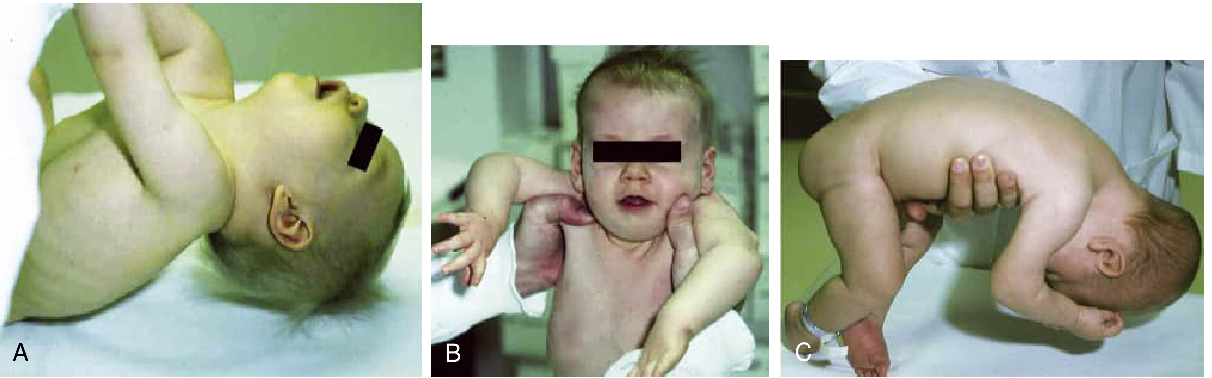
- Excessive head lag - head falls completely back (Fig. 30.2A)
- Once upright, head continues to lag or falls forward quickly
- Absence of limb flexion = appendicular hypotonia or weakness
STEP 3 - Vertical Suspension
How to perform:
- Place hands under the infant's axillae
- Lift the infant without grasping the thorax
Normal response:
- Infant remains suspended - shoulder girdle muscles hold the infant up
- Head upright in midline
- Hips and knees flexed
Hypotonic response - "Slip-through" sign (Fig. 30.2B):
- Infant slips through the examiner's hands
- Shoulders elevate passively (not held by muscle power)
- Head falls forward
- Legs hang extended
STEP 4 - Horizontal (Ventral) Suspension
How to perform:
- Hold infant prone, with the examiner's palm supporting the abdomen and chest
Normal response:
- Head above horizontal, in line with the trunk
- Limbs flexed, held off the hand
Hypotonic response - "Rag doll" sign (Fig. 30.2C):
- Head hangs down below the level of the body
- Limbs drape limply over the examiner's hand
- Infant "drapes" like a wet cloth
STEP 5 - Assessment of Primitive Reflexes
These help localize the level of the lesion:
Moro Reflex (Startle Reflex)
- Present: 28 weeks gestation to 6 months postnatal
- How to elicit: With infant supine, quickly drop the head slightly below the body level while supporting the trunk in one hand
- Normal: Initial abduction + extension of arms with open hands, followed rapidly by adduction + flexion with closed hands ("embrace" pattern)
- In central hypotonia: May be exaggerated or obligatory (persists with continued stimulus)
- In motor unit disorders: Absent or depressed (primitive reflexes are depressed in any motor unit disorder)
Asymmetric Tonic Neck Reflex (ATNR) - "Fencing Posture"
- Present: Term to ~3 months
- How to elicit: Rotate the head to one side while infant lies supine
- Normal: Extension of ipsilateral (face-side) limbs + flexion of contralateral (skull-side) limbs
- Abnormal if: Obligatory (infant cannot break the posture), absent, or asymmetric
Other Primitive Reflexes to Test:
| Reflex | How to Elicit | Normal Response | Age Range |
|---|---|---|---|
| Rooting | Stroke corner of mouth | Turns head toward stimulus | Birth to 4 months |
| Sucking | Insert gloved finger into mouth | Rhythmic sucking | Birth to 4 months |
| Palmar grasp | Press finger into palm | Finger flexion around examiner's finger | Birth to 3-4 months |
| Plantar grasp | Press thumb on ball of foot | Toe flexion | Birth to 9-12 months |
| Stepping | Hold upright, sole on surface | Alternating stepping movements | Birth to 2 months |
| Galant (trunk incurvation) | Stroke paravertebral skin in prone | Trunk curves toward stimulus side | Birth to 2 months |
STEP 6 - Deep Tendon Reflexes (DTRs)
Critical for localization:
| Finding | Interpretation |
|---|---|
| Normal or brisk reflexes with hypotonia | Central (brain) cause |
| Absent or reduced reflexes + hypotonia + weakness | Peripheral (motor unit) cause |
| Reflexes reduced disproportionate to weakness | Peripheral neuropathy (demyelinating) |
| Reflexes reduced proportionate to weakness | Myopathy or axonal neuropathy |
- Test with infant's head in the midline and limbs symmetrically positioned - any deviation produces spuriously asymmetric reflexes
STEP 7 - Additional Examination Signs
Signs favoring CENTRAL hypotonia:
- Fisting (thumbs trapped in closed hands) - early indicator of UMN involvement
- Normal or brisk DTRs
- Exaggerated/obligatory primitive reflexes
- Scissoring of legs in vertical suspension (early appendicular hypertonia emerging)
- Encephalopathy: reduced wakefulness, poor feeding, excessive irritability
- Seizures
- Dysmorphic features (face, limbs), organomegaly
- Head size/shape abnormalities
Signs favoring PERIPHERAL (Motor Unit) hypotonia:
- Weakness prominent (reduced power even with noxious stimulus)
- Absent or reduced DTRs
- Absent or depressed primitive reflexes
- Tongue fasciculations (SMA - Werdnig-Hoffmann)
- Fatigability: fluctuating ptosis, premature stopping of feeding (NMJ disorders)
- Micrognathia, high-arched palate, pectus excavatum (chronic intrauterine weakness)
- Arthrogryposis (joint contractures from reduced fetal movement)
- Sensory loss (peripheral neuropathy)
Signs of spinal cord involvement:
- Sphincter dysfunction (continuous urinary leakage, abdominal distension)
- Priapism
- Reduced response to painful stimuli below a level
- Context of difficult vaginal delivery (breech, prolonged labor)
Summary: The Localization Framework
EXAMINATION FINDINGS
        |
        |── Normal/brisk DTRs + Encephalopathy + Seizures + Fisting
        |        ↓
        |    CENTRAL (Brain) → MRI, chromosomal microarray, metabolic workup
        |
        |── Absent/reduced DTRs + Weakness + No encephalopathy
                 ↓
            PERIPHERAL (Motor Unit)
                 |
                 |── Tongue fasciculations + Areflexia → SMA (SMN1 gene test)
                 |── Fatigability + Ptosis + Fluctuating → NMJ (RNS, AChR Ab)
                 |── Constipation + Weak cry + No fever → Botulism
                 |── Elevated CK + Progressive → Muscular dystrophy/myopathy
                 └── Reduced NCS velocities + Sensory loss → Neuropathy
Reference video demonstrations:
Source: Bradley and Daroff's Neurology in Clinical Practice, Chapter 30, pp. 519-523
This is a shared conversation. Sign in to Orris to start your own chat.