I now have all the content needed for a thorough, well-illustrated answer. Here is the complete classification with MOA:
Antihypertensive Drug Classification with Mechanism of Action
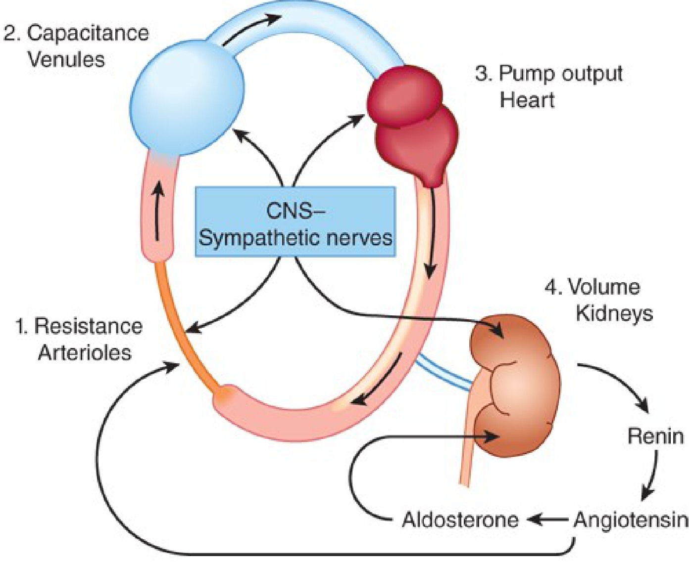
Blood pressure = Cardiac Output (CO) × Peripheral Vascular Resistance (PVR). All antihypertensive drugs act by modifying one or more of the four anatomic control sites: resistance arterioles, capacitance venules, the heart (pump output), and the kidney (volume).
Class 1 - Diuretics
These lower BP primarily by depleting body sodium stores, reducing blood volume and cardiac output. After 6-8 weeks, CO returns toward normal and peripheral vascular resistance declines. Sodium also contributes to vascular stiffness by altering sodium-calcium exchange (increasing intracellular calcium), which is reversed by diuretics.
| Subclass | Examples | Key MOA |
|---|
| Thiazides | Hydrochlorothiazide, Chlorthalidone, Indapamide | Inhibit Na+/Cl- cotransporter in distal convoluted tubule |
| Loop diuretics | Furosemide, Torsemide | Inhibit Na+/K+/2Cl- cotransporter in thick ascending limb; used in renal impairment |
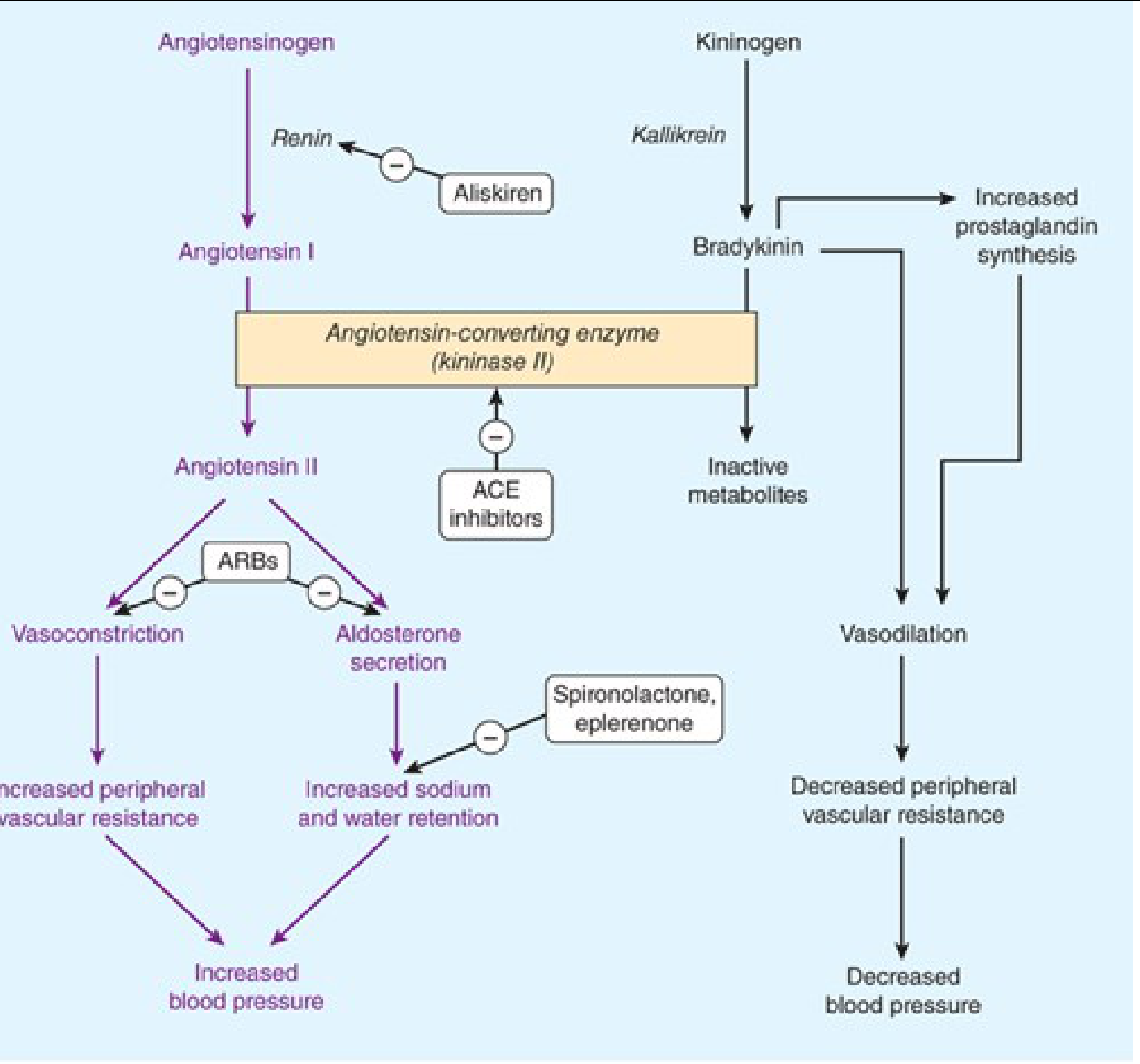
| Potassium-sparing | Spironolactone, Eplerenone, Amiloride | Spironolactone/Eplerenone: aldosterone receptor antagonists; Amiloride/Triamterene: block ENaC sodium channels in collecting duct |
Thiazides are first-line for most uncomplicated hypertension; effective in lowering BP 10-15 mmHg. Loop diuretics preferred when GFR <30 mL/min.
Class 2 - RAAS Inhibitors
2A. ACE Inhibitors (ACEi)
MOA: Block angiotensin-converting enzyme (ACE/kininase II), preventing conversion of angiotensin I → angiotensin II. This reduces vasoconstriction and aldosterone secretion (less sodium/water retention). As a side effect, ACE also degrades bradykinin - inhibiting ACE accumulates bradykinin, causing the characteristic dry cough and, rarely, angioedema.
Examples: Captopril, Enalapril, Lisinopril, Ramipril, Perindopril
2B. Angiotensin Receptor Blockers (ARBs)
MOA: Competitively block angiotensin II AT1 receptors, preventing vasoconstriction and aldosterone release. Do NOT affect bradykinin degradation, so no cough.
Examples: Losartan, Valsartan, Telmisartan, Irbesartan, Olmesartan
2C. Direct Renin Inhibitor
MOA: Aliskiren directly inhibits renin, the rate-limiting step of the RAAS cascade, blocking conversion of angiotensinogen → angiotensin I. This reduces all downstream angiotensin effects.
Class 3 - Calcium Channel Blockers (CCBs)
MOA: Block voltage-gated L-type calcium channels in vascular smooth muscle and/or cardiac muscle, reducing intracellular Ca²+ availability. This leads to:
- Vasodilation (all CCBs) → reduced PVR
- Reduced heart rate and contractility (non-dihydropyridines only)
| Subclass | Examples | Selectivity | MOA emphasis |
|---|
| Dihydropyridines (DHP) | Amlodipine, Nifedipine, Felodipine, Lercanidipine | Vascular > cardiac | Primarily arteriolar vasodilation; minimal cardiac effect |
| Non-DHP: Phenylalkylamine | Verapamil | Cardiac > vascular | Reduces HR, AV conduction, and contractility; less vasodilation |
| Non-DHP: Benzothiazepine | Diltiazem | Intermediate (cardiac + vascular) | Moderate heart rate reduction + vasodilation |
DHPs cause reflex tachycardia and are preferred for isolated systolic hypertension and angina of vasospastic type. Verapamil/diltiazem are useful in hypertension with supraventricular arrhythmias.
Class 4 - Sympatholytic (Sympathoplegic) Agents
These reduce BP by reducing PVR, inhibiting cardiac function, and increasing venous pooling. Subdivided by site of action in the sympathetic arc:
4A. Central Alpha-2 Agonists
MOA: Stimulate presynaptic α2 receptors in the vasomotor center of the medulla (nucleus tractus solitarius), reducing central sympathetic outflow → decreased CO and PVR.
Examples: Methyldopa, Clonidine, Moxonidine (imidazoline receptor agonist)
Methyldopa is the drug of choice in pregnancy-related hypertension.
4B. Beta-Blockers (β-Blockers)
MOA: Block β1 adrenergic receptors on the heart → reduce heart rate and contractility (decreased CO). Also suppress renin release from juxtaglomerular cells. With chronic use, peripheral vascular resistance eventually decreases as well.
| Subclass | Examples | Notes |
|---|
| Non-selective (β1 + β2) | Propranolol, Nadolol | Block both β1 (heart) and β2 (bronchi, vessels) |
| Cardioselective (β1) | Atenolol, Metoprolol, Bisoprolol, Nebivolol | Preferred in asthma/COPD; Nebivolol also releases NO → vasodilation |
| With α-blocking activity | Carvedilol, Labetalol | Block α1 (vasodilation) + β; reduce both CO and PVR; useful in hypertensive emergencies (Labetalol IV) |
4C. Alpha-1 Blockers (Peripheral Sympatholytics)
MOA: Block postsynaptic α1 receptors on vascular smooth muscle → vasodilation → reduced PVR. Cause first-dose hypotension (orthostatic).
Examples: Prazosin, Doxazosin, Terazosin
Also used in BPH (relax bladder neck). First-dose effect is a clinical caveat.
4D. Ganglionic Blockers (Obsolete)
MOA: Block nicotinic receptors at autonomic ganglia → interrupts both sympathetic and parasympathetic transmission. Largely obsolete due to severe side effects (marked postural hypotension, dry mouth, constipation, urinary retention).
Example: Trimethaphan (IV only, hypertensive emergencies historically)
4E. Adrenergic Neuron Blockers (Obsolete)
MOA: Deplete norepinephrine stores from sympathetic nerve terminals.
Examples: Reserpine (depletes catecholamines), Guanethidine (prevents NE release)
Class 5 - Direct Vasodilators
MOA: Directly relax arteriolar smooth muscle → reduce PVR. Because they do NOT block the sympathetic baroreflex, they trigger reflex tachycardia and sodium/water retention - hence they must be combined with a beta-blocker (to block tachycardia) and a diuretic (to block sodium retention).
| Drug | Specific MOA |
|---|
| Hydralazine | Increases cGMP in smooth muscle (possibly via NO); arteriolar > venular dilation |
| Minoxidil | Active metabolite (minoxidil sulfate) opens ATP-sensitive K+ channels in smooth muscle → membrane hyperpolarization → less contraction |
| Sodium nitroprusside | Releases NO spontaneously → activates guanylyl cyclase → increased cGMP → vasodilation of arteries AND veins; used IV in hypertensive emergencies |
| Diazoxide | Opens K+ channels in smooth muscle; IV use in emergencies |
Class 6 - Nitrates
MOA: Metabolized to nitric oxide (NO) → activates soluble guanylyl cyclase → increases cGMP → dephosphorylates myosin light chain → venous (predominantly) and arteriolar relaxation.
Examples: GTN (glyceryl trinitrate), Isosorbide dinitrate - primarily for angina, but reduce preload and afterload.
Class 7 - Newer/Miscellaneous Agents
| Drug | Class | MOA |
|---|
| Sacubitril/Valsartan (Entresto) | ARB + Neprilysin inhibitor | Valsartan blocks AT1; Sacubitril inhibits neprilysin (degrades natriuretic peptides) → elevated ANP/BNP → natriuresis, vasodilation |
| Eplerenone | Selective mineralocorticoid antagonist | More selective than spironolactone; blocks aldosterone at renal tubule; fewer anti-androgenic side effects |
| Bosentan / Ambrisentan | Endothelin receptor antagonists | Block ET-1 receptors → vasodilation; used in pulmonary arterial hypertension |
| Sildenafil / Tadalafil | PDE-5 inhibitors | Prevent degradation of cGMP → sustained vasodilation; primarily for pulmonary hypertension |
Summary Table
| Class | Primary Site of Action | Net Effect on BP |
|---|
| Diuretics | Kidney (volume) | ↓ Blood volume → ↓ CO |
| ACEi / ARB / DRI | RAAS pathway | ↓ Vasoconstriction + ↓ Na/H2O retention |
| CCBs (DHP) | Arteriolar smooth muscle (L-Ca²+) | ↓ PVR |
| CCBs (non-DHP) | Heart + arterioles | ↓ HR, ↓ CO, ↓ PVR |
| Beta-blockers | Heart + JGA (renin) | ↓ CO, ↓ renin |
| Alpha-1 blockers | Arteriolar smooth muscle | ↓ PVR |
| Central alpha-2 agonists | CNS vasomotor center | ↓ Sympathetic outflow |
| Direct vasodilators | Arteriolar smooth muscle | ↓ PVR (reflex tachycardia) |
Source: Katzung's Basic and Clinical Pharmacology, 16th Edition, Chapter 11 - Antihypertensive Agents