
Sarcoidosis pathophysiology (chart) , cutaneous manifestations in details, other manifestations in detals , diagnosis, management
lupus pernio cutaneous sarcoidosis face violaceous plaques
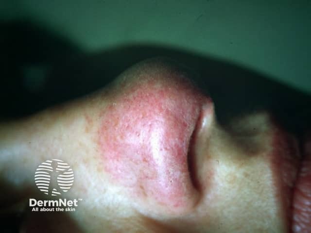
This high-resolution clinical photography shows cutaneous lupus pernio, the characteristic violaceous, indurated plaques of sarcoidosis involving the nasal dorsum and surrounding integument. Acquisition modality: bedside clinical photography, full-face anterior view, with close-up focus on the nose (nasal pyramid, alar rims) to document contour irregularities and color. The lesion appears as erythematous-violaceous plaques with firm, raised induration, subtle scaling, and blending erythema into adjacent normal skin. Dermal granulomatous inflammation in lupus pernio typically yields palpable plaques and nodules; in this image, the lesion is non-ulcerated, symmetric along the nasal region, with mild edema and vascular prominence. In the clinical context, lupus pernio strongly suggests sarcoidosis and often indicates chronic systemic involvement, particularly of the lungs and upper airways; dermatologic findings may precede or accompany pulmonary disease. Differential diagnoses include granulomatous rosacea, tuberculoid granulomatous lesions, and other granulomatous dermatoses; biopsy would show noncaseating granulomas with tight epithelioid granulomas and multinucleated giant cells. This image supports diagnostic correlation, educational case illustration, and differential diagnosis discussion for dermatology, rheumatology, pulmonology, and pathology. It has clinical relevance for prognosis and monitoring response to therapy, including corticosteroids or steroid-sparing agents, and for documenting treatment outcomes. Correlation with biopsy and systemic evaluation remains essential for definitive diagnosis.
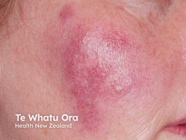
This is a high‑resolution clinical photograph of a single violaceous cutaneous plaque on the malar/cheek region. Imaging modality: clinical photography of the skin, near-frontal view of the zygomatic area under white light, without dermoscopic enhancement. Anatomical localization: integumentary system, facial skin; cheek. Visual features: a well‑circumscribed, indurated plaque with a purple‑violaceous hue, smooth to slightly glossy surface, mild surrounding erythema, and subtle texturing. The lesion geometry is oval to irregular, with a slightly lighter center suggesting mild central atrophy or edema. Medical impression: the violaceous, persistent plaque on the face is highly suggestive of cutaneous sarcoidosis presenting as lupus pernio, a prototypical cutaneous manifestation linked to systemic sarcoidosis. Alternative considerations include granulomatous rosacea, chronic infectious granulomas (mycobacterial or fungal), and other granulomatous dermatitis. Diagnostic significance: if lupus pernio is suspected, correlate with chest imaging, serum ACE levels, and biopsy showing noncaseating granulomas for confirmation. Clinical relevance: this morphology warrants multidisciplinary evaluation for systemic involvement, and consideration of dermatology, pulmonology, and rheumatology follow‑up. Potential clinical use: educational reference for dermatology training, sarcoidosis recognition, and differential diagnosis practice with facial violaceous plaques. In documentation, annotate lesion margins, color, and texture changes over time to assist monitoring and therapeutic response assessment and prognosis.
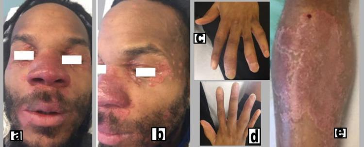
This composite clinical photograph displays the multi-systemic cutaneous and musculoskeletal manifestations of chronic sarcoidosis. Figures (a) and (b) show the patient's face with characteristic lupus pernio, presenting as indurated, violaceous (purplish) plaques and nodules affecting the nose, cheeks, and periorbital regions. Figure (e) illustrates a large, well-demarcated, erythematous to purplish indurated plaque on the medial aspect of the right leg. Figures (c) and (d) demonstrate clinical dactylitis ('sausage digits') of the hands, characterized by diffuse swelling of the fingers involving the proximal interphalangeal (PIP) and distal interphalangeal (DIP) joints. The image serves as an educational resource for identifying the classic visual signs of sarcoidosis, specifically the association between lupus pernio skin lesions and sarcoid dactylitis or inflammatory arthritis. It is intended for medical students and clinicians specializing in dermatology and rheumatology.
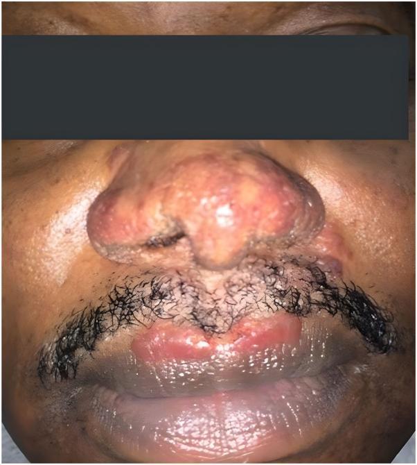
This clinical photograph displays a front-on view of the mid-face of a 56-year-old male, highlighting prominent dermatological manifestations of cutaneous sarcoidosis, specifically lupus pernio. The nasal region is significantly involved, showing diffuse, pinkish-red to violaceous infiltration with multiple confluent, shiny nodules that distort the nasal contour. The surface appears irregular and indurated. In the perioral area, reddish-brown, shiny, well-demarcated plaques are visible along the vermilion border of the upper lip and extending into the philtrum and mustache region. The affected skin shows a characteristic waxy or translucent texture. This image serves as a classic educational representation of chronic sarcoidosis, where granulomatous infiltration typically affects the 'central face' (nose, lips, and cheeks). Clinical significance lies in recognizing these specific morphologies, which are often refractory to treatment and can be associated with underlying systemic sarcoidosis or upper respiratory tract involvement.
sarcoidosis treatment guideline
Sarcoidosis
1. Pathophysiology (Flow Chart)
UNKNOWN ANTIGEN(S) (mycobacterial proteins e.g. mKatG, ESAT-6; beryllium;
inorganic dust; possibly other environmental triggers)
            │
            ▼
Genetically susceptible host (HLA-DRB1, HLA-DQB1*0201/DRB1*0301 → good
prognosis/Löfgren; BTNL2, ANXA11, CCR5 polymorphisms)
            │
            ▼
Antigen-presenting cells (macrophages/dendritic cells) process & present
antigen via HLA class II ─────────────► CD4+ T-helper (Th1) lymphocytes
            │                                          │
            ▼                                          ▼
Macrophages secrete IL-12, IL-18          T cells secrete IFN-γ, IL-2,
(drive Th1 polarization)                  TNF-α, chemotactic factors
            │                                          │
            └───────────────────┬──────────────────────┘
                                 ▼
        Recruitment & activation of monocytes/macrophages
        at the site of antigen persistence
                                 ▼
        Epithelioid histiocyte transformation, macrophage
        fusion (IFN-γ driven) → multinucleated giant cells
                                 ▼
        NON-CASEATING GRANULOMA formation
        (compact granulomas with epithelioid histiocytes,
        giant cells ± Schaumann bodies/asteroid bodies,
        rim of lymphocytes, NO central necrosis)
                                 ▼
        ┌────────────────────────┴─────────────────────────┐
        ▼                                                   ▼
Peripheral anergy / lymphopenia                 Organ infiltration & fibrosis
(circulating Treg expansion, cutaneous          (lung, skin, eye, heart, CNS,
delayed-type hypersensitivity suppressed        liver, kidney, bone, spleen)
while granuloma-forming response persists
in tissue - the classic "sarcoid paradox")
                                 ▼
        Ectopic 1α-hydroxylase activity in activated
        macrophages → ↑1,25-(OH)2 vitamin D →
        hypercalciuria/hypercalcemia, nephrolithiasis
2. Cutaneous Manifestations (in detail)
Non-specific lesions
- Erythema nodosum (EN) - the most common non-specific finding. Tender, warm, red subcutaneous nodules on the anterior shins, often with fever, polyarthralgia, uveitis, and bilateral hilar adenopathy = Löfgren syndrome (common in Scandinavian whites, rare in Black patients). ESR is often markedly elevated (>50 mm/hr). EN carries a good prognosis - spontaneous resolution within 2 years in ~80% of patients; its absence is actually a risk factor for chronic disease.
Specific lesions (contain non-caseating granulomas on biopsy)
- Papular sarcoidosis - the most common morphology; small (<1 cm) papules, especially on the face, eyelids, neck, shoulders ("miliary sarcoid" when generalized). Papules on the knee may occur in isolation. In African Americans, papules along the alar rim can be the first sign of lupus pernio and predict a poor prognosis.
- Annular sarcoidosis - coalesced papules forming red-brown annular plaques on the head/neck, with central clearing, hypopigmentation, atrophy, scarring, and sometimes alopecia.
- Plaque sarcoidosis - larger, indurated, violaceous-to-brown plaques; associated with chronic, systemic disease.
- Lupus pernio - the most distinctive and prognostically important lesion: disfiguring, violaceous, indurated plaques/nodules over the nose, cheeks, ears, and lips. Strongly associated with chronic, fibrotic pulmonary disease, granulomatous infiltration of the upper airway/nasal mucosa (epistaxis, crusting), bone cysts, and a generally poor, treatment-refractory course.
- Subcutaneous sarcoidosis (Darier-Roussy) - firm subcutaneous nodules without epidermal change.
- Scar sarcoidosis - granulomas infiltrating old scars or tattoos, causing them to become raised, inflamed, and discolored - a useful diagnostic clue.
- Less common patterns: hypopigmented macules/patches (especially in darker skin), ulcerative, verrucous/hyperkeratotic, ichthyosiform, erythrodermic, lichenoid, and alopecic (scarring) scalp sarcoidosis; nail dystrophy; mucosal/genital lesions.
3. Other (Extracutaneous) Manifestations in Detail
| System | Manifestations |
|---|---|
| Pulmonary/thoracic (>90% of cases) | Bilateral hilar lymphadenopathy, perilymphatic/bronchovascular nodules, upper-lobe predominant interstitial disease; staged radiographically by Scadding stages 0-IV (0 = normal; I = hilar adenopathy alone; II = adenopathy + parenchymal disease; III = parenchymal disease alone; IV = fibrosis). Progressive fibrosis can cause honeycombing, cavitation, chronic pulmonary aspergillosis, and pulmonary hypertension - the leading cause of sarcoidosis mortality in Western populations. |
| Ocular (~25%) | Anterior/posterior uveitis (most common), conjunctival granulomas, keratoconjunctivitis sicca, optic neuropathy, retinal vasculitis; can cause blindness if untreated. Heerfordt syndrome: facial nerve palsy + fever + parotid enlargement + anterior uveitis. |
| Cardiac | Granulomatous infiltration causing conduction block (a major cause of unexplained AV block/sudden death in young adults), ventricular arrhythmia, dilated/restrictive cardiomyopathy, pericardial effusion. More prevalent and more often fatal in Japanese patients. |
| Neurologic (neurosarcoidosis) | ~5-10%; predilection for the base of the brain; facial nerve palsy is most common, also aseptic meningitis, mass lesions, myelopathy, peripheral neuropathy; can occur without systemic disease. |
| Endocrine/Calcium | Hypercalciuria (more common) and hypercalcemia from macrophage 1α-hydroxylase converting 25(OH)D to active 1,25(OH)2D; can cause nephrocalcinosis/nephrolithiasis and renal impairment. |
| Renal | Granulomatous interstitial nephritis, nephrocalcinosis/stones from hypercalcemia. |
| Hepatic | Common histologically (granulomas), usually asymptomatic; ~33% have elevated alkaline phosphatase; rarely progresses to a primary biliary cirrhosis-like picture with portal hypertension. |
| Musculoskeletal | Acute polyarthralgia/arthritis (part of Löfgren syndrome), chronic arthropathy, cystic bone lesions (especially with lupus pernio), dactylitis ("sausage digits"), myopathy. |
| ENT/Upper airway | Nasal/sinus granulomas (often with lupus pernio), laryngeal involvement, parotid gland enlargement. |
| Hematologic/Splenic | Splenomegaly, splenic sequestration, bone marrow granulomas causing leukopenia/thrombocytopenia; hypergammaglobulinemia. |
| Constitutional | Fatigue, fever, weight loss, night sweats - common and often disabling even without major organ threat. |
4. Diagnosis
- Compatible clinical and radiographic presentation
- Histologic demonstration of non-caseating (non-necrotizing) granulomas on biopsy of an accessible site (skin lesion, peripheral lymph node, minor salivary gland, conjunctiva, or via bronchoscopy - endobronchial/transbronchial biopsy or EBUS-guided lymph node sampling)
- Exclusion of other granulomatous diseases - mycobacterial (TB) and fungal infection, berylliosis, hypersensitivity pneumonitis, lymphoma, and reaction to foreign material must be ruled out (special stains/cultures on biopsy tissue are mandatory).
- Chest X-ray/CT - Scadding staging; CT shows perilymphatic/bronchovascular nodules, hilar/mediastinal adenopathy, septal thickening.
- Serum ACE level - elevated in ~60-80% but nonspecific (elevated in other granulomatous diseases too, ~10% false-positive rate); used more for monitoring disease activity than for diagnosis.
- Serum/urine calcium, 24-hour urine calcium, vitamin D metabolites.
- Pulmonary function tests - typically restrictive pattern; obstruction in up to a third.
- PET/CT or gallium scan - can identify occult active disease sites for biopsy targeting.
- ECG ± cardiac MRI/PET - screening for cardiac sarcoidosis given risk of sudden death.
- Ophthalmologic exam - even if asymptomatic, given risk of silent uveitis.
- Kveim-Siltzbach test - historical intradermal test using sarcoid splenic tissue homogenate; largely abandoned (not standardized/available) but of historical/pathophysiologic interest, showing ~20-40% false-negative rate.
- Baseline work-up for a new diagnosis of cutaneous sarcoidosis specifically includes: physical exam, neurologic review of systems, chest radiograph, ECG, PFTs, urinalysis, CBC, comprehensive metabolic panel, serum calcium, TB screening, and ophthalmologic exam - to look for systemic disease. (Fitzpatrick's Dermatology)
5. Management
- Prednisone (or equivalent) ~0.5 mg/kg/day, typically 20-40 mg/day for about 4 weeks, then tapered to the lowest maintenance dose controlling disease (ideally <10 mg/day, target 7.5-15 mg/day).
- Higher doses are not more effective for most indications and increase morbidity; reserve aggressive high-dose ± combination immunosuppression for organ- or life-threatening disease (CNS, vision-threatening ocular disease, extensive cardiac involvement).
- Topical/intralesional corticosteroids (e.g., clobetasol, intralesional triamcinolone) for localized cutaneous disease, including lupus pernio.
- Methotrexate - most commonly used second-line agent (7.5-20 mg/week PO or SC, with folic acid); good efficacy/side-effect profile.
- Hydroxychloroquine - useful especially for cutaneous and mild disease (200-400 mg/day); requires periodic retinal exams.
- Azathioprine - comparable efficacy to methotrexate.
- Leflunomide, mycophenolate - alternative steroid-sparing agents.
- TNF-α inhibitors - infliximab is particularly effective for severe, refractory disease including lupus pernio and neurosarcoidosis; adalimumab also used. Recent systematic reviews/meta-analyses (2024-2025) support biologic efficacy in refractory pulmonary and cutaneous sarcoidosis, though golimumab showed only a non-significant trend of benefit.
- Manage hypercalcemia: avoid excess vitamin D/sun exposure, hydration, corticosteroids.
- Cardiac sarcoidosis: antiarrhythmics, ICD/pacemaker as indicated, immunosuppression.
- End-stage organ failure (lung, heart, liver, kidney): organ transplantation; disease can recur in the graft but rarely causes graft failure.
- Regular surveillance (PFTs, ophthalmology, ECG) to catch progression early.