
Hemorrhage
Reading File
The query "Hemorrhage" is broad. Let me clarify before diving into research.
Asking for Preferences
What aspect of hemorrhage are you interested in?
Searching sources...
Finding Sources
Finding Sources
Good - I can see forensic pathology and Harrison's have relevant content. Let me now pull the hemorrhage classification sections directly.
Finding Sources
Finding Sources
Robbins Basic Pathology is the ideal source for hemorrhage classification. Let me read that section plus the forensic pathology book.
Reading File
Finding Sources
Excellent content found. Let me now read the forensic medicine section on hemorrhage classification and fetch the Robbins images.
Reading File
Reading File
I now have rich content from both Robbins & Kumar Basic Pathology and the Forensic Medicine textbook. Let me also fetch the key Robbins image showing petechial hemorrhages and intracerebral hemorrhage.
Loading Image
Loading Image
I now have everything needed for a comprehensive answer. Let me compile it.
Hemorrhage - Types & Classification
Hemorrhage is defined as the escape (extravasation) of blood from the cardiovascular system into tissues, tissue layers, or body cavities. It results from vessel damage and is worsened by defects in clotting.
1. Classification by Origin (Source Vessel)
| Type | Characteristics |
|---|---|
| Capillary | Oozing from small vessels; common in contusions, congestion |
| Venous | Steady, dark red flow; less pressure than arterial |
| Arterial | Bright red, pulsatile, forceful; most dangerous |
| Cardiac | From heart itself (e.g., rupture of ventricle post-MI) |
- The Essentials of Forensic Medicine and Toxicology (36th ed.), p. 299
2. Classification by Location
External Hemorrhage
Blood exits the body surface. Dangerous if a large vessel is involved - partial transection of an artery prevents vessel retraction, so bleeding is often worse. Sudden loss is more dangerous than slow loss, as the body cannot compensate quickly enough.
Internal Hemorrhage
Blood accumulates inside body cavities or tissues. Named by location:
| Term | Location |
|---|---|
| Hemothorax | Pleural cavity |
| Hemopericardium | Pericardial sac |
| Hemoperitoneum | Peritoneal cavity |
| Hemarthrosis | Joint space |
| Hematoma | Localized clotted blood in a tissue (any site) |
Internal bleeds do not cause iron deficiency anemia because iron is efficiently recycled from phagocytosed red cells.
- Robbins & Kumar Basic Pathology, p. 82
3. Classification by Cause
| Type | Definition |
|---|---|
| Traumatic hemorrhage | Bleeding due to wounds (lacerations, fractures, penetrating injuries) |
| Spontaneous hemorrhage | Bleeding without trauma - due to hemorrhagic diatheses, vascular disease, neoplasm, inflammation |
| Primary hemorrhage | Occurs at the time of injury |
| Secondary (Reactionary) hemorrhage | Delayed - occurs hours to >24 hours after injury due to: rise in BP on recovery from shock, muscular movement dislodging a clot, or vessel wall erosion from infection |
4. Classification by Morphology / Size
This is the most clinically used morphological classification:
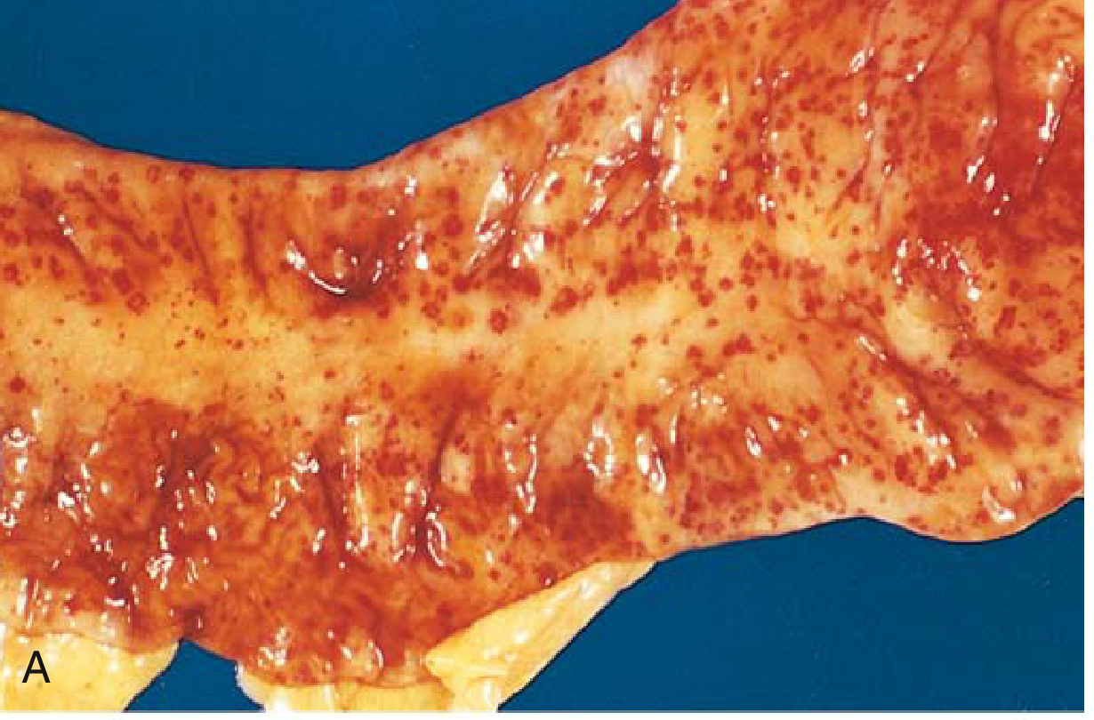
Petechiae
- Size: 1-2 mm
- Pin-point hemorrhages into skin, mucous membranes, or serosal surfaces
- Caused by: thrombocytopenia, platelet dysfunction, loss of vascular wall support (e.g., scurvy)
- Origin: capillary or venular
Purpura
- Size: 3-5 mm
- Caused by: same as petechiae, plus trauma, vasculitis, increased vascular fragility
Ecchymoses
- Size: 1-2 cm
- Subcutaneous hematomas - colloquially "bruises"
- Color changes over time reflect hemoglobin breakdown:
- Red-blue (fresh) → hemoglobin
- Blue-green → biliverdin/bilirubin
- Golden-brown → hemosiderin
- Phagocytosed and degraded by macrophages
Hematoma
- A localized, usually clotted collection of blood within a tissue forming a tumor-like swelling
- Range from trivial (bruise) to fatal (retroperitoneal hematoma from ruptured aortic aneurysm)
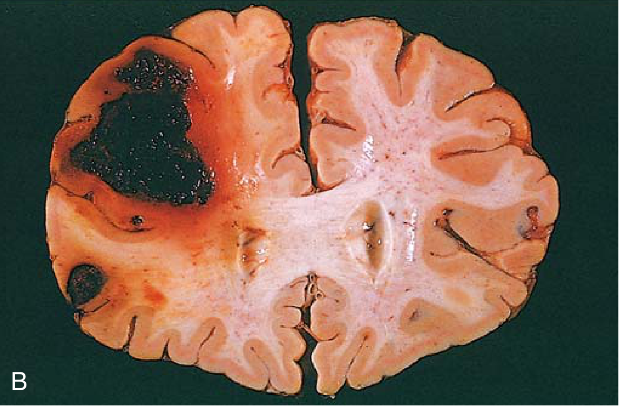
Apoplexy
- Large effusion of blood into an organ
- Term commonly used for cerebral hemorrhage
5. Factors Determining Clinical Impact
The significance of a hemorrhage depends on four key variables:
- Volume lost - rapid loss of >20% blood volume causes hypovolemic shock even in healthy adults; up to 20% may be tolerated well
- Rate of bleeding - sudden loss is more dangerous than equivalent slow loss
- Location - trivial in subcutaneous tissue, fatal in the brain (as shown above)
- Health of the individual - those with heart/lung disease decompensate at lower volumes
Summary Diagram of Classification
HEMORRHAGE
│
├── By VESSEL SOURCE → Capillary / Venous / Arterial / Cardiac
│
├── By LOCATION → External / Internal (Hematoma, Hemothorax, Hemopericardium,
│                                       Hemoperitoneum, Hemarthrosis)
│
├── By CAUSE → Traumatic / Spontaneous
│              Primary / Secondary (reactionary)
│
└── By MORPHOLOGY → Petechiae (1-2mm) → Purpura (3-5mm)
                    → Ecchymosis (1-2cm) → Hematoma → Apoplexy
Sources:
- Robbins & Kumar Basic Pathology - pp. 82-83 (morphological classification, clinical impact)
- The Essentials of Forensic Medicine and Toxicology, 36th ed. (2026) - p. 299 (vessel-source classification, primary/secondary hemorrhage)
This is a shared conversation. Sign in to Orris to start your own chat.