
Can i share photo with yoy?
Where is the option
- Look for a paperclip icon (📎) near the message input box
- Or a "+" button next to the text field
- Some interfaces have an image/photo icon (🖼️) directly in the toolbar
- Tap the attachment icon or "+" next to the text bar
- Choose "Photo Library" or "Camera"
- Look for an attachment or upload button near where you type
- You can also try dragging and dropping an image directly into the chat window
My one eye has redness.. when moving eyeball it has discomfort what could be it and what is the treatment?
red eye discomfort pain when moving eyeball causes treatment
red eye scleritis episcleritis conjunctivitis
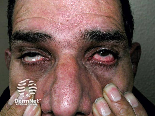
Imaging modality: Digital clinical photography of the ocular region, frontal facial view focusing on the eyelids, periorbital tissues, and anterior segment. The subject shows bilateral red eyes with conspicuous conjunctival injection and scleral/episcleral erythema; surrounding eyelid and lid-margin tissues appear mildly swollen. Anatomical context centers on the anterior ocular structures, including conjunctiva, episclera, and sclera, within the orbit; the view highlights vascular dilation and diffuse hyperemia consistent with inflammatory processes rather than infectious discharge. The differential includes scleritis, episcleritis, conjunctivitis, and iritis; distinguishing features may include depth of scleral involvement, tenderness, and pain intensity not fully captured in a still image. In relapsing polychondritis, ocular inflammation often accompanies systemic cartilage involvement and can herald flares; thus, clinicians should correlate with ENT, rheumatology findings, and systemic inflammatory markers. Diagnostic significance lies in recognizing autoimmune ocular involvement that necessitates systemic immunomodulatory therapy rather than topical antibiotics alone. Potential clinical utilities include documenting disease activity, guiding treatment escalation, educating learners about RP-associated eye findings, and serving as a reference for case-based discussions on autoimmune uveitis spectrum. This image illustrates how RP can present with red eyes due to scleritis/episcleritis/conjunctivitis/iritis, aiding differential diagnosis and interdisciplinary management. Representative RP ocular involvement warrants early rheumatology collaboration.
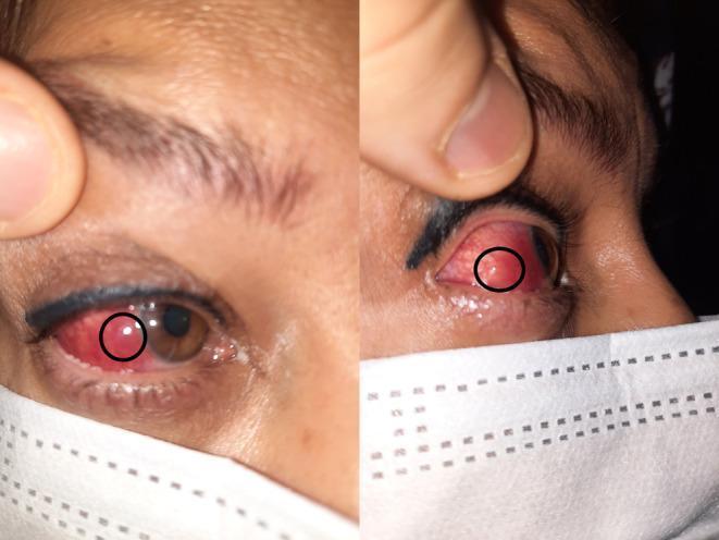
Content Type: Clinical Photograph. This composite image shows two views of a patient's right eye, highlighting inflammatory ocular signs. The left panel shows a frontal view of the eye with significant violaceous (purplish) scleral hyperemia and a distinct, circumscribed nodule (circled) on the temporal sclera. The right panel provides a lateral view, further illustrating the elevation and localized nature of the nodular lesion against the hyperemic scleral background. The purplish hue is characteristic of deep episcleral or scleral vessel involvement, often seen in scleritis. The iris appears brown and the cornea remains clear in these views. The images demonstrate the clinical presentation of nodular scleritis, an inflammatory condition of the eye's outer protective layer. These findings are pedagogically useful for distinguishing between superficial episcleritis (which typically presents with a brighter red hue) and deeper scleral inflammation (which exhibits a darker, violaceous color).
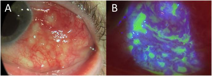
Educational slit-lamp clinical photographs showing infectious scleritis or conjunctivitis in the temporal region of the left eye. Image A (white light) demonstrates significant conjunctival and scleral injection (hyperemia) with engorged blood vessels. Centered in the inflamed area are multiple, yellowish-white multiloculated abscesses associated with active purulent drainage, indicating a localized infectious process. Image B (cobalt blue light with fluorescein staining) of the same ocular region highlights extensive epithelial disruption. The bright green fluorescence reveals multiple small, irregular conjunctival erosions and larger areas of epithelial loss corresponding to the sites of inflammation and abscess formation. These images serve as a diagnostic reference for identifying severe ocular surface infections and assessing tissue integrity through fluorescein staining.
Most Likely Causes
1. Episcleritis (Most Common, Milder)
- Inflammation of the episclera (the thin tissue layer between the conjunctiva and sclera)
- Presents as a red, sore eye - usually mild discomfort, not severe pain
- Typically affects young adults, more often women
- Eye looks bright red (not violaceous/purple)
- Blood vessels blanch when phenylephrine drops are applied
- Often resolves on its own in days to weeks
- May recur
2. Scleritis (Less Common, More Serious)
- Inflammation of the sclera (the white "wall" of the eye) - a deeper, more painful process
- Classic feature: boring, severe eye pain that worsens with eye movement and is tender to touch
- Pain may radiate to the forehead, brow, or jaw and can wake you at night
- Eye has a characteristic violaceous (purplish-red) hue, best seen in natural daylight
- Up to 50% of cases are linked to systemic autoimmune diseases (rheumatoid arthritis, lupus, vasculitis)
3. Conjunctivitis (Pink Eye)
- Inflammation of the conjunctiva - usually causes redness, discharge, gritty feeling
- Pain with eye movement is less typical unless there is significant swelling
- Can be viral (most common), bacterial (with pus-like discharge), or allergic (itching)
4. Optic Neuritis (Less Likely, but Important)
- Inflammation of the optic nerve
- Causes pain with eye movement (in 19-92% of cases) along with reduced vision
- If your vision in that eye feels blurry or dimmed, this needs urgent attention
Treatment
| Condition | Treatment |
|---|---|
| Episcleritis (mild) | Artificial tears, cold compresses; often self-resolving |
| Episcleritis (persistent) | Topical NSAID drops or mild topical steroid drops; or oral NSAIDs (ibuprofen 200-600 mg 3-4x/day, or naproxen 250-500 mg twice daily) for 10-14 days |
| Scleritis (diffuse/nodular) | Oral NSAIDs first (flurbiprofen, naproxen, indomethacin); if no improvement, oral prednisone 60-80 mg/day with taper |
| Scleritis (severe/necrotizing) | IV methylprednisolone + immunosuppressives (methotrexate, azathioprine, biologics) |
| Viral conjunctivitis | Supportive - cold compresses, artificial tears; resolves on its own in 1-2 weeks |
| Bacterial conjunctivitis | Antibiotic eye drops (e.g., ciprofloxacin, tobramycin) |
| Optic neuritis | IV methylprednisolone under specialist care; investigate for MS |
When to See a Doctor Urgently
- Significant pain (not just mild discomfort) with eye movement
- Any decrease or blurring of vision
- High sensitivity to light (photophobia)
- A purple/violaceous hue to the eye redness
- Pus or thick discharge
- Symptoms lasting more than a week
Give me best topical nsaid drops and artificial tears available in India
best topical NSAID eye drops brands available India 2024 diclofenac ketorolac nepafenac
best artificial tears eye drops brands India 2024 carboxymethylcellulose sodium hyaluronate
Topical NSAID Eye Drops Available in India
| Drug | Brand Names (India) | Dosing | Notes |
|---|---|---|---|
| Diclofenac 0.1% | Diclsure, Naclof, Voltaren Ophthalmic, Dicloflam | 1 drop 3-4x/day | Most widely available & affordable; first choice for episcleritis |
| Ketorolac 0.5% | Acular, Ketorol Eye Drops, Toradol | 1 drop 4x/day | Good for allergic itch + inflammation |
| Nepafenac 0.1% | Nevanac, Nepfenac | 1 drop 3x/day | Prodrug - better corneal penetration; preferred post-surgery |
| Bromfenac 0.09% | Xibrom, Bromday | 1-2 drops/day | Convenient once or twice daily dosing |
| Flurbiprofen 0.03% | Ocuflur | 1 drop 4x/day | Mainly used to prevent intraoperative miosis |
Best for your situation (episcleritis/red eye with discomfort): Diclofenac 0.1% (e.g., Naclof or Diclsure) is the most commonly prescribed and available option in India.
Artificial Tears Available in India
Carboxymethylcellulose (CMC) - Based
| Brand | Composition | Approx. Price | Notes |
|---|---|---|---|
| Refresh Tears (Allergan) | CMC 0.5% | ₹150-200 | Most trusted brand; widely available |
| Lubistar (Sun Pharma) | CMC 0.5% | ₹80-100 | Affordable generic |
| I-Kul Tears (Ajanta) | CMC 0.5% | ₹140-170 | Good value |
| Moisol (Sun Pharma) | CMC 0.5% + Glycerin | ₹100-130 | Added glycerin for extra moisture |
Sodium Hyaluronate (HA) - Based
| Brand | Composition | Approx. Price | Notes |
|---|---|---|---|
| Hylo Fresh / Hylo Comod (Ursapharm) | Sodium Hyaluronate 0.03-0.1% | ₹300-500 | Preservative-free; excellent for frequent use |
| I-Vizz (FDC) | Sodium Hyaluronate 0.18% | ₹200-250 | Popular in India |
| Vismed | Sodium Hyaluronate 0.18% | ₹250-350 | Preservative-free option |
HPMC (Hydroxypropyl Methylcellulose) - Based
| Brand | Composition | Approx. Price | Notes |
|---|---|---|---|
| Tears Naturale II (Alcon) | HPMC + Dextran | ₹120-150 | Classic, affordable |
| Systane Ultra (Alcon) | HPMC-based | ₹250-350 | Long-lasting, dual-layer protection; excellent for screen users |
| i-Dew | HPMC-based | ₹80-120 | Budget option for mild dryness |
Quick Recommendations for Your Case
- Short-term relief: Start with Refresh Tears or Systane Ultra (artificial tears) 4-6x/day - soothing, safe, no prescription needed
- If inflammation persists >3-4 days: Ask your doctor for Naclof (Diclofenac 0.1%) - 1 drop 3-4x/day for 7-10 days
- Preservative-free preference: Go for Hylo Fresh if you plan to use drops frequently (>4x/day) - preservatives can irritate an already red eye
Important: Do not use topical NSAIDs for more than 2 weeks without medical supervision. Avoid topical steroids without a doctor's advice as they can raise eye pressure and worsen certain infections.
No give combination of nasaid and steroids
combination NSAID steroid eye drops brands India diclofenac dexamethasone prednisolone available 2024
Combination NSAID + Steroid Eye Drops in India
Important: These are prescription-only medications. Steroids in eye drops can raise intraocular pressure, worsen infections, and cause cataract with prolonged use. Use only as directed by your ophthalmologist.
Diclofenac + Dexamethasone Combinations
| Brand Name | Composition | Manufacturer | Notes |
|---|---|---|---|
| Dexdiclof | Diclofenac 0.1% + Dexamethasone 0.1% | Various | Very widely prescribed |
| Diclozone | Diclofenac 0.1% + Dexamethasone 0.1% | — | Common in post-op care |
| Ocufen-D | Diclofenac 0.1% + Dexamethasone 0.1% | — | Widely available |
| Diclodex | Diclofenac 0.1% + Dexamethasone 0.1% | — | Generic, affordable |
Diclofenac + Prednisolone Combinations
| Brand Name | Composition | Notes |
|---|---|---|
| Diclomox-P / Diclopred | Diclofenac 0.1% + Prednisolone 0.5% or 1% | Prednisolone is a softer steroid option |
| Omnipred + Diclofenac | Often co-prescribed separately | When individual dose titration needed |
Ketorolac + Steroid Combinations
| Brand Name | Composition | Notes |
|---|---|---|
| Ketorol-DM / Acular-D | Ketorolac 0.5% + Dexamethasone 0.1% | Used post-operatively |
| Kerolong-D | Ketorolac 0.4% + Dexamethasone 0.1% | Available in some pharmacy chains |
Triple Combinations (Antibiotic + NSAID + Steroid)
| Brand Name | Composition | Notes |
|---|---|---|
| Moxifree-DK / Moxiflodex | Moxifloxacin + Diclofenac + Dexamethasone | Most popular post-op triple combo |
| Zymoxen-D | Ofloxacin + Diclofenac + Dexamethasone | Older but still used |
| Tobramycin + Diclofenac + Dexa combinations | Various brands | Budget options |
Which One for Your Condition?
| Severity | Recommended Combo |
|---|---|
| Mild | Diclofenac 0.1% + Dexamethasone 0.1% (e.g., Dexdiclof) - 1 drop 3-4x/day |
| Moderate | Same combo but 4-6x/day for first 2-3 days, then taper |
| With infection risk | Triple combo (Moxifloxacin + Diclofenac + Dexa) |
Key Safety Points
- Do not use steroid eye drops for more than 1-2 weeks without IOP (intraocular pressure) monitoring
- Check IOP after 1 week if on dexamethasone - it can cause steroid-induced glaucoma
- Avoid if you have herpes eye infection - steroids can make it catastrophically worse
- Prednisolone is considered safer than dexamethasone for IOP risk (fluorometholone is even safer)
- These drops are prescription only in India - visit your ophthalmologist before starting
And give me some artificial tears
Artificial Tears Available in India
💧 CMC (Carboxymethylcellulose) - Most Common & Affordable
| Brand | Composition | Price | Best For |
|---|---|---|---|
| Refresh Tears (Allergan) | CMC 0.5% | ₹150-200 | Most trusted, widely available |
| Lubistar (Sun Pharma) | CMC 0.5% | ₹80-100 | Budget-friendly |
| I-Kul Tears (Ajanta) | CMC 0.5% | ₹140-170 | Good value |
| Moisol (Sun Pharma) | CMC 0.5% + Glycerin | ₹100-130 | Extra moisture |
💎 Sodium Hyaluronate (HA) - Best Quality, Longer Lasting
| Brand | Composition | Price | Best For |
|---|---|---|---|
| Hylo Fresh / Hylo Comod | HA 0.03-0.1% | ₹300-500 | Preservative-free, frequent use |
| I-Vizz (FDC) | HA 0.18% | ₹200-250 | Popular in India |
| Vismed | HA 0.18% | ₹250-350 | Sensitive eyes |
🌿 HPMC - Budget, Widely Available
| Brand | Composition | Price | Best For |
|---|---|---|---|
| Systane Ultra (Alcon) | HPMC-based | ₹250-350 | Screen users, long-lasting |
| Tears Naturale II (Alcon) | HPMC + Dextran | ₹120-150 | Classic, affordable |
| i-Dew | HPMC-based | ₹80-120 | Mild dryness, budget option |
Quick Pick for Your Red Eye
- Best choice: Hylo Fresh (preservative-free HA) - preservatives in regular drops can further irritate an already red/inflamed eye
- Budget choice: Refresh Tears (CMC 0.5%) - gentle, widely available at any chemist
- Use 4-6 times/day alongside your NSAID/steroid drops, spacing them at least 5 minutes apart
Tip: Always instill artificial tears first, wait 5 min, then put the medicated (NSAID/steroid) drop. This ensures the medication isn't washed out.
Give some brand names in India
All Brand Names - Available at Indian Chemists
Artificial Tears
| Brand | Type | Price |
|---|---|---|
| Refresh Tears | CMC 0.5% | ₹150-200 |
| Lubistar | CMC 0.5% | ₹80-100 |
| I-Kul Tears | CMC 0.5% | ₹140-170 |
| Moisol | CMC + Glycerin | ₹100-130 |
| Systane Ultra | HPMC | ₹250-350 |
| Tears Naturale II | HPMC + Dextran | ₹120-150 |
| Hylo Fresh | Sodium Hyaluronate | ₹300-500 |
| I-Vizz | Sodium Hyaluronate | ₹200-250 |
NSAID Eye Drops (Prescription)
| Brand | Drug |
|---|---|
| Naclof | Diclofenac 0.1% |
| Nevanac | Nepafenac 0.1% |
| Acular | Ketorolac 0.5% |
| Ocuflur | Flurbiprofen 0.03% |
NSAID + Steroid Combos (Prescription)
| Brand | Drug |
|---|---|
| Dexdiclof | Diclofenac + Dexamethasone |
| Ocufen-D | Diclofenac + Dexamethasone |
| Diclodex | Diclofenac + Dexamethasone |
| Moxifree-DK | Moxifloxacin + Diclofenac + Dexa |
OTC (no prescription needed): Artificial tears only Prescription required: All NSAID and steroid drops