
Entropion and ectropion
entropion ectropion eyelid
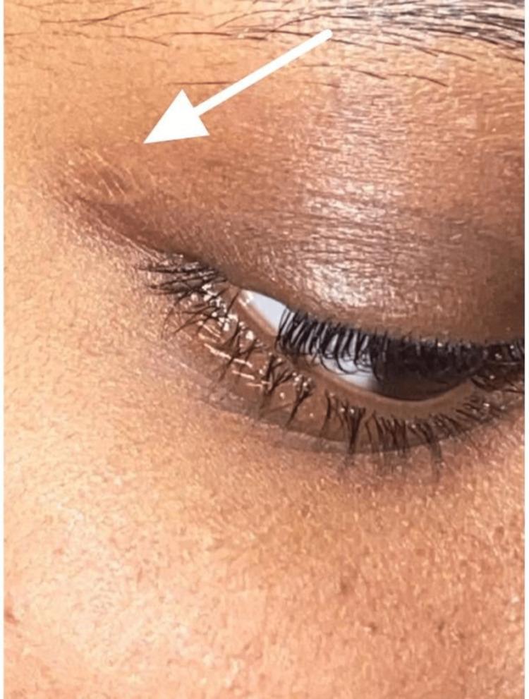
This clinical photograph shows a close-up, lateral view of a patient's right eye and upper eyelid following the surgical excision of a molluscum contagiosum (MC) lesion. The focus is on the eyelid's surface, which demonstrates a successful post-operative result. A white arrow points to the lateral aspect of the upper eyelid where the excision was performed, highlighting a small, slightly raised area near the lash line that is nearly indistinguishable from the surrounding skin texture. The skin exhibits a normal tone without significant hyperpigmentation, erythema, or visible scarring. The eyelid margin is well-aligned and preserved, showing no evidence of ectropion, entropion, or mechanical distortion. The eyelashes are intact, and the conjunctiva appears healthy. This image is used to illustrate the satisfactory cosmetic and functional outcomes that can be achieved through simple unroofing and curettage for pedunculated eyelid lesions, confirming the absence of residual pathology or surgical complications in a common dermatological viral infection case.
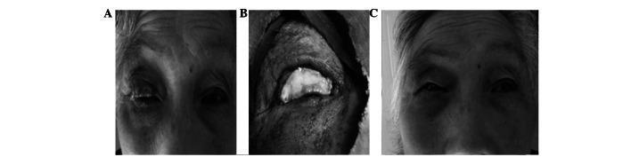
A sequence of clinical photographs documenting the surgical management of an eyelid neoplasm. (A) Preoperative image showing a large neoplasm on the right upper eyelid causing significant distortion of the eyelid margin and obscuring the globe. (B) Intraoperative view following full-thickness excision of the neoplasm, resulting in a large tarsal plate and eyelid margin defect. A transplant involving a trimmed tarsal plate and palpebral conjunctiva is shown being integrated into the surgical defect for reconstruction. (C) Postoperative follow-up at 3 months showing successful healing and functional recovery. The eyelid demonstrates effective closure with no evidence of entropion or ectropion. Notable findings include a minor palpebral fissure height and length discrepancy (greater than 2mm) compared to the contralateral side and mild marginal notching. This sequence serves as an educational tool for eyelid reconstruction techniques using tarsoconjunctival grafts and illustrates outcomes in ocular oncology and oculoplastic surgery.
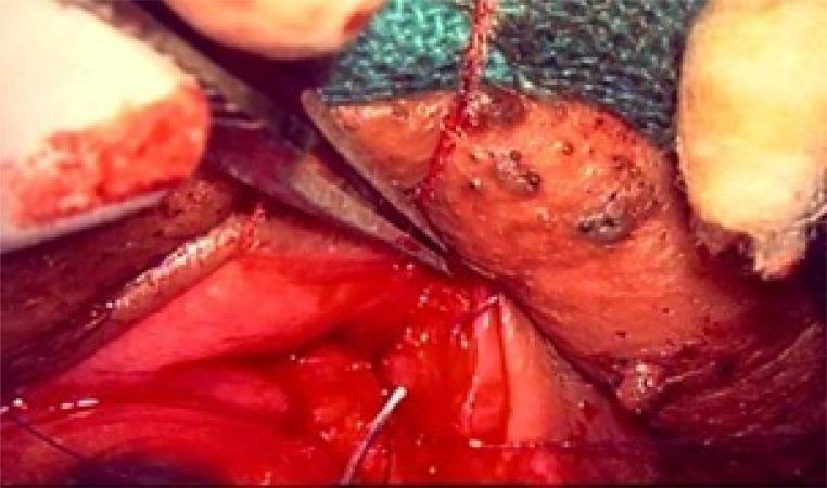
This clinical photograph captures an intraoperative view of an ophthalmic surgical procedure, specifically a Transconjunctival Retractor Plication (TRP) combined with a Lateral Tarsal Strip (LTS) for eyelid malposition correction. The image shows the lower eyelid anatomy with focus on the tarsal plate and retractors. Metallic surgical instruments, including a clamp or needle holder, are visible manipulating the tissue. The surgical field displays the red, vascularized posterior surface of the eyelid and the lower edge of the tarsus. Fine white or translucent sutures are placed through the tissue to secure the reattachment of the eyelid retractors. The surrounding area includes surgical drapes and swabs, highlighting the sterile clinical environment. This visual provides educational insight into oculoplastic techniques used for treating conditions like involutional entropion or ectropion by demonstrating the precise surgical handling and suturing of delicate periocular structures.
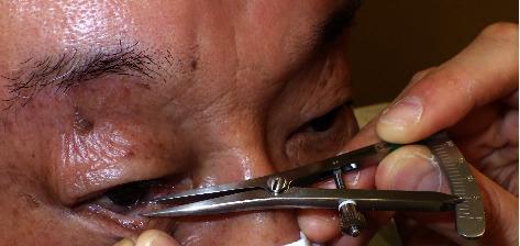
This clinical photograph demonstrates the objective assessment of horizontal eyelid laxity using the pinch test on a patient's lower eyelid. A metallic ophthalmic caliper is utilized to measure the degree of distensibility. The lower eyelid margin is being manually pinched and pulled anteriorly away from the globe. The caliper's tips are positioned to measure the precise distance between the eyeball (specifically the corneal limbus below the pupil) and the displaced lower eyelid margin. The surrounding periocular skin shows signs of chronological aging, including rhytids and solar lentigines, and a small pigmented lesion is noted near the lateral canthus. This procedural image illustrates a key diagnostic step in the evaluation of conditions such as involutional ectropion or entropion, where assessing the integrity of the medial and lateral canthal tendons and the overall tightness of the eyelid is essential for surgical planning.
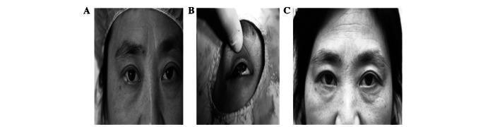
A series of three clinical photographs (A, B, C) documenting the surgical management of a right lower eyelid neoplasm. Panel A shows the preoperative frontal view of a patient with a localized lesion on the right lower eyelid margin. Panel B provides an intraoperative close-up of the right eye following tumor excision, revealing a large tarsal defect approximately half the length of the palpebral margin with visible sutures in the tarsal palpebral conjunctiva for reconstruction. Panel C displays the postoperative result three months after surgery. The image demonstrates successful anatomical restoration with preserved palpebral fissure symmetry (less than 2 mm difference in height and length compared to the contralateral side), normal eyelid closure, and the absence of complications such as entropion or ectropion. This clinical sequence illustrates effective ophthalmic reconstructive surgery following eyelid malignancy excision, focusing on functional and cosmetic outcomes in oculoplastics.
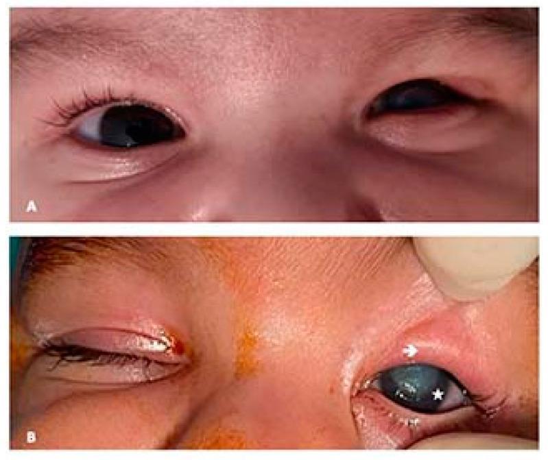
Two-panel clinical photograph (A and B) illustrating congenital upper eyelid entropion in a pediatric patient. Panel A shows the initial clinical presentation with the right eye appearing normal and the left upper eyelid exhibiting inward rotation. Panel B shows a preoperative view with annotations. The upper eyelid of the left eye demonstrates severe entropion, with an arrow indicating hypertrophy of the upper pretarsal orbicularis muscle. The ciliary margin shows a notable absence of cilia (eyelashes) due to chronic irritation or congenital defect. A star marks a central corneal leucoma (opacification) on the left eye, which is a secondary complication resulting from the persistent inward turning of the eyelid and subsequent corneal rubbing. This image serves as a clinical reference for pediatric ophthalmology, highlighting the diagnostic features of primary upper eyelid entropion and its associated corneal pathology.
entropion ectropion eyelid
Entropion and Ectropion
ECTROPION
Types
1. Involutional (Age-related) Ectropion - Most Common
- Affects the lower lid of elderly individuals
- Symptoms: Epiphora (tear overflow), ocular surface disease, cosmetically poor red exposed conjunctiva
- Long-standing cases: Tarsal conjunctiva becomes chronically inflamed, thickened, and keratinized
- Aetiology (3 laxity factors):
- Horizontal lid laxity - lid can be pulled 8+ mm from the globe and fails to snap back without blinking
- Lateral canthal tendon laxity - rounded lateral canthus; lower lid can be pulled medially >2 mm
- Medial canthal tendon laxity - pulling lid laterally displaces the inferior punctum; mild laxity = punctum reaches limbus; severe = punctum reaches pupil
- Generalized ectropion: Lateral tarsal strip procedure (shortens and reattaches the lower canthal tendon to the lateral orbital rim); alternative is tarsoconjunctival pentagon excision
- Medial ectropion: Medial conjunctival diamond excision (medial spindle procedure), often combined with tarsal strip or lateral canthal sling
2. Cicatricial Ectropion
- Caused by scarring/contracture of skin pulling the eyelid away from the globe
- Both upper and lower lids may be involved
- Key sign: pushing skin over the orbital margin with a finger relieves the ectropion; opening the mouth accentuates the eversion
- Causes: burns, trauma, dermatitis, ichthyosis
- Treatment: Mild localized - excision of scar tissue + vertical skin lengthening (Z-plasty); severe generalized - transposition flaps or free skin grafts (from upper lids, posterior auricular, preauricular, supraclavicular areas)
3. Paralytic Ectropion
- Caused by ipsilateral facial nerve palsy (CN VII)
- Associated with: upper and lower lid retraction, brow ptosis
- Complications: Exposure keratopathy (lagophthalmos), epiphora (malpositioned punctum, failed lacrimal pump, increased tear production from corneal exposure)
- Treatment:
- Temporary: High-viscosity lubricants, lid taping during sleep, botulinum toxin into levator to induce temporary ptosis, temporary tarsorrhaphy (especially with poor Bell's phenomenon)
- Permanent (after 6-12 months no improvement or irreversible nerve damage): Medial canthoplasty (if canthal tendon intact), platinum weight implant in upper lid to correct lagophthalmos
4. Mechanical Ectropion
- Due to a mass or lesion weighing down the lower lid
ENTROPION
Types
1. Involutional (Age-related) Entropion - Most Common
- Affects mainly the lower lid of elderly
- Consequences: Constant lash-cornea contact (pseudotrichiasis) causes:
- Irritation
- Corneal punctate epithelial erosions
- In severe cases: pannus formation and corneal ulceration
- Aetiology (4 factors):
- Horizontal lid laxity - stretching of canthal tendons and tarsal plate
- Vertical lid instability - attenuation, dehiscence or disinsertion of lower lid retractors (recognized by decreased lower lid excursion in downgaze)
- Over-riding of pretarsal by preseptal orbicularis during lid closure - moves the lower border of the tarsal plate anteriorly and tips the lid margin inward
- Orbital septum laxity with prolapse of orbital fat into the lower lid
- Temporary protection: Lubricants, taping, soft bandage contact lenses, botulinum toxin chemodenervation of orbicularis
- Surgical:
- Transverse everting sutures - quick, easy; corrects for several months; useful in confused/frail patients
- Wies procedure - durable correction; full-thickness horizontal lid splitting + everting sutures; scar creates barrier between preseptal and pretarsal orbicularis
- Lower lid retractor reinsertion - direct exposure and advancement of retractors; can be primary or for recurrence
- Lateral canthal sling (tarsal strip) or full-thickness lateral pentagon excision - corrects horizontal lid laxity, retains lid against globe
2. Cicatricial Entropion
- Scarring of the palpebral conjunctiva rotates the lid margin toward the globe
- Causes: cicatrizing conjunctivitis, trachoma (most important globally), trauma, chemical injuries
- Treatment:
- Mild: Tarsal fracture (transverse tarsotomy) with anterior rotation of the lid margin
- Severe: Replacement of deficient/keratinized conjunctiva; composite grafts to replace scarred and contracted tarsal plate
- Horizontal lid laxity corrected with lateral tarsal sling
3. Congenital Entropion
- Rare; may affect upper or lower lid
- Can cause corneal ulceration/leucoma if untreated (especially upper lid - constant corneal rubbing)
- Associated with hypertrophy of the pretarsal orbicularis
Comparison Summary
| Feature | Ectropion | Entropion |
|---|---|---|
| Direction | Lid everts outward | Lid inverts inward |
| Commonest type | Involutional (lower lid) | Involutional (lower lid) |
| Main symptom | Epiphora, exposed conjunctiva | Trichiasis, corneal irritation |
| Corneal risk | Exposure keratopathy | Punctate erosions, ulceration |
| Key test (laxity) | Snap-back / distraction test | Decreased excursion in downgaze |
| Temporary fix | Lubrication, taping | Botulinum toxin, taping |
| Definitive surgery | Lateral tarsal strip | Wies procedure / retractor reinsertion |
Explain blow in and blow out fractures of orbit
orbital blowout fracture CT scan floor medial wall
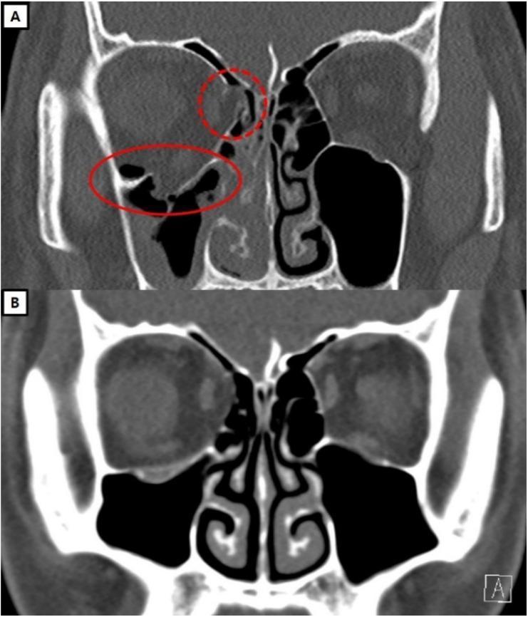
This Comparison Chart consists of two coronal CT scans (A and B) showing a human skull at the level of the orbits, illustrating the surgical management of facial trauma. Scan A (Preoperative) demonstrates a significant blowout fracture of the right orbit. Red annotations highlight two primary defects: a dashed circle indicates a fracture of the medial orbital wall (lamina papyracea) with soft tissue herniation into the ethmoid sinus, and a solid oval indicates a large fracture of the orbital floor with prolapse of orbital fat and inferior rectus muscle into the maxillary sinus. Scan B (Postoperative, one-year follow-up) shows the same anatomical section after reconstructive surgery. The orbital floor and medial wall are restored to their anatomical positions, displaying a continuous bony or implant-supported barrier. The soft tissue contents are fully repositioned within the orbital cavity, and symmetry is restored relative to the contralateral left orbit. This visual aids in teaching clinical radiology and maxillofacial surgery, specifically the identification of blowout fractures and the evaluation of post-surgical reduction and orbital volume restoration.
This diagnostic image consists of two side-by-side coronal slices from a facial computerized tomography (CT) scan (bone window) demonstrating surgical reconstruction of an orbital blowout fracture. The left orbit (viewer's right) exhibits a fracture involving the inferomedial wall. A hyperdense, radio-opaque implant, identified as a titanium-reinforced porous polyethylene (TR-PPE) plate, is seen positioned along the left medial orbital floor and lower medial wall to reconstruct the defect. A small, focal, high-density point (marked by a white arrow) indicates a single titanium screw used to fixate the plate to the inferior orbital rim. Anatomical landmarks include the bilateral maxillary sinuses, ethmoid air cells, and the nasal cavity. The left orbit shows some soft tissue herniation and postoperative changes compared to the intact right orbit. This image illustrates maxillofacial trauma management and the use of alloplastic materials in orbital floor reconstruction to restore orbital volume and prevent enophthalmos.
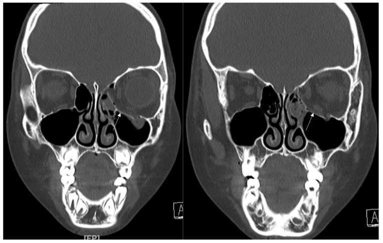
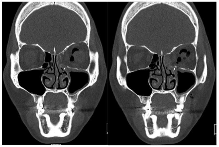
This diagnostic image consists of two coronal slices from a non-contrast facial CT scan of a pediatric patient, demonstrating a left-sided orbital blowout fracture. The images highlight the mid-facial anatomy, including the frontal bone, orbits, nasal cavity, ethmoid air cells, and maxillary sinuses. On the patient's right side, the orbital floor and medial wall are intact with clear sinus aeration. Conversely, the left side displays a significant discontinuity in the inferomedial orbital wall (white arrows), representing the fracture site. Visible pathology includes the displacement of bony fragments and the herniation of orbital contents (soft tissue) into the superior portion of the left maxillary sinus. There is also opacification of the adjacent left ethmoid air cells, suggestive of post-traumatic hemorrhage or mucosal edema. This imaging is clinically significant for diagnosing complex orbital fractures and assessing potential extraocular muscle entrapment in cases of facial trauma.
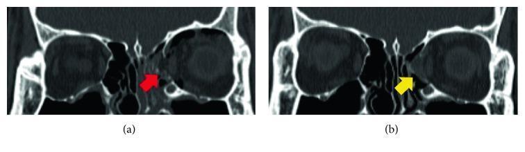
Coronal non-contrast CT scans of the orbits demonstrating a comparison between an acute medial orbital wall fracture and its surgical reconstruction. In image (a), a red arrow indicates a significant bony defect and discontinuity of the left medial orbital wall (lamina papyracea), characteristic of a blowout fracture with potential herniation of orbital contents into the ethmoid sinus. Image (b) shows the same anatomical region postoperatively, where a yellow arrow identifies a combined implant consisting of a resorbable meshed plate and allogenic bone substitute. The implant is seen bridging the previous defect, restoring the structural integrity and contour of the medial orbital wall. The reconstructed area exhibits a hybrid density lower than cortical bone but higher than surrounding soft tissue, indicating successful reduction and placement of the graft material. This comparison illustrates common diagnostic findings in maxillofacial trauma and the appearance of bioresorbable implants in orbital floor and wall repair.
orbital blowout fracture
Orbital Blow-Out and Blow-In Fractures
BLOW-OUT FRACTURE
Definition
Mechanism - Two Theories
Anatomy of Weakness
- Most common site: posteromedial orbital floor (maxillary bone), medial to the infraorbital neurovascular bundle
- Second most common: medial wall (lamina papyracea into the ethmoid sinus)
- The lateral wall and roof are more robust and less commonly fractured in pure blow-outs
- The orbital floor is the shortest wall and is roughly equilateral-triangular in cross-section
Clinical Features
- Periorbital ecchymosis ("black eye") and oedema
- Subconjunctival haemorrhage
- Subcutaneous emphysema - crackling sensation on palpation, especially after blowing the nose (due to air entering from the fractured sinus)
- Infraorbital nerve hypoesthesia - numbness of the lower lid, cheek, side of the nose, upper lip, upper teeth and gums (the fracture commonly involves the infraorbital canal)
- Restriction of upgaze (and sometimes downgaze) is the hallmark - caused by:
- Mechanical entrapment of the inferior rectus or inferior oblique muscles (or their surrounding connective tissue/fat) within the fracture
- Haemorrhage and oedema tightening the septa connecting the inferior rectus to the periorbita
- Forced duction test positive - cannot passively rotate the eye upward
- Intraocular pressure rises by 1-15 mmHg on upgaze compared to primary position - suggests inferior rectus entrapment
- Enophthalmos - sinking of the globe due to increased orbital volume from herniation of fat into the sinus; may be masked initially by oedema and only apparent after 2-4 weeks
- Hypoglobus - inferior displacement of the globe
- Corneal abrasion, traumatic iritis, hyphema, lens dislocation/subluxation, retinal dialysis/detachment, commotio retinae
"White-Eye" Blow-Out Fracture (Paediatric Trapdoor)
- Children's bones are more elastic (greenstick-type fracture)
- A small floor fracture opens like a trapdoor, orbital contents herniate through, then the bone snaps back, trapping the muscle (trapdoor mechanism)
- Classically little external bruising or subconjunctival haemorrhage - hence "white eye"
- Acute nausea, vomiting, bradycardia = oculocardiac (oculovagal) reflex - a triad indicating tight muscle entrapment
- This is a surgical emergency - irreversible muscle ischaemia and fibrosis can occur within hours
- CT may appear near-normal (minimal floor displacement)
Investigations
- CT with coronal sections - gold standard; shows floor defect, "tear-drop sign" (soft tissue prolapsing into the maxillary antrum), muscle entrapment, and extent of fracture
- Hess chart - maps the restriction of eye movements; shows restricted left upgaze and restriction on downgaze (inferior rectus) with secondary overaction of the contralateral eye
- Visual acuity must be documented and monitored
Treatment
- Ice packs, oral antibiotics (cephalexin 250-500 mg QDS for 10 days - sinus contamination risk)
- Nasal precautions: do not blow nose, no sneezing with closed mouth, no sucking through a straw (prevents forcing infected sinus contents into orbit or worsening emphysema)
- Nasal decongestants
- Systemic steroids if severe orbital oedema compromises the optic nerve
- Observe for 7-10 days for oedema and haemorrhage to settle
- Symptomatic persistent diplopia + positive forced ductions + CT evidence of entrapment with no improvement over 1-2 weeks
- Early enophthalmos ≥3 mm (or >2 mm per Kanski)
- Significant globe ptosis (hypoglobus)
- Floor defect involving >50% of orbital floor (high risk of late enophthalmos)
- Associated rim and facial fractures
- Blow-in fracture (bone fragments may compromise vision - see below)
- Oculocardiac reflex signs (intractable nausea, vomiting, bradycardia) = emergent surgery
- Herniation of globe into maxillary sinus = emergent surgery
- Small cracks without herniation - no treatment needed
- Fractures up to half the floor with no herniation, no enophthalmos, and improving diplopia - can be observed
- In patients with enophthalmos <2 mm, outcomes are similar whether surgery is done at 2 weeks or 6 months
- Preferably repair within 2 weeks of injury
- Delayed repair (>6 weeks) becomes progressively more difficult due to fibrosis and contracture
- Transconjunctival incision (preferred) or subciliary approach
- Periosteum elevated from orbital floor
- Entrapped orbital contents freed
- Bony defect repaired with a synthetic implant (titanium-reinforced porous polyethylene plate, resorbable mesh, etc.)
BLOW-IN FRACTURE
Definition
Key Distinguishing Features
| Feature | Blow-Out | Blow-In |
|---|---|---|
| Fragment direction | Outward into sinus | Inward into orbit |
| Orbital volume | Increases | Decreases |
| Globe position | Enophthalmos | Exophthalmos (proptosis) |
| Rim status | Intact | Usually involved (impure fracture) |
| Most common wall | Floor | Orbital roof (most common type of roof fracture) |
Types
- Pure blow-in: Only the orbital roof is fractured without the orbital rim
- Impure blow-in: Involves both orbital roof and rim - this is the most commonly encountered type of orbital roof fracture
Clinical Features (orbital roof blow-in)
- Upper eyelid haematoma (may expand late, from intracranial or intraorbital haemorrhage decompressing into the superior orbit)
- Proptosis (secondary to prolapsed intracranial contents or reduced orbital volume)
- Forehead hypoesthesia (supraorbital/supratrochlear nerve)
- Contour deformity of the forehead and supraorbital ridge
- CSF rhinorrhea (if dura violated)
- Neurological deficits
- Pulsatile proptosis (if there is a communication with intracranial contents)
Management
- Interdisciplinary - ENT, Neurosurgery, and Ophthalmology mandatory
- Intracranial injury takes precedence, then globe injury
- Non-displaced/minimally displaced fractures not involving the frontal sinus may be observed in asymptomatic patients (monitor for pulsatile proptosis)
- Blow-in fracture of the floor/wall is itself a surgical indication - bone fragments may directly compress the optic nerve or globe and compromise vision
- Displaced fractures with visible bony contour deformity require open reduction and internal fixation
Summary Comparison
| Feature | Blow-Out | Blow-In |
|---|---|---|
| Mechanism | Hydraulic pressure or buckling | Direct trauma to orbital rim |
| Fragment direction | Into sinus (outward) | Into orbit (inward) |
| Orbital rim | Intact | Usually fractured |
| Orbital volume | Expanded | Reduced |
| Globe position | Enophthalmos / hypoglobus | Exophthalmos / proptosis |
| Most common site | Orbital floor (posteromedial) | Orbital roof |
| Key sign | Restricted upgaze + diplopia | Proptosis + possible CSF rhinorrhea |
| CT sign | "Tear-drop" into maxillary sinus | Fragment in orbital cavity |
| Urgency | Urgent if trapdoor/oculocardiac reflex | Often requires neurosurgical input |
- Kanski's Clinical Ophthalmology: A Systematic Approach, 10th Ed., pp. 924-927
- K.J. Lee's Essential Otolaryngology, 11th Ed., pp. 1119-1121, 1128
- Bailey & Love's Short Practice of Surgery, 28th Ed., p. 434
- Tintinalli's Emergency Medicine, p. 1590
- The Wills Eye Manual, 8th Ed., pp. 104-105