
Pnh
I'll cover PNH (Paroxysmal Nocturnal Hemoglobinuria) from the medical library. Let me pull up the relevant content.
Reading File
Searching sources...
Finding Sources
Finding Sources
Good - I have multiple sources. Let me read the most comprehensive ones in parallel.
Reading File
Reading File
Reading File
Reading File
Let me get the Goldman-Cecil and Harrison's content plus the flow cytometry diagnosis content and a recent PubMed update.
Reading File
Reading File
Searching PubMed
paroxysmal nocturnal hemoglobinuria treatment complement inhibitor
I now have comprehensive content from multiple authoritative sources. Let me also fetch the PNH flow cytometry image and one treatment diagram.
Loading Image
Loading Image
I now have everything needed for a comprehensive answer. Here is the full review:
Paroxysmal Nocturnal Hemoglobinuria (PNH)
Definition
PNH is a rare, acquired clonal stem cell disorder in which somatic mutation of the PIGA gene causes deficiency of GPI-anchored complement regulatory proteins on blood cell surfaces, resulting in complement-mediated intravascular hemolysis, thrombosis, and variable cytopenias. It is the only hemolytic anemia caused by an acquired genetic defect.
- Robbins & Kumar Basic Pathology, p. 605
- Goldman-Cecil Medicine, Ch. 146
Pathogenesis
The PIGA Mutation
The PIGA gene on the short arm of the X chromosome encodes an enzyme essential for synthesizing glycosylphosphatidylinositol (GPI), a specialized phospholipid that anchors many proteins to the cell membrane. Because PIGA is X-linked, a single somatic mutation in any hematopoietic stem cell (HSC) is sufficient to eliminate GPI synthesis in all progeny of that clone - red cells, white cells, and platelets alike.
- Nearly 150 different PIGA mutations have been identified
- Most inactivate PIGA completely → PNH III cells (fully deficient)
- Partial deficiency → PNH II cells (3-5x normal complement sensitivity)
- PNH III cells: 15-25x normal complement sensitivity
Why Does the Clone Expand?
Small numbers of PIGA-deficient cells exist in normal individuals (~1 in 50,000 RBCs) but never expand. In PNH patients, a second step - possibly autoimmune attack against GPI-anchored antigens - provides a selective advantage to the mutant clone, allowing it to expand. This explains the frequent association between PNH and aplastic anemia (an autoimmune marrow failure syndrome).
GPI-Linked Proteins Lost in PNH
| Protein | Function |
|---|---|
| CD59 (MIRL, protectin) | Most important - inhibits C3 convertase; blocks C9 polymerization (the final MAC assembly step) |
| CD55 (DAF) | Decay-accelerating factor; breaks down C3/C5 convertases |
| C8-binding protein | Homologous restriction factor |
| CD58, CD14, CD24, CD16a | Additional GPI-linked surface proteins |
| Acetylcholinesterase, LAP | Membrane-associated enzymes |
With CD55 and CD59 absent, the membrane attack complex (C5b-9/MAC) assembles unimpeded on red cell surfaces → intravascular hemolysis.
Clinical Features
Classic Triad
- Hemolytic anemia - chronic intravascular, often with reticulocytosis less than expected
- Venous thrombosis - in unusual sites
- Cytopenias (variable) - neutropenia in 3/5, thrombocytopenia in 2/3 at some point
Hemoglobinuria
The classic "paroxysmal nocturnal" presentation occurs in only ~25% of cases. The rest present with chronic hemolysis without dramatic hemoglobinuria. The nocturnal pattern is attributed to a mild drop in blood pH during sleep (CO2 retention), which activates complement. However, this relationship is not firmly confirmed.
Triggers of hemolysis: infection, surgery, blood transfusion, contrast dye injection, severe exercise, fever, acidosis, hypoxia.
Thrombosis - The Major Killer
- Affects ~40% of patients
- 85% venous, often in unusual sites:
- Hepatic veins (Budd-Chiari syndrome)
- Portal vein
- Cerebral veins
- Abdominal veins
- Mechanism: CD59 absence on platelets → phosphatidylserine externalization → prothrombinase complex assembly; also, free hemoglobin scavenges nitric oxide (NO), causing vasoconstriction and platelet activation
- Abdominal pain in ~1/3 of patients is linked to NO scavenging by free hemoglobin (also causes dysphagia, erectile dysfunction)
Iron Deficiency
Chronic loss of iron in urine as hemosiderinuria → iron deficiency → hypochromic microcytic anemia superimposed on hemolysis. Hemosiderinuria is almost constantly present.
Epidemiology
- Incidence: ~2-5 per million/year
- Any age, most common 10-50 years; mean age at diagnosis ~34 years
- Female:male ratio ~1:1
- Median survival ~20 years after diagnosis
PNH Categories
- Classic PNH - hemolysis predominant, large clone
- PNH in the setting of another bone marrow disorder (aplastic anemia, MDS) - cytopenias predominant; PNH clone usually small
- Subclinical PNH - small GPI-deficient clone, no clinical hemolysis
- Found in 50-60% of aplastic anemia patients
- Found in 15-20% of MDS patients
Diagnosis
Gold Standard: Flow Cytometry (FLAER-based)
The loss of GPI-linked proteins is detected by flow cytometry of peripheral blood. At least two different cell lineages must be evaluated.
Recommended panel:
- Neutrophils: FLAER + CD24 + CD15 + CD45
- Monocytes: FLAER + CD14 + CD64 + CD45
- Red blood cells: CD235a / CD59 (two-color)
FLAER (fluorescent derivative of bacterial proaerolysin) binds the GPI anchor itself directly - the key reagent for high-sensitivity testing.
Flow cytometry findings in PNH:
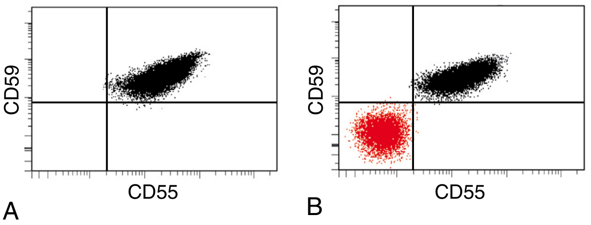
Panel A (normal): All red cells express CD55 and CD59 normally. Panel B (PNH): A large population (red) is negative for both CD55 and CD59 - the PNH clone.
Other Lab Findings
| Finding | Explanation |
|---|---|
| Normocytic anemia (or hypochromic/microcytic) | Hemolysis + iron deficiency |
| Reticulocytosis (less than expected) | Marrow suppression may coexist |
| Hemosiderinuria | Chronic iron loss in urine |
| Negative direct antiglobulin (Coombs) test | Distinguishes PNH from autoimmune hemolysis |
| Elevated LDH | Intravascular hemolysis |
| Low haptoglobin | Free hemoglobin binding |
| Pancytopenia | Common during disease course |
Treatment
Anti-Complement Therapy (Disease-Modifying)
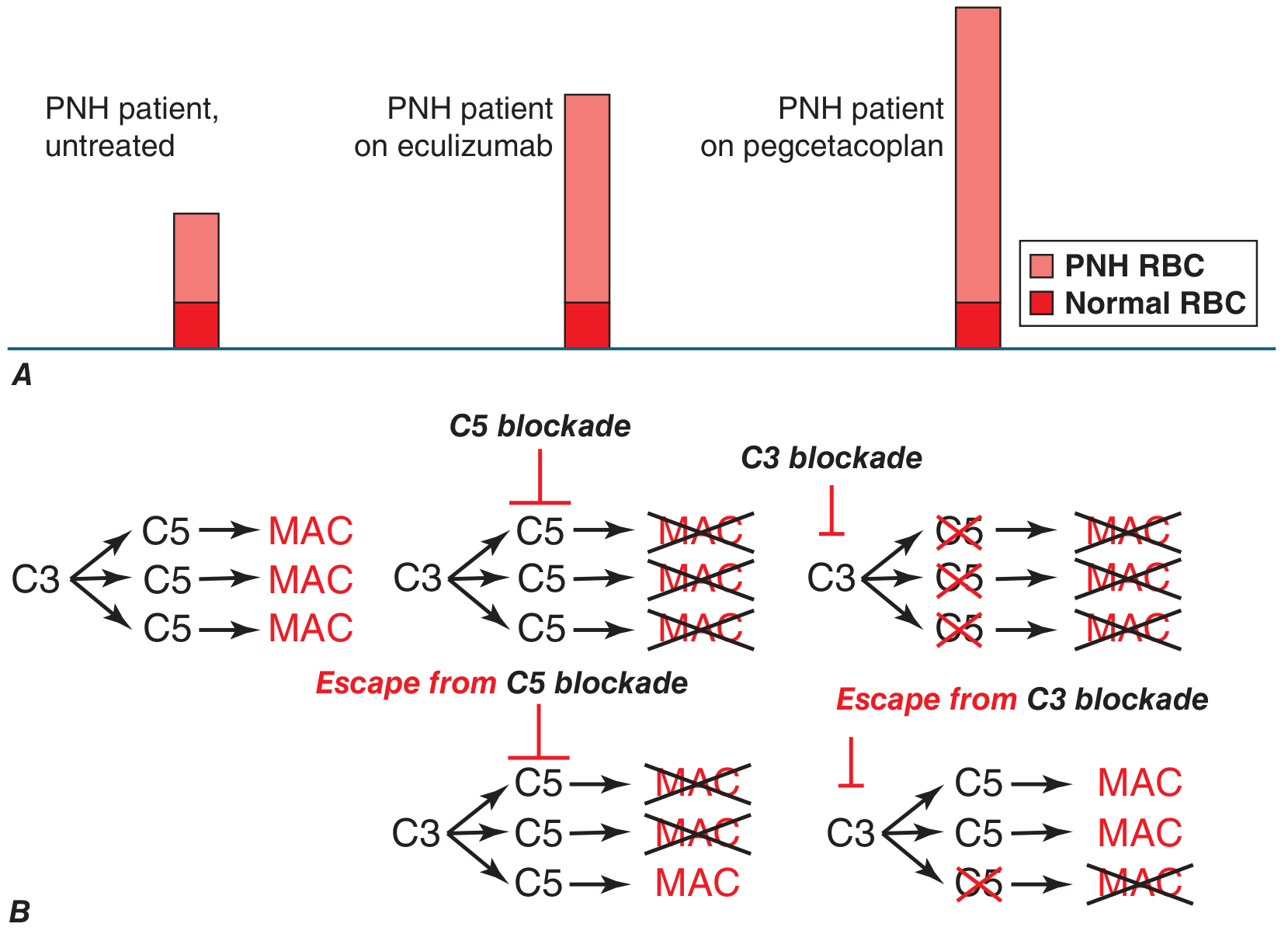
The major advance in PNH management came with eculizumab (approved 2007) - a humanized monoclonal antibody that blocks C5, preventing MAC formation.
Mechanism comparison:
Current complement inhibitors:
| Drug | Target | Notes |
|---|---|---|
| Eculizumab | C5 | First approved; IV every 2 weeks; abolishes intravascular hemolysis; reduces thrombosis risk |
| Ravulizumab | C5 | Long-acting; IV every 8 weeks |
| Crovalimab | C5 | Subcutaneous; self-administered; 2026 systematic review confirms efficacy |
| Pegcetacoplan | C3 | Proximal inhibitor; prevents both intravascular AND extravascular hemolysis; corrects anemia more completely |
| Iptacopan | Factor B (alternative pathway) | Oral; targets upstream |
Key limitation of C5 inhibitors: PNH red cells, now protected from MAC lysis, still accumulate C3b fragments on their surface → become opsonized → iatrogenic extravascular hemolysis (Coombs test becomes positive). Pegcetacoplan, acting at C3, prevents this.
Breakthrough hemolysis risk: Incomplete C5 blockade → some MAC forms. With C3 blockade, if incomplete, the downstream enzymatic cascade amplifies - potentially more severe breakthrough. Clinicians must maintain adequate drug levels.
Meningococcal Vaccination - Mandatory
All patients on complement inhibitors must be vaccinated against Neisseria meningitidis (and should receive prophylactic antibiotics) because blockade of the terminal complement pathway removes a key defense against encapsulated organisms, especially meningococcus.
Allogeneic BMT (Stem Cell Transplant)
The only curative option. Reserved for:
- Severe aplastic anemia with PNH
- Refractory disease
- Significant cytopenias
- Carries nonnegligible procedure-related risks
Supportive Care
- Iron supplementation (for hemosiderinuria-related deficiency)
- Folate supplementation
- Anticoagulation for thrombosis (not clearly effective prophylactically in untreated patients; complement inhibitors reduce thrombosis risk more effectively)
- Transfusions when needed (washed RBCs preferred to avoid additional complement triggers)
- Corticosteroids may reduce some hemolytic episodes
Key Associations
- Aplastic anemia - bidirectional; PNH may evolve from AA; AA may follow PNH; autoimmune basis explains the link
- MDS - small subclinical PNH clones in 15-20% of MDS
- Budd-Chiari syndrome - think PNH in any young patient with hepatic vein thrombosis
- Iron deficiency with hemolytic markers - unusual combination that should raise PNH suspicion
Summary Mnemonics
PNH = PIGA mutation → No GPI → No CD55/CD59 → MAC attacks RBCs
Classic triad: Hemolysis + Thrombosis + Cytopenias
Remember: Coombs negative (unlike autoimmune hemolytic anemia), hemosiderinuria always present, nocturnal hemoglobinuria in minority only.
Sources:
- Robbins & Kumar Basic Pathology, p. 605-606
- Robbins, Cotran & Kumar Pathologic Basis of Disease, p. 606
- Goldman-Cecil Medicine, Ch. 146
- Harrison's Principles of Internal Medicine 22E (2025), p. 848-849
- Henry's Clinical Diagnosis and Management by Laboratory Methods, p. 693, 798
Recent literature: A 2026 systematic review and meta-analysis (PMID 41299829) confirmed the efficacy and safety of crovalimab (subcutaneous C5 inhibitor) in PNH, supporting its use as an alternative to IV anti-C5 therapy. A 2025 systematic review (PMID 40244439) examined PNH in pregnancy, where eculizumab is considered safe and is the treatment of choice given the high thrombotic risk.
Spleen anatomy
Searching sources...
Finding Sources
Finding Sources
Finding Sources
Reading File
Reading File
Reading File
Reading File
Good content - now let me fetch the anatomical diagrams and get the immunology/radiology content.
Loading Image
Loading Image
Reading File
Excellent - I have comprehensive content and diagrams. Let me fetch one more key image (the splenic microanatomy) and compile the full answer.
Loading Image
Here is the complete overview of spleen anatomy:
Spleen Anatomy
Position and Size
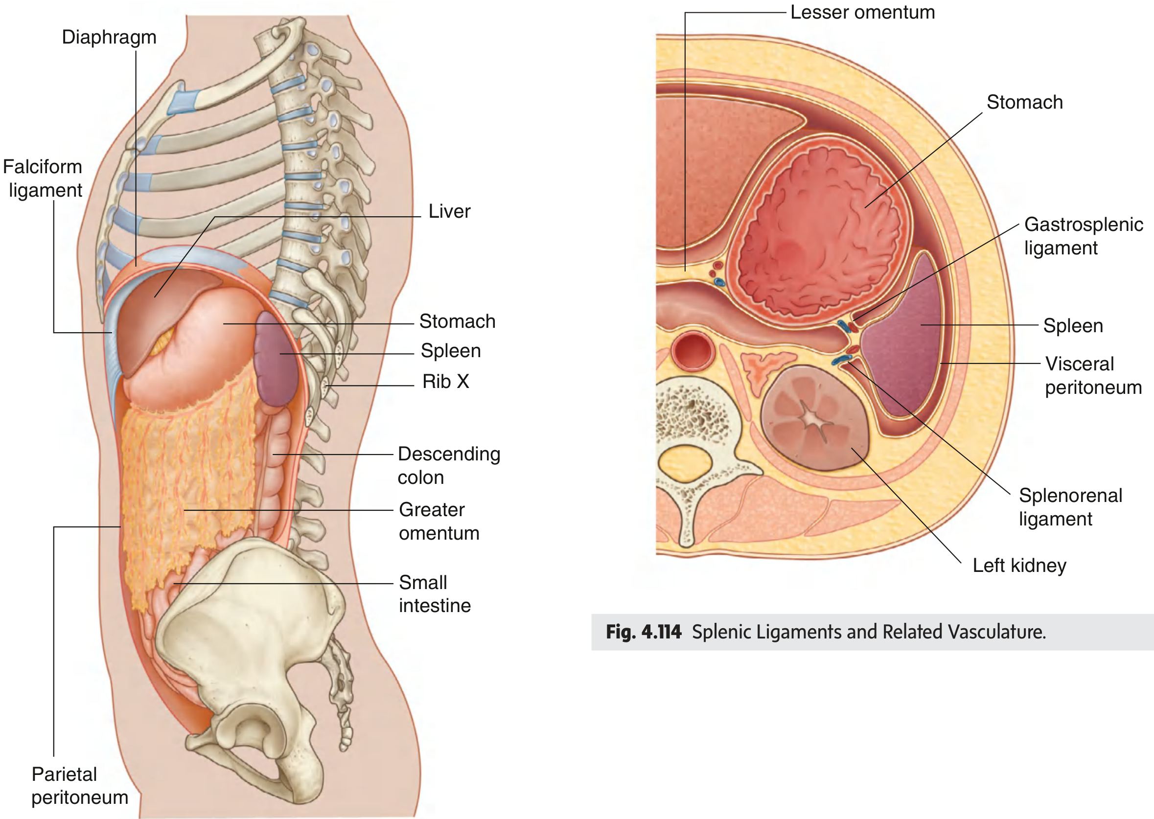
The spleen is the largest lymphoid organ in the body. It lies in the left upper quadrant (left hypochondrium) of the abdomen, against the diaphragm, at the level of ribs IX-X (protected by ribs 9-12). In a healthy adult it is 10-12 cm in length and weighs approximately 100-150 g. It is entirely surrounded by visceral peritoneum except at the hilum.
Development
The spleen develops as part of the vascular system within the dorsal mesentery that suspends the developing stomach from the body wall. This explains why its ligaments are actually folds of the greater omentum.
Surfaces and Poles
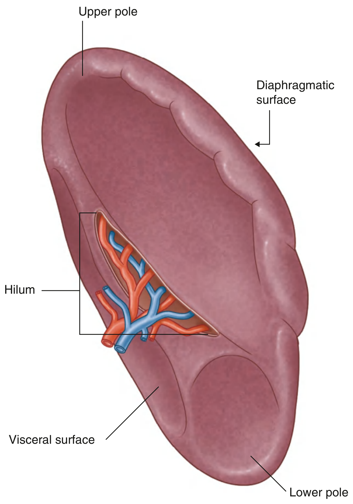
| Surface/Pole | Description |
|---|---|
| Diaphragmatic surface | Convex; faces the diaphragm superolaterally |
| Visceral surface | Concave; faces the stomach, left kidney, and tail of pancreas |
| Upper pole | Pointed; directed superomedially |
| Lower pole | Rounded; directed inferolaterally |
| Hilum | On the visceral surface; entry/exit point for splenic vessels; occasionally touched by the tail of the pancreas |
Visceral Relations
| Structure | Relation |
|---|---|
| Stomach | Anteromedial (separated by gastrosplenic ligament) |
| Left kidney | Posteromedial (separated by splenorenal ligament) |
| Tail of pancreas | May reach the hilum |
| Diaphragm | Posterosuperior |
| Splenic flexure of colon | Inferior |
Peritoneal Ligaments
The spleen is held in place by four ligaments - the first two are parts of the greater omentum:
| Ligament | Contents | Connects |
|---|---|---|
| Gastrosplenic | Short gastric vessels + left gastro-omental (gastroepiploic) vessels | Stomach (greater curvature) → Spleen |
| Splenorenal (lienorenal) | Splenic artery and vein; tail of pancreas | Left kidney → Spleen |
| Phrenicosplenic (part of phrenocolic ligament) | Usually avascular | Diaphragm → Spleen (superiorly) |
| Splenocolic | Usually avascular | Spleen → Splenic flexure of colon (inferiorly) |
The phrenicosplenic and splenocolic ligaments are typically avascular and can be safely divided during splenectomy.
Blood Supply
Arterial
- Splenic artery - from the celiac trunk (occasionally arises from the aorta, SMA, middle colic, left gastric, or left/right hepatic arteries)
- Two branching patterns:
- Distributed type (more common) - multiple branches spread out before entering the hilum
- Magistral type - long trunk that divides closer to the hilum into 3-4 compact branches (makes vessel dissection harder surgically)
- The splenic trunk also gives off the left gastroepiploic artery (runs along the greater curvature of the stomach) and short gastric arteries (supply the fundus)
- Inferior polar arteries arise from the left gastroepiploic artery
Venous
- Splenic vein exits the hilum → joins the superior mesenteric vein to form the portal vein
- Venous drainage goes into the portal circulation
Microscopic (Internal) Anatomy
The spleen is enclosed by a thin connective tissue capsule covered by peritoneal epithelium. The capsule sends projections called trabeculae into the parenchyma, most anchored to the hilum, dividing the organ into segments. The parenchyma between capsule and trabeculae is the splenic pulp - vascularized reticular connective tissue.
Red Pulp (~75-80% of volume)
- Contains large amounts of blood
- Made of pulp cords (of Billroth) and splenic sinusoids between them
- Sinusoidal walls are lined by spindle-shaped longitudinal endothelium with slit-like openings (allowing RBCs to squeeze through)
- Discontinuous basement membrane; lined by circumferential reticulin fibers
- Contains macrophages - critical for filtering the blood (removing aged RBCs, opsonized bacteria, immune complexes)
White Pulp (lymphoid tissue)
Organized around central arteries (branches of the splenic artery):
| Structure | Cell Type | Function |
|---|---|---|
| Periarteriolar lymphoid sheaths (PALS) | Mainly T lymphocytes | T cell zone; surrounds central arteries |
| Lymphoid follicles | B lymphocytes | B cell zone; lie between marginal sinus and PALS |
| Marginal zone | B cells + specialized macrophages | Boundary between red and white pulp; samples blood-borne antigens via DCs |
The PALS in the spleen are the equivalent of the paracortex in lymph nodes. Follicles can develop germinal centers during active immune responses.
Marginal Zone
Surrounds the marginal sinus. Contains marginal zone B cells (functionally distinct from follicular B cells, with limited antigen specificities) and macrophages. Blood-borne antigens are delivered here by circulating dendritic cells or sampled directly by marginal zone macrophages.
Vascular Architecture (the key to understanding splenic function)
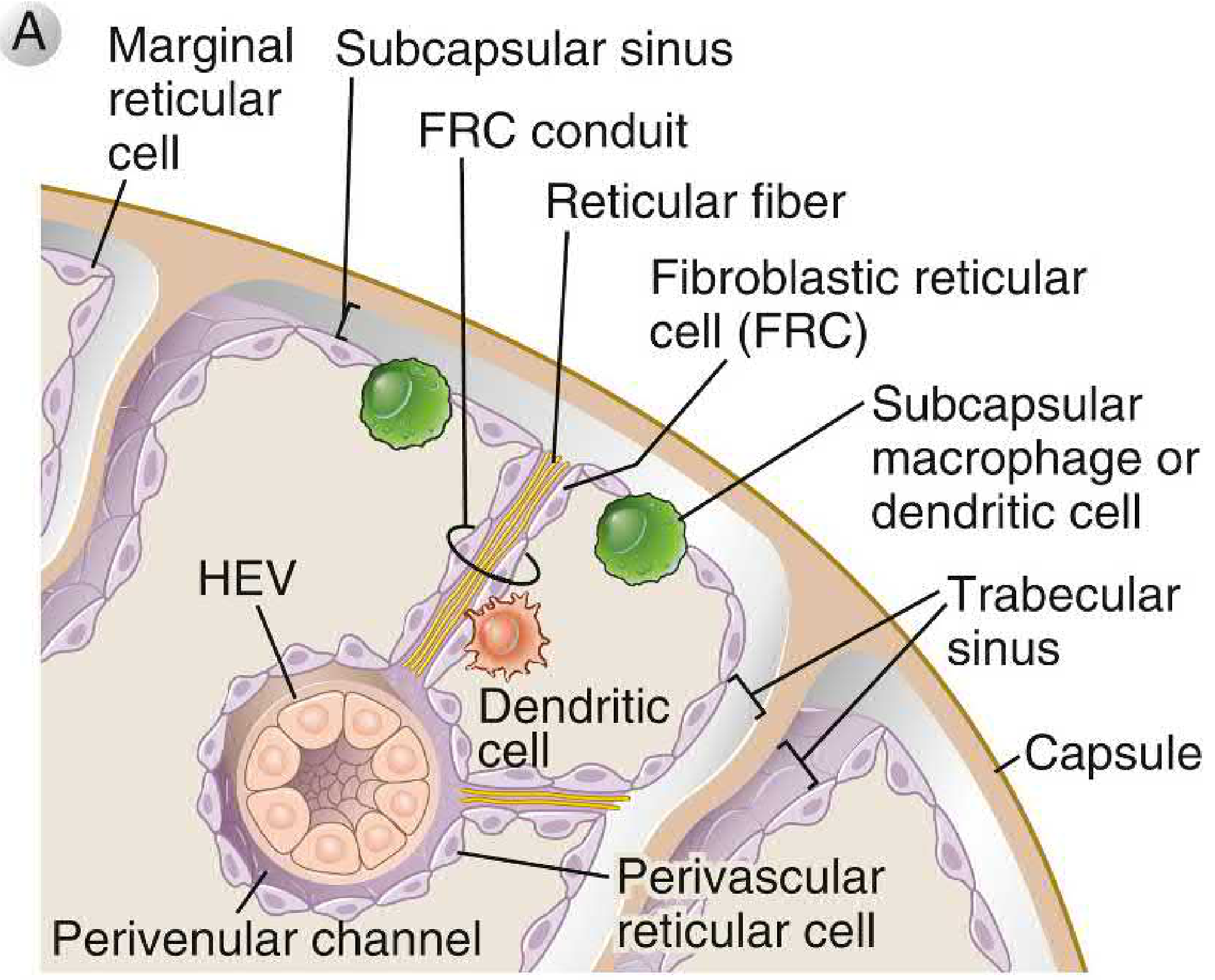
Blood flow through the spleen:
Splenic artery → Trabecular arteries (in trabeculae) → Pulp arteries → Central arteries (surrounded by PALS) → Penicillar arterioles (~50 per central artery) → Sheathed capillaries (Schweigger-Seidel ellipsoids; surrounded by macrophages) → Arterial capillaries
Then two routes:
- Open circulation (~majority): capillaries empty into perisinusoidal cords → blood filters through slit-like openings in sinusoidal walls → splenic sinusoids
- Closed circulation (~minority): capillaries open directly into sinusoids
Splenic sinusoids → Pulp veins → Trabecular veins → Splenic vein
The slit-like openings in sinusoidal walls act as a mechanical "filter" - rigid or aged RBCs cannot deform to pass through and are trapped and destroyed by macrophages.
Functions (Anatomy-Function Correlation)
| Compartment | Function |
|---|---|
| Red pulp macrophages | Remove aged/damaged RBCs, opsonized bacteria (esp. encapsulated organisms), immune complexes |
| White pulp (PALS) | T cell activation against blood-borne antigens |
| White pulp (follicles) | B cell activation; antibody production (esp. IgM against encapsulated bacteria) |
| Marginal zone | First-line sampling of blood-borne antigens |
| Fetal spleen | Extramedullary hematopoiesis (can resume in adults with marrow failure) |
| Overall | Blood reservoir; platelet storage |
Clinical Correlates
- Splenomegaly: palpable below the left costal margin; normal spleen is not palpable. The spleen must enlarge ~2-3x before becoming palpable
- Splenectomy / asplenia: loss of filtration for encapsulated bacteria (pneumococci, meningococci, H. influenzae) → OPSI (overwhelming post-splenectomy infection). Vaccination against these organisms is mandatory
- Splenic rupture: most common from blunt abdominal trauma; protected by ribs 9-12 but vulnerable. Blood collects in left upper quadrant. Managed non-operatively in stable patients (especially children)
- Splenic infarct: most commonly from emboli (e.g., in infective endocarditis, sickle cell disease); presents with left upper quadrant pain radiating to the left shoulder (Kehr's sign - diaphragmatic irritation)
- Accessory spleen (splenunculi): found in ~10-15% of people, most commonly at the splenic hilum or in the gastrosplenic ligament. Clinically important if splenectomy is performed for haematological disease (residual accessory spleen can proliferate and cause disease relapse)
- Wandering spleen: hypermobile spleen due to lax ligaments; can undergo torsion
Sources:
- Gray's Anatomy for Students, p. 399-400
- Cellular and Molecular Immunology (Abbas), p. 120-122
- Color Atlas of Human Anatomy, Vol. 2 - Internal Organs, p. 662
- Current Surgical Therapy 14e, p. (Surgical Anatomy of the Spleen)
- Bailey and Love's Short Practice of Surgery 28e
Spleen injury in surgery
Searching sources...
Finding Sources
Finding Sources
Finding Sources
Reading File
Reading File
Reading File
Excellent content. Let me get the operative management section and Bailey & Love's grading table.
Reading File
Reading File
Now let me get the operative management and NOM (non-operative management) content from Maingot's, plus the radiology section.
Reading File
Reading File
Let me get the NOM and operative management content from Maingot's block 12, and the CT trauma imaging section.
Reading File
Reading File
Now let me get the operative management (NOM vs splenectomy decision-making) from Maingot's block 12, specifically around line 900+.
Reading File
Let me specifically get the trauma/NOM management section from Maingot's block 12 (around lines 550-640 which precedes what I've already read).
Reading File
I now have enough comprehensive material. Let me compile the full answer.
Splenic Injury in Surgery
Overview and Epidemiology
The spleen is the most commonly injured solid organ in abdominal trauma. It is vulnerable despite rib cage protection (ribs 9-12) because of its highly vascular, fragile parenchyma and relatively fixed position. Splenic injury may occur from:
- Blunt trauma - most common; motor vehicle collisions, falls, sporting injuries, assault
- Penetrating trauma - stab wounds, gunshot wounds; may traverse the anterior/posterior abdominal wall, flank, or transthoracically through the pleura and diaphragm
- Iatrogenic injury - operative trauma during adjacent procedures (especially colectomy, gastric surgery)
- Spontaneous rupture - rare; from underlying disease (neoplastic 30%, infectious 27%, inflammatory 20%, drug-related 9%)
Iatrogenic splenic injury deserves special mention: In a 16-year Mayo Clinic review of ~14,000 colectomies, the risk of splenic injury requiring splenectomy or repair was 0.4%. The majority occurred during mobilization of the splenic flexure, though 10% had no splenic flexure mobilization at all - likely from tension on the colon. Patients with incidental splenectomy during colorectal cancer surgery had significantly higher 30-day morbidity (34%) and mortality (15%), and poorer long-term cancer outcomes.
AAST Spleen Injury Scale (Grading)
The American Association for the Surgery of Trauma (AAST) grading system guides management decisions. CT grading has sensitivity and specificity >95%.
| Grade | Injury Type | Description |
|---|---|---|
| I | Haematoma | Subcapsular, <10% surface area |
| Laceration | Capsular tear, <1 cm parenchymal depth | |
| II | Haematoma | Subcapsular, 10-50% surface area; intraparenchymal <5 cm diameter |
| Laceration | 1-3 cm parenchymal depth, not involving trabecular vessel | |
| III | Haematoma | Subcapsular >50% surface area or expanding; ruptured subcapsular/parenchymal haematoma; intraparenchymal ≥5 cm or expanding |
| Laceration | >3 cm parenchymal depth or involving trabecular vessels | |
| IV | Laceration | Involving segmental or hilar vessels producing major devascularisation (>25% of spleen) |
| V | Laceration | Completely shattered spleen |
| Vascular | Hilar injury devascularising the spleen |
Advance one grade for multiple injuries up to Grade III.
Diagnosis
Clinical
- Left upper quadrant pain, left shoulder tip pain (Kehr's sign - referred pain from diaphragmatic irritation by blood)
- Haemodynamic instability in significant injury
- Decreasing serial haematocrit (ongoing intraperitoneal haemorrhage)
- WBC often rises >15,000/mm³
- Plain films may show: fractured ribs, elevated left hemidiaphragm, enlarged splenic shadow, medial gastric displacement, widening between splenic flexure and preperitoneal fat
Imaging
CT (gold standard)
- Multiphase contrast-enhanced CT: sensitivity and specificity >95%
- Parenchymal laceration: linear low-attenuation defects, almost always with haemoperitoneum
- Haematoma: may be hyperdense or isodense on unenhanced CT; fails to enhance with IV contrast
- Vascular blush (focal area of contrast enhancement within a haematoma): indicates active arterial bleeding - strongly predicts failure of non-operative management and signals need for angioembolisation
- Subcapsular haematoma: crescentic well-defined lesion along splenic margin
- A laceration traversing both capsular surfaces = splenic fracture
Ultrasound (FAST exam)
- Used in initial resuscitation to detect free fluid
- Not sensitive enough to reliably exclude significant splenic injury
- Useful for follow-up of haematoma resolution
Note: Lateral clefts are normal splenic variants that can simulate lacerations - important to recognise on imaging.
Management
Decision Framework
The key question is: Haemodynamically stable or unstable?
SPLENIC INJURY
│
├─ Haemodynamically UNSTABLE → Emergency laparotomy
│
└─ Haemodynamically STABLE
│
├─ Grades I-III (most) → Non-operative management (NOM)
├─ Grade IV-V → Consider NOM if no blush; angioembolisation if blush
└─ Blush on CT → Angioembolisation
1. Non-Operative Management (NOM)
NOM has become the standard of care for haemodynamically stable patients with splenic injury, and is successful in ~80-90% of adults and >95% of children.
Prerequisites for NOM:
- Haemodynamic stability
- No peritoneal signs requiring laparotomy
- No associated injuries mandating surgery
- Access to ICU monitoring, serial clinical examination, blood products, and operating theatre
Protocol:
- ICU admission and close haemodynamic monitoring
- Serial haematocrit and clinical examination
- Bed rest (duration varies by grade: Grade I-II ~2 days, Grade III ~3-5 days)
- Avoid NSAIDs and anticoagulants
- Activity restriction after discharge (avoid contact sports for weeks to months based on grade)
- Follow-up CT or ultrasound recommended - risk of splenic pseudoaneurysm after trauma is unrelated to injury severity
Failure of NOM (requiring operative intervention) indicated by:
- Haemodynamic deterioration
- Increasing transfusion requirements
- Signs of peritonitis
- Other intra-abdominal injuries identified
Children: NOM is especially preferred. The paediatric spleen has a thicker capsule and greater splenic resilience. Most isolated splenic injuries in children are managed non-operatively regardless of grade. Surgery is reserved for haemodynamic instability or failure of NOM.
2. Angioembolisation
- Indicated for haemodynamically stable patients with vascular blush on CT, or high-grade injuries (Grade III-V) with ongoing concern for failure
- Can be performed as proximal (main splenic artery) or distal (branch vessel) embolisation
- Preserves splenic tissue and function (especially immune function) better than splenectomy
- Reduces failure rate of NOM in high-grade injuries
- Complication: Post-embolisation pancreatitis if the pancreatica magna vessel is occluded
3. Operative Management
Indications for surgery:
- Haemodynamic instability not responding to resuscitation
- Failed NOM
- Peritonitis or other injuries requiring laparotomy
- Penetrating splenic injury (relative - consider NOM if stable)
A. Splenorrhaphy (Splenic Repair/Conservation)
Spleen-preserving surgery is desirable to maintain immunological function (especially in children and young adults). Options:
| Technique | Use |
|---|---|
| Direct suture haemostasis | Minor capsular tears, Grade I-II |
| Topical haemostatic agents (oxidised cellulose, fibrin glue) | Surface ooze |
| Argon beam coagulation | Surface bleeding |
| Partial splenectomy | Polar injuries (upper or lower pole) |
| Mesh splenorrhaphy | Encasing the spleen in absorbable mesh to tamponade diffuse parenchymal bleeding |
Repair is attempted in ~50% of cases but many ultimately require splenectomy, particularly in the compromised patient with multiple injuries or ongoing haemorrhage.
B. Splenectomy
Definitive treatment when:
- Grade V injury (shattered spleen, hilar devascularisation)
- Failure of repair
- Haemodynamic instability
- Multiple intra-abdominal injuries or physiological compromise
Open splenectomy technique:
- Midline laparotomy (best access)
- Manual compression / packing to control haemorrhage
- Divide the phrenicosplenic and splenocolic ligaments (avascular - safe to cut)
- Divide short gastric vessels (gastrosplenic ligament) - careful not to injure the stomach
- Expose the splenic hilum within the splenorenal ligament
- Ligate and divide splenic artery first (reduces blood loss), then splenic vein
- Check for accessory spleens (present in ~10-15%) - must remove if operating for haematological disease, though irrelevant in trauma
Laparoscopic splenectomy: Standard for elective splenectomy (haematological disease), not typically used in acute trauma due to haemodynamic instability and limited exposure.
4. Post-Splenectomy Care
Following splenectomy there are significant (though often transient) changes:
- Thrombocytosis - platelet count rises, may mimic sepsis clinically
- Leucocytosis - WBC rises transiently
- Howell-Jolly bodies appear on blood film (nuclear remnants no longer pitted by spleen)
- Overwhelming Post-Splenectomy Infection (OPSI) - lifelong risk; most dangerous in first 2 years
OPSI:
- Most commonly caused by encapsulated bacteria: Streptococcus pneumoniae (most common), Haemophilus influenzae type b, Neisseria meningitidis
- Presents as rapidly progressive sepsis with high mortality
- Prevention:
- Vaccination (pneumococcus, meningococcus, H. influenzae) - ideally given 2 weeks pre-operatively for elective; 2-3 weeks post-op if emergency (by which time immune system has partly recovered)
- Lifelong prophylactic penicillin (especially in children and for first 2 years post-splenectomy)
- Patient education: seek urgent medical attention for any febrile illness
5. Delayed Splenic Rupture
- Occurs days to weeks after initial trauma, classically after an apparently minor injury
- Mechanism: initial subcapsular haematoma expands and then ruptures through the capsule
- Classic presentation: patient discharged after conservative management, returns with sudden cardiovascular collapse
- Pseudoaneurysm may form and rupture - hence follow-up imaging is recommended regardless of injury severity
- Underscores importance of patient education after NOM and follow-up CT
Splenic Injury in Specific Surgical Contexts
During Colectomy / Splenic Flexure Mobilisation
- Most common iatrogenic splenic injury
- Caused by traction on splenocolic ligament, inadvertent avulsion
- Prevention: gentle retraction, early identification of splenorenal ligament, avoid excessive traction on the colon near the splenic flexure
- If identified intra-operatively: attempt topical haemostasis → splenorrhaphy → splenectomy if uncontrollable
During Gastric Surgery / Bariatric Surgery
- Short gastric vessel avulsion during fundoplication or sleeve gastrectomy
- Traction on gastrosplenic ligament → capsular tear or parenchymal injury
- Splenic haematoma may be managed conservatively if haemodynamically stable (as illustrated in Maingot's: a large splenic haematoma after gastric bypass resolved without intervention)
During Left Nephrectomy / Adrenalectomy
- Spleen is immediately adjacent to the left adrenal and upper pole of the left kidney
- Retractor injury or inadvertent capsular tear possible
Splenic Artery Aneurysm (related surgical consideration)
- Most common visceral artery aneurysm
- Higher rupture risk in pregnancy - rupture of aneurysms <2 cm reported; maternal mortality 22%, fetal mortality 15%; 69% rupture in third trimester
- Surgical indications: all symptomatic aneurysms; asymptomatic aneurysms >2 cm; all aneurysms detected in pregnancy
- Proximal lesions: resection ± end-to-end anastomosis or ligation (spleen preserved via collaterals)
- Distal/hilar lesions: laparoscopic splenectomy with aneurysm resection
- Endovascular options available but <90% success rate with risks of pancreatitis and abscess
Sources:
- Maingot's Abdominal Operations, Ch. 62 (Splenic Surgery)
- Bailey and Love's Short Practice of Surgery 28e, Table 19.1
- Schwartz's Principles of Surgery 11e
- Grainger & Allison's Diagnostic Radiology (CT of Splenic Trauma)
Why cect
Great question - this is asking why contrast-enhanced CT (CECT) is used for splenic injury specifically. Let me pull the detailed reasoning from the textbooks.
Finding Sources
Finding Sources
Reading File
Finding Sources
Reading File
Let me fetch the CT image showing active contrast extravasation in splenic laceration.
Loading Image
I have everything needed for a precise, focused answer.
Why CECT (Contrast-Enhanced CT) for Splenic Injury?
This comes down to one core principle: the spleen is a highly vascular organ, and the most critical information in splenic trauma - what is bleeding, how much, and where - is only visible when contrast is given. Plain/unenhanced CT alone is insufficient.
Here is the full reasoning, broken down:
1. The Spleen Is Vascular - Contrast Reveals Its Architecture
The spleen receives 250-300 mL of blood per minute (5% of cardiac output). Its parenchyma is entirely made up of vascular sinusoids - it has no hollow lumen to rupture, no air shadow, no calcification to detect injury. On unenhanced CT, the spleen is simply a homogeneous soft-tissue density, 5-10 HU lower than the liver. A haematoma in the early phase may be hyperdense, isodense, or hypodense to normal parenchyma - and critically, isodense haematomas are invisible without contrast.
With IV contrast:
- Normal splenic parenchyma enhances brightly and uniformly in the venous phase
- Injured/non-perfused areas fail to enhance - standing out as dark defects
- This contrast between enhancing normal tissue and non-enhancing injured tissue is what makes lacerations, haematomas, and devascularisation visible
2. The Vascular Blush - The Most Important Finding
The single most clinically critical reason for giving contrast is to detect the vascular blush (also called active extravasation):
"Active bleeding can be identified by visualizing extravasation of contrast material (i.e., high-density blush or accumulation of contrast-laden blood). At times, this extravasation will be free into the peritoneal space or contained within an intraparenchymal pseudoaneurysm."
- Sabiston Textbook of Surgery
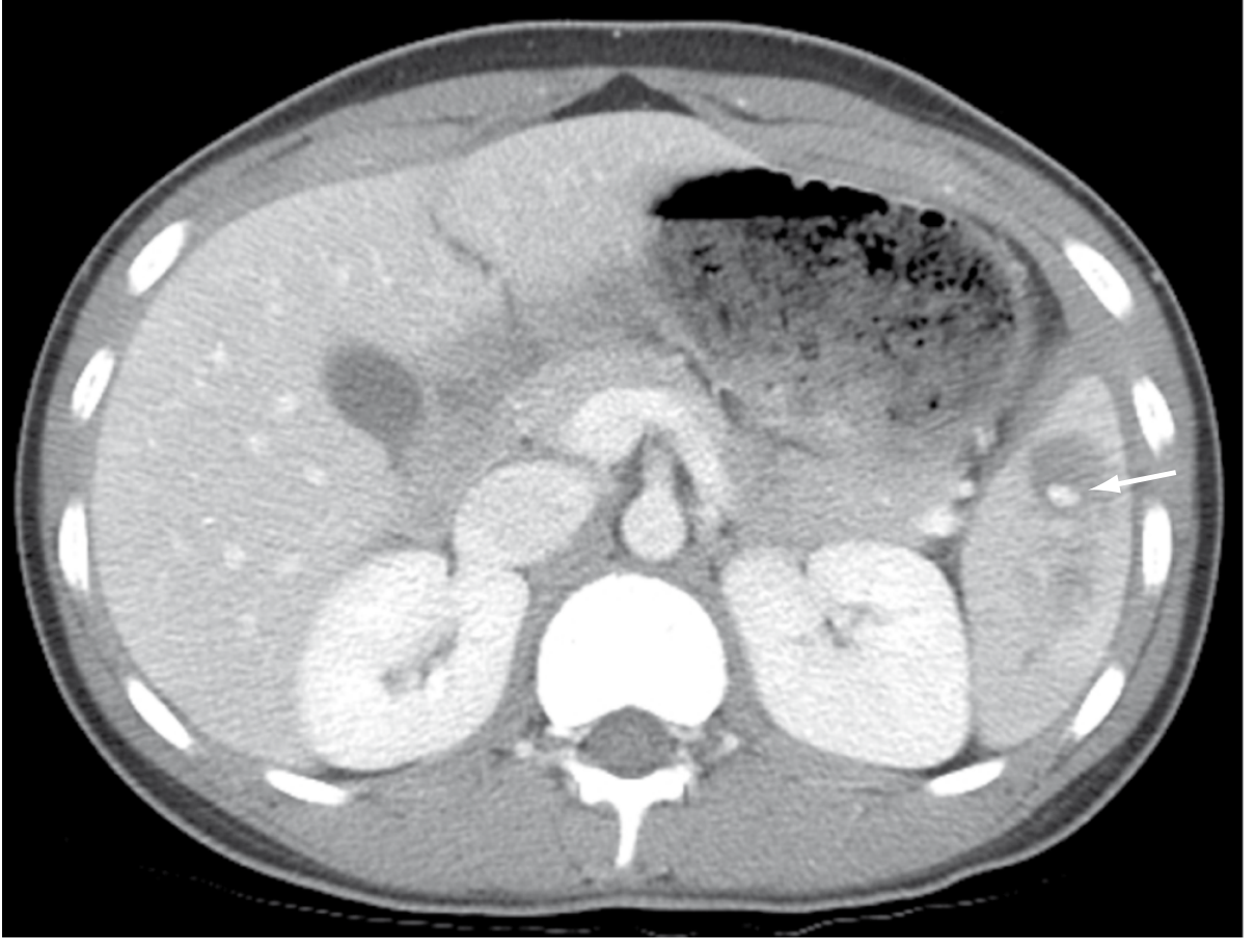
What the blush means: The bright spot/focus of high attenuation = contrast-laden blood leaking from a torn vessel in real time. This tells you:
- Active arterial haemorrhage is occurring right now
- Non-operative management (NOM) is likely to fail without intervention
- Angioembolisation is needed urgently
- If free into peritoneum → may need operative intervention
Without contrast, this critical finding is completely invisible - there is no way to distinguish active bleeding from surrounding haematoma on plain CT.
3. Multiphase CECT - What Each Phase Adds
CECT is ideally performed in multiple phases, each contributing specific information:
| Phase | What it shows in splenic trauma |
|---|---|
| Unenhanced | Baseline density; acute haematoma may be hyperdense (fresh clot); calcifications |
| Arterial phase (~25-35 sec) | Active arterial extravasation (blush); pseudoaneurysm; splenic artery injury |
| Portal venous phase (~70-80 sec) | Best for parenchymal laceration, haematoma extent, free fluid, grade assignment; uniform splenic enhancement makes defects most visible |
| Delayed phase (~3-5 min) | Distinguishes true extravasation (enlarges/changes shape) from pseudoaneurysm (stable pool); identifies venous bleeding |
The characteristic striped heterogeneous enhancement seen in the arterial phase is a normal spleen finding (from fast and slow vascular channels in the red pulp cords) - not to be mistaken for injury. It resolves to homogeneous enhancement in the venous phase.
4. Why Not Ultrasound?
Ultrasound (FAST exam) is used in the primary survey to rapidly detect free fluid (haemoperitoneum) at the bedside. But:
- Not sensitive enough to exclude significant splenic injury reliably
- Cannot characterise the injury or assign a grade
- Cannot detect active bleeding / vascular blush
- Operator-dependent, limited by body habitus, bowel gas, rib shadowing
FAST is a screening tool for haemoperitoneum. CECT is the definitive characterisation tool.
5. CECT Drives the Management Decision
The CECT findings directly determine what you do next:
| CECT Finding | Management |
|---|---|
| Grade I-II, no blush, haemodynamically stable | NOM - bed rest, monitoring |
| Grade III, no blush, stable | NOM with close monitoring; repeat imaging |
| Any grade, vascular blush present, stable | Angioembolisation |
| Grade IV-V, stable | CTA/angiography within 24-96 hours even without blush |
| Grade V (shattered/devascularised) or unstable | Operative splenectomy |
| Pseudoaneurysm | Angioembolisation |
This is why the Grainger & Allison radiology text states: "Multiphase contrast-enhanced CT has a sensitivity and specificity above 95% and is the established imaging technique for splenic injury diagnosis and grading."
Without contrast, you get anatomy. With contrast, you get physiology - you see whether the spleen is bleeding, how fast, from where, and how much parenchyma is viable.
6. Specific Contrast-Related Findings to Know
| Finding on CECT | Significance |
|---|---|
| Vascular blush (focal high attenuation in parenchyma) | Active arterial haemorrhage - NOM likely to fail |
| Contrast pooling free in peritoneum | Active bleeding into peritoneal cavity |
| Intraparenchymal pseudoaneurysm (contained contrast pool) | Risk of delayed rupture - needs embolisation |
| Non-enhancing parenchyma | Devascularisation / infarction (Grade IV-V) |
| Subcapsular haematoma (crescentic, no enhancement) | Tamponaded by capsule - can manage conservatively if stable |
| Perisplenic haematoma | Capsular disruption |
| Haemoperitoneum (free fluid in abdomen) | Indicates significant injury regardless of visible laceration |
In Summary
CECT is used because:
- Unenhanced CT misses isodense haematomas and gives no vascular information
- The vascular blush - the single most important finding that changes management - is only detectable with contrast
- It grades the injury precisely (Grades I-V) to guide NOM vs. embolisation vs. surgery
- It detects pseudoaneurysms that would be missed and can cause delayed rupture
- It shows associated injuries (liver, pancreas, kidney, mesentery, bowel) which may themselves mandate surgery
- Sensitivity/specificity 96-100% - no other modality comes close in the stable patient
Sources:
- Sabiston Textbook of Surgery 21e, Ch. 36 (Splenic Injuries)
- Grainger & Allison's Diagnostic Radiology (Splenic Trauma section)
- Maingot's Abdominal Operations, Ch. 62
This is a shared conversation. Sign in to Orris to start your own chat.