
Differentiated thyroid cancer
"differentiated thyroid cancer" AND management
papillary thyroid carcinoma histology nuclear features
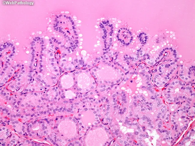
Thyroid gland histology section examined under light microscopy with Hematoxylin and Eosin stain. The thyroid follicles show marked epithelial hyperplasia with crowding of nuclei, producing papillary-like folds into the colloid. This pattern occurs in Graves disease and reflects hormonally driven follicular hyperplasia rather than neoplastic papillary thyroid carcinoma. Notable features include focal papillary infoldings with absence of malignant nuclear characteristics such as grooves, inclusions, or clearing. Clinical context typically reveals hyperthyroidism, with low TSH and elevated thyroid hormones. The changes are autoimmune-mediated, driven by thyroid-stimulating immunoglobulin (TSI) stimulating TSH receptors, leading to diffuse follicular enlargement and increased colloid turnover. In the absence of carcinoma-associated nuclear morphology, these findings should not be misinterpreted as papillary thyroid carcinoma. The image highlights how architectural changes can mimic neoplasia while lacking PTC cytology. Differential considerations include papillary carcinoma, nodular goiter, and thyroiditis; the decisive factors are epithelial architecture and nuclear phenotype. This histology is clinically significant to avoid overtreatment and guide management such as antithyroid therapy, radioiodine, or surgery based on clinical and biochemical context. Educationally, it supports pathology training, radiology-pathology correlation, and recognition of Graves-associated thyroid changes in suspected hyperthyroid patients. Correlate with serology and imaging for comprehensive patient assessment. With clinical correlation.
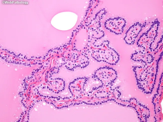
This is a brightfield, hematoxylin and eosin stained histology slide of thyroid tissue examined by light microscopy at high magnification. The specimen is thyroid gland parenchyma showing diffuse follicular hyperplasia characteristic of Graves disease. Follicles appear crowded with epithelial cells lining irregular, papillary-like folds protruding into the lumen, creating pseudo-papillary architectures that could mimic papillary thyroid carcinoma if evaluated in isolation. The nuclei within the thyrocytes are hyperplastic and dense but lack the distinctive nuclear features of papillary carcinoma, such as crowding with nuclear grooves, inclusions, Orphan Annie eye clearing, and powdery chromatin. Colloid is variably present or scant; stroma is vascular, and the overall architecture is polyfollicular with ingrowths into luminal spaces. This pattern reflects autoimmune-driven stimulation by TSH receptor autoantibodies (e.g., thyroid-stimulating immunoglobulin), resulting in diffuse goiter with hyperthyroid state. Clinically, Graves disease presents with weight loss, tachycardia, heat intolerance, and sometimes exophthalmos; histology aids diagnosis when correlated with serology and uptake studies. Key diagnostic significance: avoid mislabeling as papillary thyroid carcinoma; rely on clinical context and absence of malignant nuclear features. Potential educational uses include differential diagnosis training for thyroid pathology, immune-mediated thyroid hyperplasia vs neoplasia, and radiology-pathology correlation. This image supports teaching clinics and pathology review conferences.
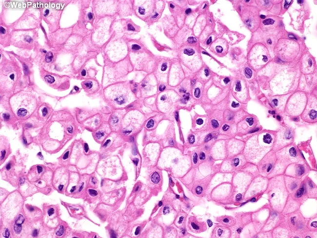
This is a histopathology slide of thyroid tissue prepared by hematoxylin and eosin staining, examined under brightfield light microscopy at high magnification. The specimen demonstrates epithelial neoplasm consistent with papillary thyroid carcinoma (PTC). The neoplastic cells are arranged in sheets and, where present, papillary fronds with fibrovascular cores are recognizable. The predominant cytoplasm is finely reticular and eosinophilic, with blander nuclear features. The nuclei show characteristic raisinoid appearance with nuclear grooves, giving an Orphan Annie eye look; the nuclear membranes are distinct and often thickened. Perinuclear halos, i.e., clear nuclear clearing around the chromatin, are evident. Some tumor cells exhibit voluminous cytoplasm, and in the plane of section, nuclei may be absent in those cells, a phenomenon of sectioning rather than true anuclear cells. The cytoplasmic clearing and grooves are diagnostic, aiding differentiation from benign nodules. The histology carries diagnostic significance, with PTC carrying generally favorable prognosis but potential for lymphatic metastasis; differential includes follicular carcinoma, medullary carcinoma, Hashimoto-related changes, and benign adenomas. Clinically, presence of these features correlates with occult metastasis risk and guides surgical planning, post-operative radioiodine therapy considerations, and molecular testing (e.g., BRAF V600E). This image is relevant for medical education, pathology teaching files, digital atlases, and research on thyroid cancer morphology.
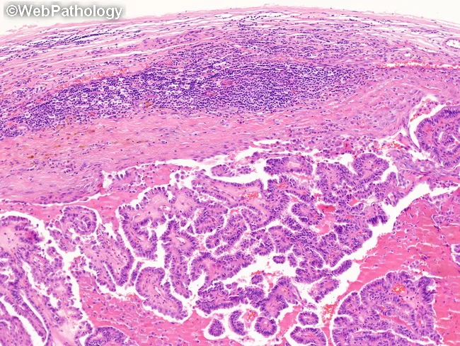
Light microscopy of a cervical lymph node with metastatic papillary thyroid carcinoma. Specimen: Lymph node from the neck region submitted for metastatic carcinoma assessment. The node shows effacement of normal architecture by a papillary thyroid carcinoma metastasis that forms arborizing papillary fronds with fibrovascular cores. Tumor cells display characteristic nuclear features of papillary carcinoma, including optically clear (ground-glass) nuclei, nuclear grooves, and intranuclear pseudoinclusions. The metastatic deposit replaces most of the nodal parenchyma while a rim of residual lymphoid tissue remains at the periphery. Cystic degeneration is frequently observed within metastatic nodal deposits, which can mimic a branchial cleft cyst. Psammoma bodies may be present in some sections. The lesion is typically ipsilateral to the primary thyroid lesion, with possible bilateral involvement in a minority of cases; mediastinal spread is less common. The histology underscores the importance of recognizing PTC features even within lymph nodes to avoid misdiagnosis as benign cystic lesions. Differential considerations include branchial cleft cyst, metastatic squamous cell carcinoma, and other papillary thyroid carcinoma variants. Diagnostic significance includes confirmation of nodal metastasis, staging implications (N stage), and impact on surgical planning and adjuvant therapy. Clinically, this image supports educational, diagnostic, and research use in head and neck oncology, thyroid pathology, and FNA-histopathology correlation.
Differentiated Thyroid Cancer (DTC)
1. Classification
| Type | Frequency | Key Feature |
|---|---|---|
| Papillary (PTC) | ~84% | Lymphatic spread; "Orphan Annie" nuclei |
| Follicular (FTC) | ~5% | Hematogenous spread; requires histology for diagnosis |
| Oncocytic (OCA) | ~5% | ≥75% oncocytic cells; less RAI-avid |
| NIFTP* | Rare | Previously encapsulated fvPTC; now low-risk, not malignant |
2. Pathology
Papillary Thyroid Carcinoma (PTC)
- Branching papillae with fibrovascular stalks covered by cuboidal epithelium
- "Orphan Annie eye" ground-glass nuclei - finely dispersed chromatin giving an optically clear/empty appearance
- Nuclear grooves and pseudoinclusions (cytoplasmic invaginations) - diagnostic even without papillary architecture
- Psammoma bodies - concentrically calcified structures; virtually absent in FTC and medullary carcinoma
- Lymphatic invasion common; vascular invasion uncommon in small tumors
- Cervical lymph node metastases in up to 50% of cases
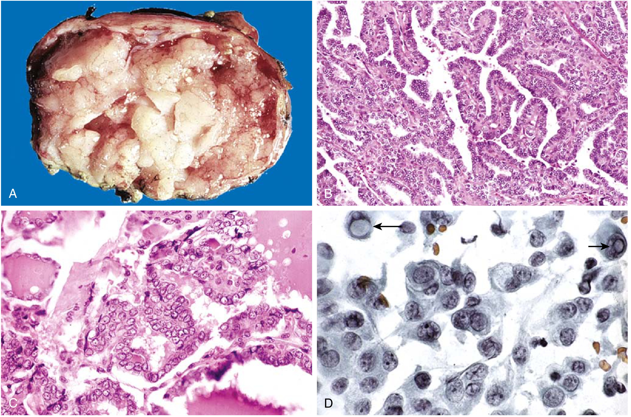
- Classic & follicular variant - indolent; favorable prognosis
- Tall cell, columnar cell, hobnail, diffuse sclerosis - aggressive subtypes (<1% of PTC); require intensive treatment and close follow-up
- NIFTP - encapsulated follicular variant WITHOUT capsular/vascular invasion; reclassified as non-malignant in 2016
Follicular Thyroid Carcinoma (FTC)
- Diagnosis requires histology (not FNA): capsular and/or vascular invasion on histology, since follicular adenoma and carcinoma have identical cytology
- Minimally invasive (capsular invasion only): very low metastatic risk; lobectomy alone may suffice
- Angioinvasive FTC: aggressive; spreads hematogenously to bone, lung, and CNS
- More common in iodine-deficient regions
- Peak incidence: 4th-6th decades; female predominance (3:1)
- Lymph node metastases in <10% (contrast with PTC)
- Poor prognostic features: distant metastases, age >55, tumor >4 cm, marked vascular invasion
Oncocytic Carcinoma (OCA)
- 2022 WHO classification replaced "Hurthle cell carcinoma"
- Defined as invasive follicular neoplasm with ≥75% oncocytic cells showing large vesicular nuclei with irregular macronucleoli
- Spreads via both lymphatic and hematogenous routes
- Distant metastases in ~20% at diagnosis
- 5-year overall survival ~85%; only 24% with distant metastases
- Less RAI-avid than PTC/FTC - harder to treat if recurrent
3. Molecular Biology & Risk Factors
- BRAF V600E mutation: present in ~60% of PTC; associated with aggressive behavior, extrathyroidal extension, and RAI resistance
- RAS mutations: more common in FTC and follicular variant PTC
- RET/PTC rearrangements: typically in PTC, especially radiation-induced
- PAX8-PPARG fusion: characteristic of FTC; present in up to one-third of encapsulated follicular variant PTC
- Prior ionizing radiation exposure (head/neck; most strongly linked to PTC and FTC)
- Female sex (3:1 F:M ratio)
- Iodine deficiency (FTC more prevalent)
- Familial syndromes: Cowden syndrome (PTEN mutation), familial adenomatous polyposis (APC), Carney complex, DICER1 syndrome
4. Clinical Presentation & Diagnosis
- Most present as a painless thyroid nodule or incidental finding on imaging
- Some present as a metastatic cervical lymph node (especially PTC)
- Rarely: dysphonia (RLN involvement), dysphagia, or stridor from invasion
- Thyroid ultrasound - assess nodule characteristics (TIRADS), cervical lymphadenopathy
- TSH - if suppressed, consider thyroid scintigraphy
- Fine-needle aspiration (FNA) - Bethesda classification guides management
- FNA can diagnose PTC (nuclear features); cannot definitively diagnose FTC (need capsular/vascular invasion on resection specimen)
- Molecular testing (e.g., ThyroSeq, Afirma) for indeterminate FNA (Bethesda III/IV)
- Serum thyroglobulin (Tg) and anti-Tg antibodies - baseline for post-treatment surveillance
5. Staging - AJCC 8th Edition
| Parameter | Details |
|---|---|
| Age cut-off | 55 years (critical divider; age <55 = max stage II) |
| Stage I | Any T, any N, M0; age <55 |
| Stage II | Any T, any N, M1; age <55 OR T3, T4 without M1; age ≥55 |
| Stage III | T4a, any N, M0; age ≥55 |
| Stage IVA | T4b, any N, M0 OR any T, N1b, M0; age ≥55 |
| Stage IVB | Any T, any N, M1; age ≥55 |
6. Management
A. Surgery
- Lobectomy is acceptable for intrathyroidal T1b-T2 tumors (>1 cm, <4 cm) without metastatic disease and normal contralateral lobe on US
- Near-total/total thyroidectomy is preferred for:
- Tumors >4 cm (T3)
- Evidence of extrathyroidal invasion
- Metastatic disease
- High-risk pathologic features (vascular invasion, aggressive subtype)
- Preoperative cervical US mandatory to assess central and lateral compartments
- Central neck dissection (level VI) for clinically node-positive disease
- Lateral neck dissection for confirmed lateral LN metastasis (FNA-proven)
B. TSH Suppression Therapy
| Risk Category | TSH Target |
|---|---|
| Low risk | 0.5-2.0 mIU/L (lower normal range) |
| Intermediate risk | 0.1-0.5 mIU/L |
| High risk / known metastatic disease | <0.1 mIU/L |
C. Radioiodine (¹³¹I) Therapy
- Stage I T1 tumors (≤2 cm): RAI offers no benefit - not recommended
- Low-risk tumors >2 cm but <4 cm: no demonstrated benefit
- High-risk features (larger tumors, aggressive variants, vascular invasion, extrathyroidal invasion, large-volume LN metastases): RAI may reduce recurrence and improve survival in older patients
D. External Beam Radiation Therapy (EBRT)
- Reserved for unresectable or gross residual disease not amenable to RAI
- Charged particle radiotherapy is under investigation for RAI-refractory disease
E. Systemic Therapy (Targeted)
- Lenvatinib (multi-kinase inhibitor) and sorafenib - approved for RAI-refractory DTC; improve progression-free survival
- Selective BRAF inhibitors (e.g., dabrafenib + trametinib) for BRAF V600E-mutated RAI-refractory DTC
- RET inhibitors (selpercatinib, pralsetinib) for RET fusion-positive DTC
7. Surveillance After Initial Treatment
| Test | Purpose | Timing |
|---|---|---|
| Serum thyroglobulin (Tg) | Tumor marker; rising Tg = recurrence | Every 6-12 months |
| Anti-Tg antibodies | Interference with Tg assay | Concurrent with Tg |
| Neck ultrasound | Detect local/regional recurrence | 6-12 months post-surgery, then annually |
| Stimulated Tg (rhTSH or withdrawal) | More sensitive detection | At ~6-12 months; periodically in intermediate/high-risk |
| Diagnostic whole-body scan | RAI-avid disease; especially if Tg rising | Selectively in high-risk |
8. Prognosis
- Overall: 10-year survival >95% (PTC); excellent for localized disease
- Stage I/II: Disease-specific survival similar to the general population
- Stage IV with distant metastases: Mortality markedly increased; this group is ~1% of patients
- FTC with angioinvasion: Less favorable than PTC; higher proportion present with stage IV disease
- OCA with distant metastases: ~24% 5-year survival
9. Special Situations
Pregnancy
- DTC found during pregnancy: surgery typically deferred to postpartum if stable on US at 24 weeks
- RAI is contraindicated in pregnancy and breastfeeding
Pediatric DTC
- More aggressive presentation (higher rates of nodal and pulmonary metastases) but excellent long-term survival
- Total thyroidectomy generally preferred; debate ongoing regarding lobectomy vs. total thyroidectomy in low-risk pediatric cases (recent meta-analysis, PMID 40915491, 2025)
NIFTP (Low-Risk)
- Thyroid lobectomy is adequate treatment
- No TSH suppression or RAI required
- Follow-up as for low-risk thyroid disease