
Ischirectal fossa
Reading File
Now let me search the medical library for the ischioanal/ischiorectal fossa:
Finding Sources
Finding Sources
Reading File
Reading File
Now let me also search for clinical aspects (abscesses, contents) and fetch the key images:
Finding Sources
Loading Image
Loading Image
Now let me get the clinical details on ischiorectal abscess from Schwartz's:
Reading File
Loading Image
Excellent. I now have comprehensive material. Here is a full, well-illustrated answer:
Ischiorectal (Ischioanal) Fossa
The term ischiorectal fossa is the older, widely used name. Modern anatomy texts (e.g., Thieme Atlas) prefer ischioanal fossa because the space is actually lateral to the anal canal, not the rectum. Both terms refer to the same space.
Shape and Location
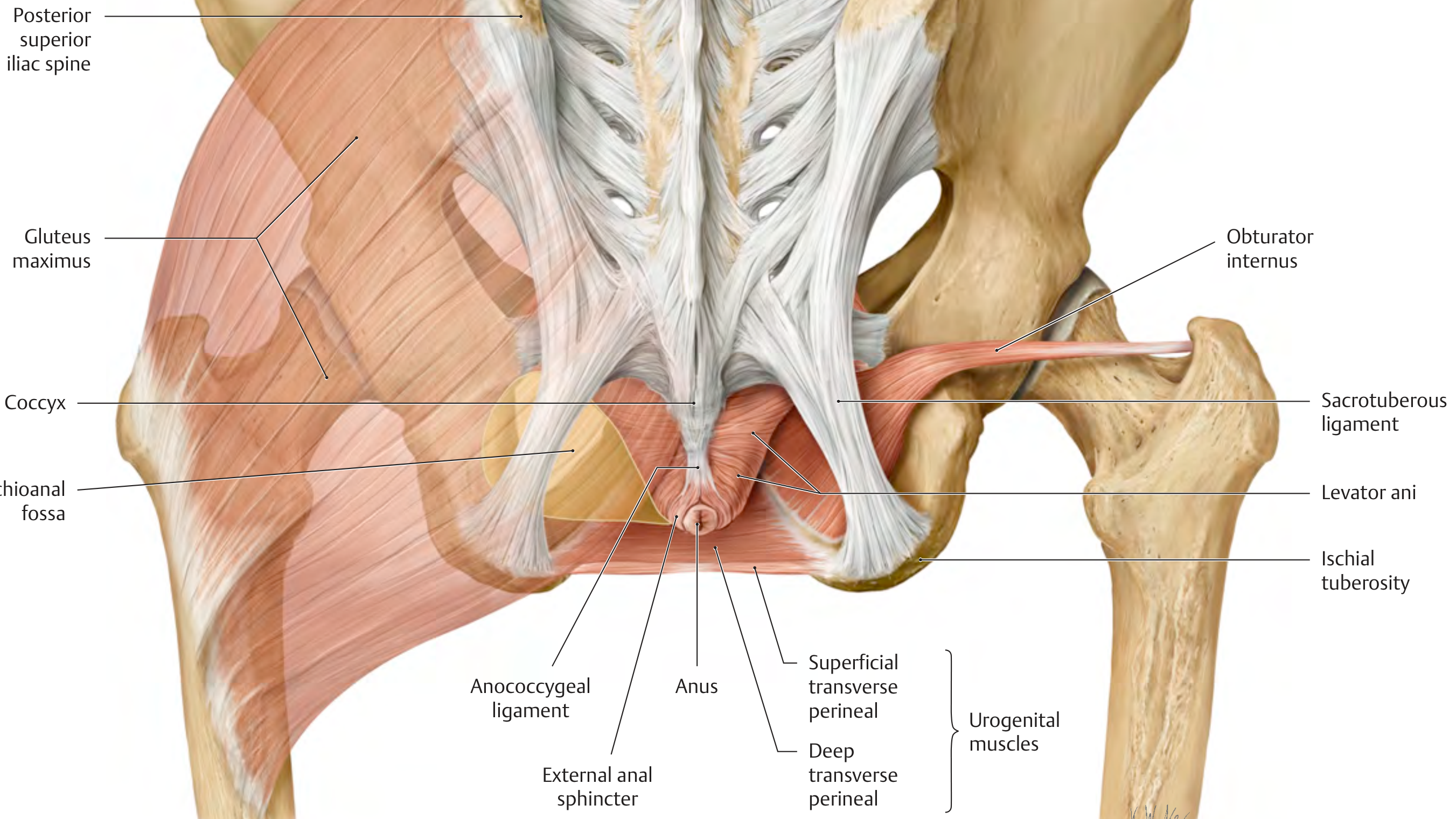
The ischioanal fossa is a pyramid-shaped (wedge-shaped) space situated on either side of the anal canal, below the pelvic floor (pelvic diaphragm). There is one fossa on each side; posteriorly the two fossae communicate across the midline via the deep postanal space (posterior to the anococcygeal body).
- The tip (apex) of the pyramid points anterosuperiorly, toward the symphysis pubis, at the junction of the levator ani and the obturator internus.
- The base faces inferiorly and is covered by perineal skin.
Boundaries
| Wall | Structure |
|---|---|
| Superomedial | Levator ani (and its inferior fascia - the inferior fascia of the pelvic diaphragm) |
| Lateral | Obturator internus muscle and its overlying obturator fascia; ischial tuberosity |
| Inferior (base) | Perineal skin |
| Posterior entrance | Gluteus maximus + sacrotuberous ligament |
| Anterior extension | Extends anteriorly to the posterior border of the urogenital diaphragm |
| Medial (inner wall) | External anal sphincter |
The fossa therefore communicates anteriorly with the anterior recess (above the urogenital diaphragm) and posteriorly it connects with the other side through the deep postanal space.
Contents
-
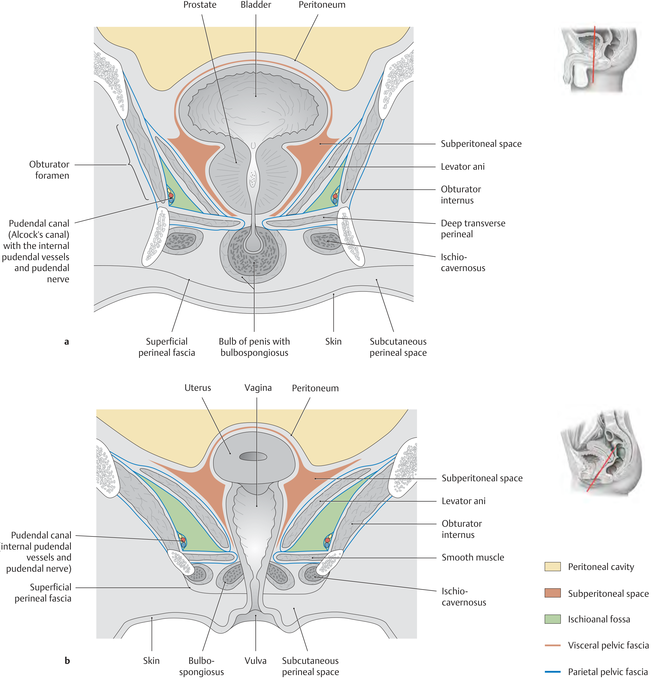
Fat pad (fat body of the ischioanal fossa) - fills most of the space; acts as a mobile cushion that slides downward and backward during defecation or parturition, allowing the anal canal and levator ani to move freely.
-
Inferior rectal (inferior haemorrhoidal) vessels and nerves - branches of the internal pudendal vessels and pudendal nerve that cross the fossa to reach the external anal sphincter and perianal skin.
-
Pudendal canal (Alcock's canal) - a fascial tunnel formed by the split obturator fascia running along the lateral wall of the fossa. It contains:
- Internal pudendal artery and veins
- Pudendal nerve (S2-S4) The canal begins just below the ischial spine and courses anteroinferiorly.
-
Perineal branch of S4 - small cutaneous branches traversing the fat.
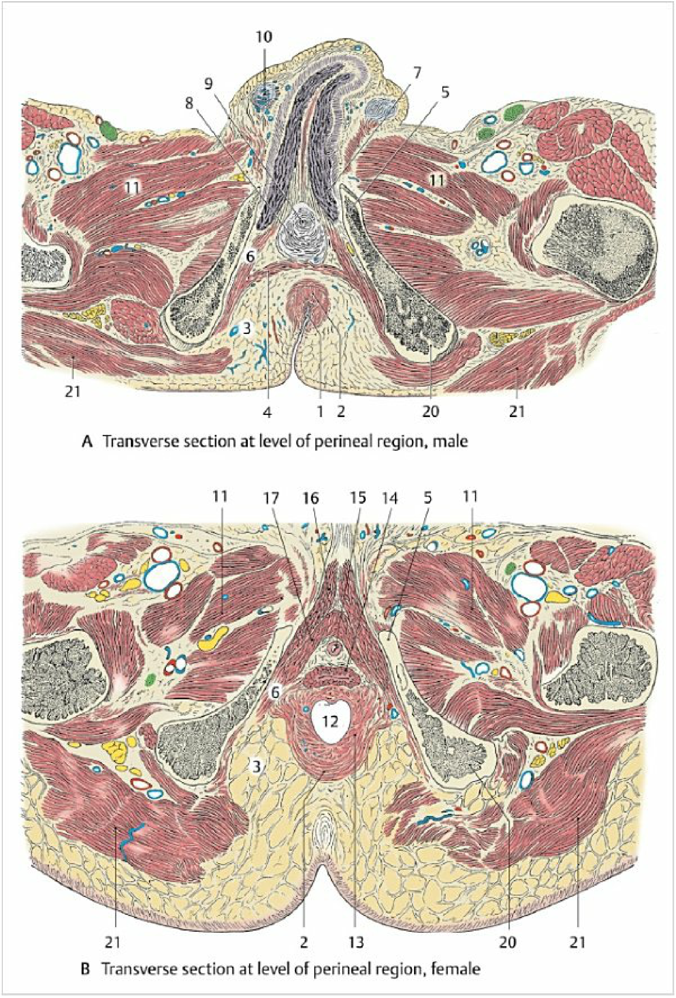
Transverse sections (male and female)
Clinical Significance
1. Ischiorectal (Perirectal) Abscess
The large fatty space of the ischioanal fossa is the most common site for perirectal abscess formation. Infection typically originates from infected anal glands (cryptoglandular theory) and spreads laterally through the external sphincter into the fossa.
- Presentation: Severe anal/perianal pain, diffuse tender swelling laterally in the fossa on digital rectal exam, fever, sometimes urinary retention or sepsis.
- Horseshoe abscess: Infection tracks posteriorly through the deep postanal space to involve both sides simultaneously. This requires drainage of the deep postanal space plus counterincisions over one or both ischiorectal spaces.
- Extension upward: Because the levator ani slopes upward, abscesses can expand superiorly without pointing through the buttock skin - they may present as a palpable bulge above the dentate line. Upward extension produces a supraelevator abscess.
- Treatment: Surgical drainage as soon as diagnosis is established. Antibiotics alone are ineffective. Antibiotics are added only if there is extensive cellulitis, immunocompromise, diabetes, or valvular heart disease.
2. Ischiorectal Fistula
Inadequately treated abscesses often result in fistula-in-ano, commonly a transsphincteric type passing through the external sphincter into the ischioanal fossa.
3. Pudendal Nerve Block
The pudendal canal in the lateral wall of the fossa is the landmark for pudendal nerve block - used for perineal analgesia in obstetrics and anorectal surgery.
4. Spread of Infection
The communication between the two fossae through the deep postanal space explains why untreated unilateral abscesses can become bilateral (horseshoe) abscesses. The anterior recess extension can also spread infection into the urogenital triangle.
Summary Table
| Feature | Detail |
|---|---|
| Shape | Pyramid/wedge |
| Apex | Anterosuperior (obturator internus + levator ani junction) |
| Base | Perineal skin |
| Superomedial boundary | Levator ani + inferior fascia of pelvic diaphragm |
| Lateral boundary | Obturator internus + ischial tuberosity |
| Main content | Ischioanal fat pad |
| Key neurovascular structure | Pudendal canal (Alcock's canal) on lateral wall |
| Clinical relevance | Ischiorectal abscess, horseshoe abscess, pudendal nerve block, fistula-in-ano |
Sources:
- General Anatomy and Musculoskeletal System (THIEME Atlas of Anatomy), pp. 581-582
- Color Atlas of Human Anatomy Vol. 2, Internal Organs, pp. 476-477
- Schwartz's Principles of Surgery, 11th ed., Chapter 29 (Anorectal Sepsis)
Perineal pouches
Finding Sources
Finding Sources
Reading File
Finding Sources
Reading File
Finding Sources
Reading File
Finding Sources
Reading File
Finding Sources
Reading File
Reading File
Now let me fetch the key diagrams:
Loading Image
Loading Image
Loading Image
I now have comprehensive material for a thorough answer. Here it is:
Perineal Pouches
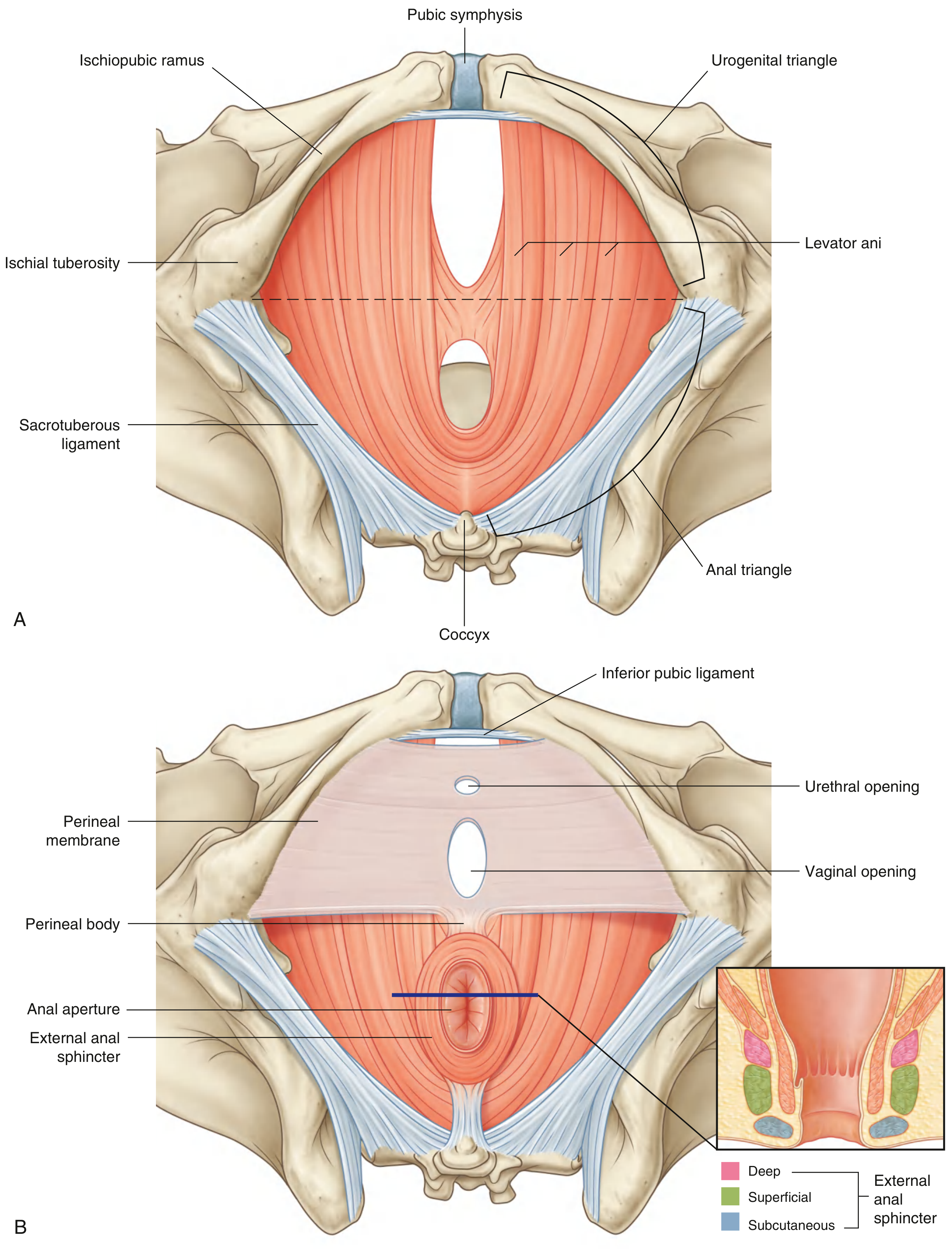
The perineum is the diamond-shaped region inferior to the pelvic diaphragm (levator ani + coccygeus). A line drawn between the two ischial tuberosities divides it into an anterior urogenital triangle and a posterior anal triangle. Within the urogenital triangle, the perineal membrane subdivides the space into two compartments called the perineal pouches.
The Perineal Membrane - The Key Dividing Structure
The perineal membrane is a thick fibrous sheet that fills the urogenital triangle. It has:
- A free posterior border anchored in the midline to the perineal body
- Lateral attachments to the pubic arch (ischiopubic rami)
- Anteriorly it ends as the transverse perineal ligament, with a gap between it and the arcuate pubic ligament for the deep dorsal vein of the penis/clitoris
The urethra (and in women, the vagina) pass through the perineal membrane to exit the pelvis.
1. Superficial Perineal Pouch
Definition
The space between the perineal membrane (above) and the membranous layer of the superficial fascia / Colles fascia (below).
Boundaries
| Wall | Structure |
|---|---|
| Floor (inferior) | Membranous layer of superficial fascia (Colles fascia) |
| Roof (superior) | Perineal membrane |
| Lateral | Fuses with fascia lata at pubic arch; Colles fascia fuses laterally to pubic arch |
| Posterior | Colles fascia sweeps beneath transverse perineal muscles and fuses with the posterior perineal membrane - the pouch is closed posteriorly |
| Anterior | Open - communicates with the potential space deep to Scarpa's fascia of the anterior abdominal wall |
Contents
Erectile structures (present in both sexes):
- Crura of the penis / clitoris - proximal attached ends of the corpora cavernosa, anchored to the ischiopubic rami
- Bulb of the penis (men) / Bulbs of the vestibule (women) - proximal erectile tissue anchored to the perineal membrane
- Greater vestibular glands (Bartholin's glands) - only in women, one on each side, lying on the perineal membrane deep to the posterior end of the bulb of the vestibule
Muscles (superficial perineal muscles - all innervated by the perineal branch of the pudendal nerve, S2-S4):
| Muscle | Origin | Insertion | Function |
|---|---|---|---|
| Ischiocavernosus | Ischial tuberosity & ramus | Crus of penis/clitoris | Forces blood from crura into body of penis/clitoris during erection |
| Bulbospongiosus | Perineal body (women); perineal body + midline raphe (men) | Bulb of vestibule/corpus spongiosum, perineal membrane | Forces blood distally into glans; in men: expels urine/semen from urethra |
| Superficial transverse perineal | Ischial tuberosity & ramus | Perineal body | Stabilizes the perineal body |
Neurovascular:
- Perineal branches of the internal pudendal artery and perineal nerve (branches of pudendal nerve S2-S4)
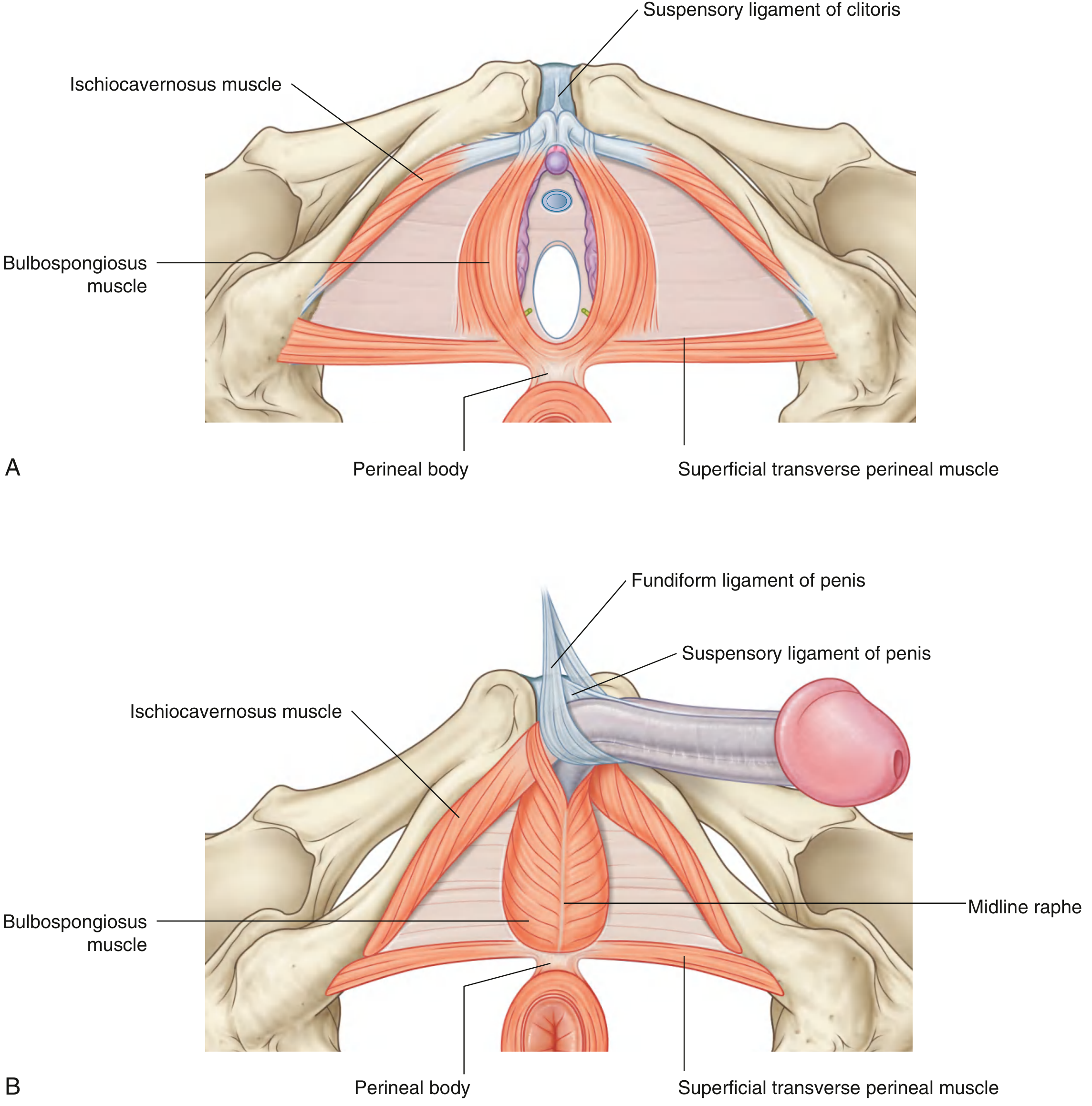
2. Deep Perineal Pouch
Definition
The space immediately superior to (above) the perineal membrane, between the perineal membrane and the superior fascia of the urogenital diaphragm. It is a thin but important region.
Contents
Muscles (deep perineal muscles - all innervated by perineal branch of pudendal nerve, S2-S4):
| Muscle | Notes |
|---|---|
| External urethral sphincter | Encircles the urethra; the somatic/voluntary sphincter of the urethra |
| Deep transverse perineal | Runs transversely; stabilizes the perineal body |
| Sphincter urethrovaginalis (women only) | Surrounds urethra AND vagina together; aids urethral closure |
| Compressor urethrae (women only) | Arises from ischiopubic rami; fibers meet anterior to urethra; aids urethral closure |
Glands:
- Bulbourethral glands (Cowper's glands) - only in men; paired, pea-sized, open into the bulbous urethra; secrete pre-ejaculatory fluid
Neurovascular:
- Branches of the internal pudendal artery and veins
- Dorsal nerve of the penis/clitoris (terminal branch of pudendal nerve)
- Parasympathetic cavernous nerves from the inferior hypogastric plexus (S2-S4) pass through the deep pouch to innervate the erectile tissues and mediate erection
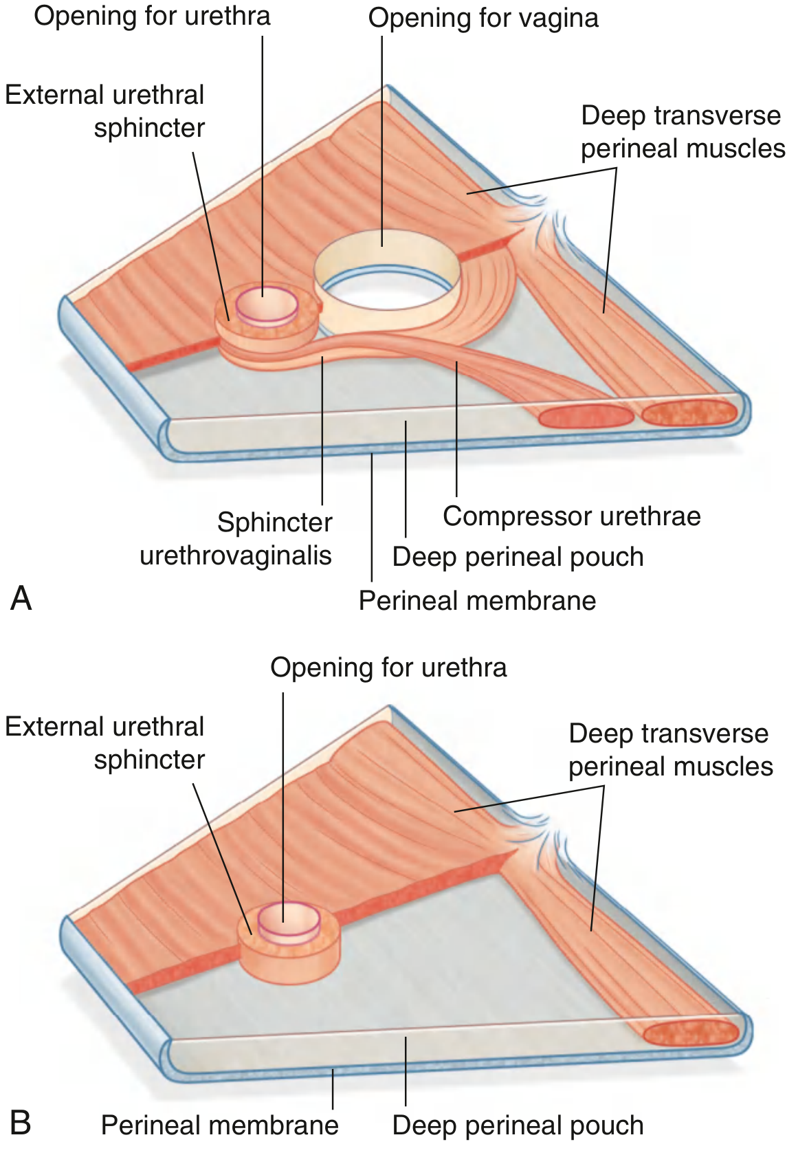
Comparison Table
| Feature | Superficial Perineal Pouch | Deep Perineal Pouch |
|---|---|---|
| Location | Below perineal membrane | Above perineal membrane |
| Floor | Colles fascia (membranous superficial fascia) | Perineal membrane |
| Roof | Perineal membrane | Superior fascia of urogenital diaphragm |
| Muscles | Ischiocavernosus, bulbospongiosus, superficial transverse perineal | External urethral sphincter, deep transverse perineal; + compressor urethrae & sphincter urethrovaginalis (females) |
| Erectile tissue | Crura, bulb of penis / bulbs of vestibule | None |
| Glands | Greater vestibular glands (females) | Bulbourethral/Cowper's glands (males) |
| Sphincter function | None | External urethral sphincter (voluntary) |
| Communication | Open anteriorly into anterior abdominal wall (Scarpa's fascia plane) | Closed |
Perineal Body
A fibromuscular node in the midline at the posterior border of the perineal membrane. 8 muscles converge on it:
- External anal sphincter
- Superficial transverse perineal (×2)
- Deep transverse perineal (×2)
- Bulbospongiosus (×2)
- Levator ani (pubococcygeus/puborectalis fibers)
It is clinically important because obstetric perineal tears disrupt the perineal body, leading to pelvic organ prolapse and urinary/fecal incontinence if not repaired.
Clinical Significance
Urethral rupture and extravasation of urine
Because the superficial perineal pouch is closed posteriorly (Colles fascia fuses with the perineal membrane) but open anteriorly, rupture of the bulbous urethra causes urine to extravasate:
- Into the superficial perineal pouch
- Up into the anterior abdominal wall (deep to Scarpa's fascia)
- Into the scrotum and penis
- NOT into the thighs (because Colles fascia fuses with fascia lata laterally)
- NOT into the perineum posteriorly (fused)
Pudendal nerve block
The pudendal nerve traverses both pouches via its branches; a pudendal nerve block at the ischial spine interrupts sensation to both pouches.
Episiotomy / Perineal tears
Obstetric injuries involve superficial pouch muscles (bulbospongiosus, superficial transverse perineal) and the perineal body.
Sources:
- Gray's Anatomy for Students, pp. 580-587 (Perineal membrane, deep and superficial perineal pouches, muscles)
- Campbell-Walsh-Wein Urology, Chapter 82 (Superficial and deep perineal spaces)
- Schwartz's Principles of Surgery, 11th ed. (Anorectal anatomy)
This is a shared conversation. Sign in to Orris to start your own chat.