
Posterior/dorsal column (Gracile and cuneate tracts) Anterior spinothalamic tract Lateral spinothalamic tract Anterior spinocerebellar tract Posterior spinocerebellar tract Spinotectal tract Spino-olivary tract Spinoreticular tract Lateral corticospinal tract Anterior corticospinal tract Rubrospinal tract Lateral vestibulospinal tract Medial vestibulospinal tract Reticulospinal tract Rubrospinal tract Each tract give best possible main points with diagram, explain in detail where it goes where it cross and what it does About each Tract Take ur time but give me the best possible notes with diagrams
Topic map
1. Ascending sensory tracts
- Dorsal columns
- Fasciculus gracilis
- Fasciculus cuneatus
- Anterolateral system
- Anterior spinothalamic tract
- Lateral spinothalamic tract
- Spinocerebellar pathways
- Posterior (dorsal) spinocerebellar tract
- Anterior (ventral) spinocerebellar tract
- Other ascending pathways
- Spinotectal tract
- Spino-olivary tract
- Spinoreticular tract
2. Descending motor tracts
- Pyramidal tracts
- Lateral corticospinal tract
- Anterior corticospinal tract
- Extrapyramidal tracts
- Rubrospinal tract
- Lateral vestibulospinal tract
- Medial vestibulospinal tract
- Reticulospinal tracts
High-yield overview
- Dorsal column-medial lemniscus pathway carries fine touch, vibration, conscious proprioception, and two-point discrimination. It crosses in the caudal medulla.
- Spinothalamic tracts carry pain, temperature, crude touch, and pressure. They cross in the spinal cord, usually within 1-2 segments through the anterior white commissure.
- Spinocerebellar tracts carry unconscious proprioceptive information to the cerebellum for coordination. Posterior spinocerebellar fibers are mainly uncrossed; anterior spinocerebellar fibers characteristically cross twice.
- Corticospinal tracts mediate voluntary skilled movement. Most fibers cross at the pyramidal decussation and form the lateral corticospinal tract.
- Brainstem extrapyramidal tracts regulate posture, balance, muscle tone, automatic movements, and head/neck orientation.
- Dorsal columns
- Spinothalamic tracts
- Spinocerebellar tracts
- Other ascending tracts
- Corticospinal tracts
- Extrapyramidal descending tracts
Ok go with top 6 with diagrams
spinal cord tracts AND neuroanatomy
labeled cross-sectional diagram of spinal cord ascending descending tracts dorsal columns spinothalamic corticospinal
NCBI Bookshelf spinal cord ascending descending tracts dorsal column spinothalamic corticospinal decussation
Spinal Cord Tracts: High-yield Detailed Notes
Orientation diagram: where the major tracts lie
              POSTERIOR / DORSAL

                  Posterior median sulcus
                           │
              ┌────────────────────────┐
              │  Cuneate | Gracile     │  ← Dorsal columns
              │ (T6 up)  | (all cord)  │
              │          │              │
              │ DSCT     │     LCST     │
              │          │ Rubrospinal  │
              │          │              │
              │          │  LSTT        │
              │          │              │
              │          │  ASCT        │
              │                          │
              │     Anterior CST         │
              │ Ant. spinothalamic       │
              └───────────┬────────────┘
                       Anterior
- DCML: dorsal column-medial lemniscus pathway
- LSTT: lateral spinothalamic tract
- ASTT: anterior spinothalamic tract
- DSCT: dorsal/posterior spinocerebellar tract
- ASCT: anterior/ventral spinocerebellar tract
- LCST: lateral corticospinal tract
1. Dorsal column-medial lemniscus pathway
Fasciculus gracilis and fasciculus cuneatus
Main function
| Tract | Body area represented | Sensations carried |
|---|---|---|
| Fasciculus gracilis | Lower trunk and lower limb, below T6 | Fine/discriminative touch, vibration, conscious proprioception, pressure, two-point discrimination, stereognosis |
| Fasciculus cuneatus | Upper trunk and upper limb, T6 and above | Same modalities from upper body |
Position in posterior column
Posterior spinal cord cross-section

            Posterior median sulcus
                     │
        ┌───────────────────────┐
        │     CUNEATUS          │  Lateral
        │   Upper limb/trunk    │
        │───────────────────────│
        │     GRACILIS          │  Medial
        │   Lower limb/trunk    │
        └───────────────────────┘
- Gracilis = lower body = medial
- Cuneatus = upper body = lateral
- Cuneate fasciculus is present only at T6 and above.
Pathway and crossing
Receptor in skin, muscle, joint, tendon
        │
        ▼
1st-order neuron: dorsal root ganglion
        │
        ▼
Enters spinal cord through dorsal root
        │
        └──── Ascends IPSILATERALLY in posterior column ────┐
                                                             │
                     Gracilis: nucleus gracilis, caudal medulla
                     Cuneatus: nucleus cuneatus, caudal medulla
                                                             │
                                                             ▼
2nd-order fibers cross as INTERNAL ARCUATE FIBERS
                 in caudal medulla
                                                             │
                                                             ▼
Contralateral MEDIAL LEMNISCUS
        │
        ▼
Ventral posterolateral nucleus, VPL, of thalamus
        │
        ▼
3rd-order neuron through posterior limb of internal capsule
        │
        ▼
Primary somatosensory cortex, postcentral gyrus
Decussation
- Crosses in the caudal medulla, not in the spinal cord.
- The crossing fibers are called internal arcuate fibers.
- After crossing, they form the medial lemniscus.
Clinical lesion pattern
| Site of lesion | Deficit |
|---|---|
| Posterior column in spinal cord | Ipsilateral loss below lesion of vibration, proprioception, discriminative touch |
| Medial lemniscus above medullary crossing | Contralateral loss of these sensations |
| Sensory cortex | Contralateral cortical sensory deficits, impaired graphesthesia and stereognosis |
Important signs of posterior-column damage
- Sensory ataxia
- Positive Romberg sign
- Stamping gait
- Loss of vibration sense, often tested at great toe or malleolus
- Pseudoathetosis due to loss of position sense
2. Spinothalamic tracts
Comparison
| Feature | Lateral spinothalamic tract | Anterior spinothalamic tract |
|---|---|---|
| Main modality | Pain and temperature | Crude/light touch and pressure |
| Site of crossing | Spinal cord | Spinal cord |
| Side of ascent | Contralateral | Contralateral |
| Final conscious relay | VPL thalamus | VPL thalamus |
| Cortex | Primary somatosensory cortex | Primary somatosensory cortex |
A. Lateral spinothalamic tract
Function
- Fast sharp pain
- Slow aching/burning pain
- Temperature
- Often itch and some crude tactile information
Pathway
Pain / temperature receptor
        │
        ▼
1st-order neuron in dorsal root ganglion
        │
        ▼
Enters dorsolateral spinal cord
        │
        ▼
May ascend or descend 1-2 spinal segments in Lissauer tract
        │
        ▼
Synapse in dorsal horn
  - Substantia gelatinosa: lamina II
  - Nucleus proprius: laminae III-IV
  - Lamina I and V also participate
        │
        ▼
2nd-order neuron crosses in ANTERIOR WHITE COMMISSURE
        │
        ▼
Ascends CONTRALATERALLY in anterolateral funiculus
        │
        ▼
VPL nucleus of thalamus
        │
        ▼
Primary somatosensory cortex
Decussation
- Fibers cross through the anterior white commissure.
- This occurs approximately 1-2 spinal segments above the level at which the sensory fiber entered.
Somatotopy
Lateral spinothalamic tract

More medial                 More lateral
Cervical → Thoracic → Lumbar → Sacral
Clinical relevance
- Contralateral loss of pain and temperature
- Beginning about 1-2 segments below the lesion.
B. Anterior spinothalamic tract
Function
- Crude touch
- Light touch
- Pressure
Pathway
Crude touch / pressure receptor
        │
        ▼
Dorsal root ganglion
        │
        ▼
Dorsal horn
        │
        ▼
Crosses in anterior white commissure
        │
        ▼
Ascends contralaterally in anterior funiculus
        │
        ▼
VPL thalamus
        │
        ▼
Primary somatosensory cortex
Clinical point
Brown-Sequard syndrome: classic tract pattern
                RIGHT hemicord lesion

RIGHT, below lesion:
- LMN signs at exact level
- UMN weakness below lesion
- Loss of vibration, position, fine touch

LEFT, beginning 1-2 levels below:
- Loss of pain and temperature
- Dorsal columns and corticospinal fibers have not crossed yet at spinal level, so their deficits are ipsilateral.
- Spinothalamic fibers crossed close to entry, so deficits are contralateral.
3. Spinocerebellar tracts
Main purpose
| Tract | Main source | Cerebellar peduncle | Net side supplied |
|---|---|---|---|
| Posterior/dorsal spinocerebellar | Lower limb and trunk | Inferior cerebellar peduncle | Ipsilateral |
| Anterior/ventral spinocerebellar | Mainly lower limb interneuronal activity | Superior cerebellar peduncle | Ipsilateral after double crossing |
A. Posterior spinocerebellar tract
Function
- Unconscious proprioception from lower limb and lower trunk
- Input from muscle spindles
- Input from Golgi tendon organs
- Information relevant to posture and coordination
Origin
- Clarke nucleus, also called nucleus dorsalis
- Present mainly from T1 to L2/L3
- Sensory fibers from lower levels can ascend in fasciculus gracilis before synapsing in Clarke nucleus.
Pathway
Muscle spindle / Golgi tendon organ / joint receptor
        │
        ▼
Dorsal root ganglion
        │
        ▼
Clarke nucleus, T1-L2
        │
        ▼
Posterior spinocerebellar tract
        │
        └──── Ascends IPSILATERALLY in lateral funiculus ────┐
                                                              │
                                                              ▼
Inferior cerebellar peduncle
        │
        ▼
Ipsilateral cerebellar cortex
  Mainly vermis and intermediate zone
Crossing
- Does not cross.
- The right side of body is represented in the right cerebellum.
Lesion
- Ipsilateral limb ataxia
- Gait incoordination
- Dysmetria may be present
- Conscious proprioception can remain relatively preserved because dorsal columns are separate.
Upper-limb equivalent: cuneocerebellar tract
- Primary fibers enter cervical cord.
- Ascend in fasciculus cuneatus.
- Synapse in accessory cuneate nucleus in medulla.
- Fibers enter cerebellum via inferior cerebellar peduncle.
- They remain ipsilateral.
B. Anterior spinocerebellar tract
Function
- Integrated activity from spinal interneuronal circuits
- Information about lower-limb movement and spinal motor activity
- A type of internal “status report” about what the spinal cord is doing
Origin
- Spinal border cells, mainly in lumbosacral cord, approximately L4-S3.
Double-crossed pathway
Lower-limb sensory and interneuronal input
        │
        ▼
Spinal border cells, lumbosacral cord
        │
        ▼
FIRST CROSSING:
anterior white commissure
        │
        ▼
Ascends on CONTRALATERAL side
in anterior/lateral funiculus
        │
        ▼
Superior cerebellar peduncle
        │
        ▼
SECOND CROSSING:
within or near cerebellum
        │
        ▼
Ends in cerebellum IPSILATERAL to original body side
Key memory point
Anterior spinocerebellar tract:
Crosses in spinal cord → crosses again in/near cerebellum

Two crossings = ends ipsilateral to its body of origin.
Lesion
4. Other ascending tracts
A. Spinotectal tract
Function
- Orienting eyes, head, and neck toward a stimulus
- Defensive responses to painful stimuli
- Reflexive attention to biologically important sensory events
Pathway
Pain / temperature input
        │
        ▼
Dorsal root ganglion
        │
        ▼
Dorsal horn
        │
        ▼
Crosses in anterior white commissure
        │
        ▼
Ascends in anterolateral system
        │
        ▼
Superior colliculus and other midbrain tectal areas
        │
        ▼
Reflex orientation of head and eyes
Main target
- Superior colliculus, part of the tectum of midbrain.
B. Spino-olivary tract
Function
Simplified pathway
Peripheral proprioceptive and cutaneous input
        │
        ▼
Dorsal horn / spinal interneurons
        │
        ▼
Spino-olivary fibers, largely crossed or bilateral
        │
        ▼
Inferior olivary nucleus in medulla
        │
        ▼
Olivocerebellar fibers cross
        │
        ▼
Contralateral cerebellum via inferior cerebellar peduncle
Main point
- Connects spinal sensory activity with the olivocerebellar system.
- Likely contributes to timing, error signaling, motor adaptation, and coordination.
- Details of its precise organization and functional importance are less settled than those of the major tracts.
C. Spinoreticular tract
Function
- Arousal and alertness
- Emotional/affective unpleasantness of pain
- Autonomic responses to pain
- Prolonged pain behavior
Pathway
Slow pain / visceral pain / nociceptive input
        │
        ▼
Dorsal horn
        │
        ▼
Bilateral ascent, usually contralateral predominance,
within anterolateral system
        │
        ▼
Reticular formation of medulla and pons
        │
        ▼
Intralaminar thalamic nuclei
        │
        ▼
Widespread cortex, limbic system, autonomic centers
Clinical meaning
- “Where is the pain and how intense is it?” is largely related to lateral spinothalamic processing.
- “How unpleasant, exhausting, alarming, or attention-demanding is it?” involves reticular, thalamic, and limbic connections, including spinoreticular pathways.
5. Corticospinal tracts
Common supraspinal course
Motor cortex and related cortical regions
  - Primary motor cortex
  - Premotor cortex
  - Supplementary motor area
  - Somatosensory cortex also contributes
        │
        ▼
Corona radiata
        │
        ▼
Posterior limb of internal capsule
        │
        ▼
Middle three-fifths of cerebral peduncle, midbrain
        │
        ▼
Basis pontis
        │
        ▼
Medullary pyramids
        │
        ├──── 85-90% cross at pyramidal decussation → LCST
        │
        └──── 10-15% remain uncrossed initially → ACST
A. Lateral corticospinal tract
Function
- Skilled voluntary movement
- Fine fractionated movement of fingers and hands
- Voluntary distal limb movement
- Especially important for contralateral limb control
Pathway
Motor cortex
        │
        ▼
Internal capsule → midbrain → pons → medullary pyramids
        │
        ▼
PYRAMIDAL DECUSSATION, caudal medulla
        │
        ▼
Descends in lateral funiculus as LCST
        │
        ▼
Synapses mainly on spinal interneurons
and, for some hand muscles, more directly on alpha motor neurons
        │
        ▼
Ventral horn motor neurons
        │
        ▼
Contralateral skeletal muscle
Crossing
- Crosses at pyramidal decussation in the caudal medulla.
- Therefore, the left motor cortex controls the right body, and vice versa.
Somatotopy
- Cervical fibers tend to lie more medially
- Sacral fibers tend to lie more laterally
Lesions
| Lesion location | Weakness side |
|---|---|
| Motor cortex, internal capsule, brainstem above pyramidal decussation | Contralateral UMN weakness |
| Spinal cord below pyramidal decussation | Ipsilateral UMN weakness below lesion |
Upper motor neuron signs
- Weakness with pyramidal distribution
- Spasticity
- Hyperreflexia
- Clonus
- Extensor plantar response, Babinski sign
- Reduced fine voluntary movement
B. Anterior corticospinal tract
Function
- Axial muscles
- Trunk muscles
- Proximal limb muscles
- Postural adjustments associated with voluntary movement
Pathway
Motor cortex
        │
        ▼
Descends through brainstem pyramids
        │
        ▼
Does NOT cross at pyramidal decussation initially
        │
        ▼
Descends ipsilaterally in anterior funiculus
        │
        ▼
At segmental spinal level:
many fibers cross through anterior white commissure
        │
        ▼
Synapses bilaterally, especially on medial motor neuron pools
        │
        ▼
Axial and proximal muscles
Crossing
- Most fibers do not cross in the medulla.
- Many cross near their spinal termination.
- There is substantial bilateral innervation.
Clinical consequence
6. Extrapyramidal descending tracts
- Posture
- Balance
- Muscle tone
- Automatic movement
- Gait
- Head and eye stabilization
- Proximal and axial muscle control
Functional division
Lateral descending system:
- Lateral corticospinal
- Rubrospinal
Mainly distal limb control

Medial/ventromedial descending system:
- Vestibulospinal
- Reticulospinal
- Anterior corticospinal
- Tectospinal
Mainly axial and proximal muscle control, posture and balance
A. Rubrospinal tract
Origin
- Red nucleus in the midbrain, mainly magnocellular region.
Function
- Facilitates flexor activity, especially upper-limb flexors
- Contributes to distal limb control
- More important in many nonhuman mammals than in humans
- In humans, the lateral corticospinal tract is the dominant pathway for skilled distal movement
Pathway
Red nucleus, midbrain
        │
        ▼
Crosses immediately in ventral tegmental decussation
        │
        ▼
Descends contralaterally in lateral funiculus
just anterior to LCST
        │
        ▼
Mostly terminates at cervical spinal levels
        │
        ▼
Interneurons and motor neurons supplying upper limb,
especially flexor muscle groups
Crossing
- Crosses in the midbrain, immediately after leaving red nucleus.
Lesion significance
B. Lateral vestibulospinal tract
Origin
- Lateral vestibular nucleus, also called Deiters nucleus.
Function
- Maintains upright posture
- Facilitates antigravity extensor muscles
- Helps maintain balance in response to vestibular information
- Influences trunk and limb extensor tone
Pathway
Vestibular apparatus
        │
        ▼
Lateral vestibular nucleus
        │
        ▼
Descends IPSILATERALLY without crossing
in anterior funiculus
        │
        ▼
Terminates at many spinal levels
        │
        ▼
Facilitates ipsilateral extensor motor neurons
and inhibits opposing flexor patterns
Crossing
- No major crossing.
- It acts mainly ipsilaterally.
Memory aid
C. Medial vestibulospinal tract
Origin
- Mainly medial vestibular nucleus, with contributions from inferior vestibular nucleus.
Function
- Coordinates head and neck position
- Stabilizes the head during body movement
- Assists vestibulocollic reflexes
- Works with eye movement systems to stabilize gaze
Pathway
Vestibular nuclei
        │
        ▼
Medial longitudinal fasciculus, MLF
        │
        ▼
Descends bilaterally in anterior funiculus
        │
        ▼
Mainly cervical and upper thoracic spinal cord
        │
        ▼
Neck and upper trunk motor neurons
Crossing
- Has bilateral projections.
- Unlike lateral vestibulospinal tract, it primarily reaches cervical and upper thoracic levels rather than the entire spinal cord.
Memory aid
D. Reticulospinal tracts
| System | Origin | Main course | Major effect |
|---|---|---|---|
| Pontine, medial reticulospinal | Pontine reticular formation | Mainly ipsilateral, anterior funiculus | Facilitates extensor tone and posture |
| Medullary, lateral reticulospinal | Medullary reticular formation | Bilateral, lateral/anterior funiculi | Modulates or inhibits extensor tone; contributes to locomotion and voluntary postural control |
Overall functions
- Automatic postural responses
- Anticipatory postural adjustment before voluntary movement
- Locomotor control
- Muscle tone regulation
- Coordination of axial and proximal muscles
- Integration of cortical, vestibular, visual, and cerebellar input
Pathway diagram
Motor cortex, cerebellum, vestibular system,
sensory inputs and limbic/autonomic centers
        │
        ▼
Reticular formation of pons and medulla
        │
        ├──── Pontine reticulospinal tract
        │       ↓ mainly ipsilateral
        │       ↓ promotes extensor tone/postural support
        │
        └──── Medullary reticulospinal tract
                ↓ bilateral influence
                ↓ modulates extensor tone and locomotion
        │
        ▼
Spinal interneurons and medial motor neuron pools
        │
        ▼
Axial and proximal muscles
Clinical significance
- Why postural control is partly preserved after some corticospinal injuries
- Spasticity after upper motor neuron lesions
- Automatic gait and postural reactions
- Abnormal flexor/extensor posturing in severe CNS injury
One-page crossing summary
| Tract | Crosses? | Where? | Final side of body representation |
|---|---|---|---|
| Fasciculus gracilis/cuneatus | Yes | Caudal medulla | Contralateral cortex |
| Lateral spinothalamic | Yes | Anterior white commissure, 1-2 segments above entry | Contralateral cortex |
| Anterior spinothalamic | Yes, largely | Anterior white commissure | Contralateral cortex, with bilateral overlap |
| Posterior spinocerebellar | No | Does not cross | Ipsilateral cerebellum |
| Anterior spinocerebellar | Yes, twice | Spinal cord then cerebellum | Ipsilateral cerebellum |
| Spinotectal | Mainly yes | Spinal cord | Mainly contralateral midbrain |
| Spino-olivary | Bilateral/variable | Complex | Cerebellar influence through inferior olive |
| Spinoreticular | Bilateral, contralateral predominance | Spinal cord | Bilateral brainstem/thalamic influence |
| Lateral corticospinal | Yes | Pyramidal decussation, caudal medulla | Contralateral body |
| Anterior corticospinal | Many cross near termination | Segmental spinal level | Mainly bilateral axial control |
| Rubrospinal | Yes | Ventral tegmental decussation, midbrain | Contralateral upper-limb influence |
| Lateral vestibulospinal | No | Does not cross | Ipsilateral extensors |
| Medial vestibulospinal | Bilateral projections | Through MLF | Bilateral neck/upper trunk control |
| Reticulospinal | Mostly bilateral, pathway-dependent | Variable | Bilateral axial/proximal control |
Best mnemonic set
Dorsal columns:
Touch, vibration, position
Cross in medulla

Spinothalamic:
Pain, temperature, crude touch
Cross in spinal cord

Posterior spinocerebellar:
Does not cross

Anterior spinocerebellar:
Crosses twice

Lateral corticospinal:
Cross in pyramids
Controls distal limbs

Anterior corticospinal:
Mostly uncrossed until spinal level
Controls trunk

Lateral vestibulospinal:
Lateral nucleus → ipsilateral limb extensors

Medial vestibulospinal:
MLF → bilateral neck muscles

Explain in this diagrams...I asked u this type of diagrams
Medical teaching diagram of the dorsal column medial lemniscus pathway. Show body receptors, dorsal root ganglion, posterior spinal cord, fasciculus gracilis medial for lower limb and fasciculus cuneatus lateral for upper limb, nuclei gracilis and cuneatus in caudal medulla, crossing as internal arcuate fibers, contralateral medial lemniscus, VPL thalamus, internal capsule, and primary somatosensory cortex. Use one clear colored continuous pathway and clear legible labels. Show the crossing point prominently.
Medical teaching diagram comparing lateral and anterior spinothalamic pathways. Include peripheral receptor, dorsal root ganglion, dorsal horn, Lissauer tract, crossing through anterior white commissure within one to two spinal segments, contralateral anterolateral funiculus, VPL thalamus and primary somatosensory cortex. Clearly distinguish lateral spinothalamic pain and temperature from anterior spinothalamic crude touch and pressure. Use clear labels and arrows.
Medical teaching diagram comparing posterior dorsal and anterior ventral spinocerebellar tracts. Show receptors, dorsal root ganglion, Clarke nucleus T1 to L2, uncrossed posterior spinocerebellar tract to inferior cerebellar peduncle, and anterior spinocerebellar tract from spinal border cells with two crossings and entry through superior cerebellar peduncle. Clearly mark posterior tract does not cross and anterior tract crosses twice. Clear labeled arrows.
Medical teaching diagram of corticospinal pathways. Show motor cortex, corona radiata, posterior limb of internal capsule, cerebral peduncle, pons, medullary pyramids, pyramidal decussation. Then show lateral corticospinal tract after crossing in lateral spinal cord to distal limb muscles, and anterior corticospinal tract descending uncrossed in anterior cord then crossing segmentally with bilateral axial muscle connections. Use clear labels and arrows.
Medical teaching diagram of brainstem extrapyramidal spinal tracts. Show red nucleus to contralateral rubrospinal tract, lateral vestibular nucleus to ipsilateral lateral vestibulospinal tract and extensor muscles, medial vestibular nucleus through medial longitudinal fasciculus bilaterally to neck muscles, and pontine and medullary reticular formation to reticulospinal pathways for posture and gait. Include spinal cord cross sections and clearly mark where each pathway crosses or remains uncrossed.
Medical teaching diagram of other ascending spinal tracts: spinotectal, spino-olivary and spinoreticular. Show dorsal horn origin, their anterolateral ascent, spinotectal to superior colliculus for orienting head and eyes, spino-olivary to inferior olivary nucleus then cerebellum, and spinoreticular to medullary and pontine reticular formation then intralaminar thalamus and limbic cortex for arousal and affective pain. Use clear labeled arrows and state crossing patterns simply.
1. Dorsal column-medial lemniscus pathway
Fasciculus gracilis and fasciculus cuneatus
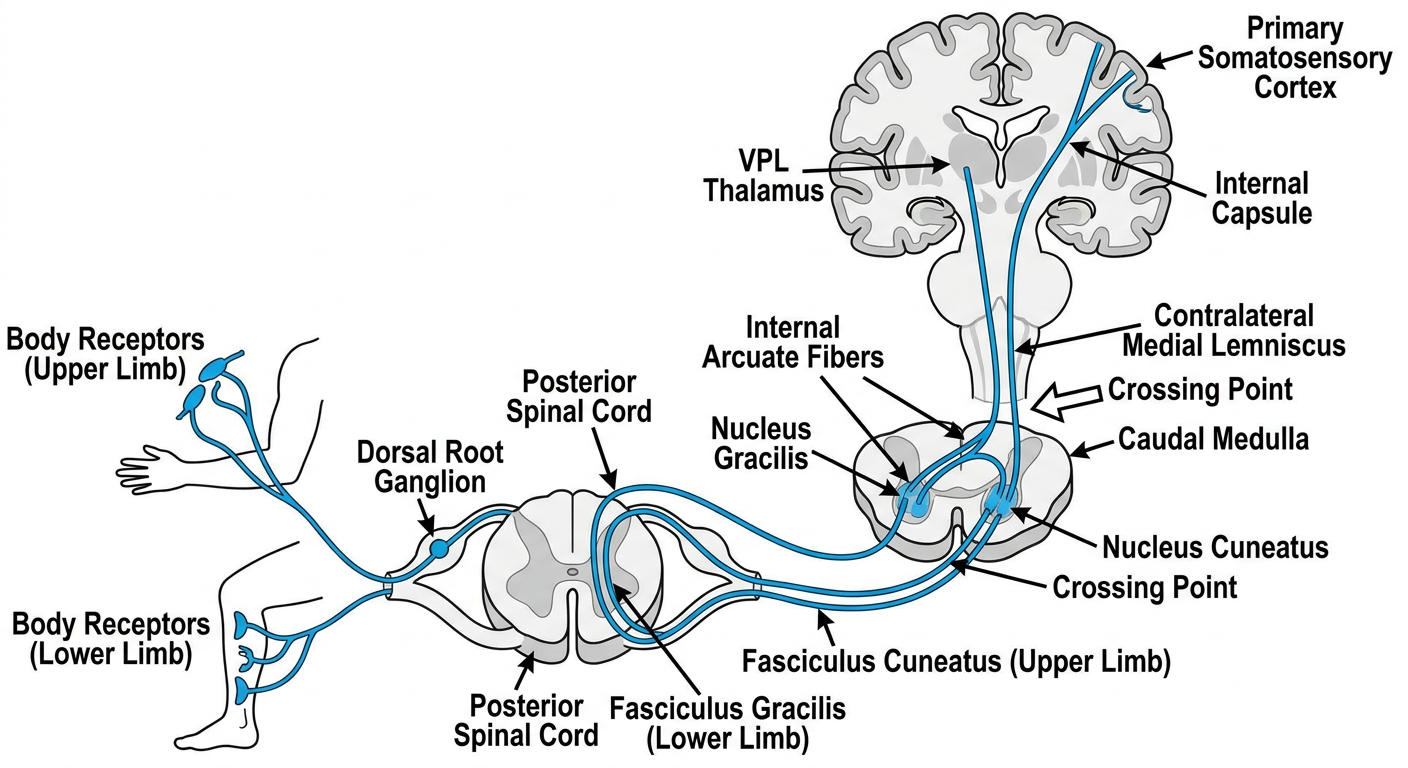
Carries
- Fine/discriminative touch
- Vibration
- Conscious proprioception or joint-position sense
- Two-point discrimination
- Stereognosis, identifying an object by touch
How to read the diagram
- A receptor in the skin, muscle, tendon, or joint detects the stimulus.
- The first-order neuron cell body lies in the dorsal root ganglion.
- It enters the posterior side of the spinal cord and ascends on the same side.
- From the leg and lower trunk, it ascends in fasciculus gracilis.
- From the upper trunk and arm, it ascends in fasciculus cuneatus.
- It synapses in the nucleus gracilis or nucleus cuneatus in the caudal medulla.
- The second-order neuron now crosses in the medulla as the internal arcuate fibers.
- After crossing, it ascends as the medial lemniscus.
- It synapses in the VPL nucleus of thalamus.
- The third-order neuron passes through the posterior limb of internal capsule to the primary somatosensory cortex.
Crossing
Spinal cord: does NOT cross
Caudal medulla: crosses
Above medulla: information is contralateral
Very important arrangement
Posterior column in spinal cord

Medial                         Lateral
Gracile tract                  Cuneate tract
Lower limb + lower trunk       Upper limb + upper trunk
Lesion
- Lesion of right posterior column in spinal cord:
- Right-sided loss of vibration, position sense, and fine touch below lesion.
- Lesion above the medullary crossing:
- Contralateral sensory loss.
2. Anterolateral system
Lateral and anterior spinothalamic tracts
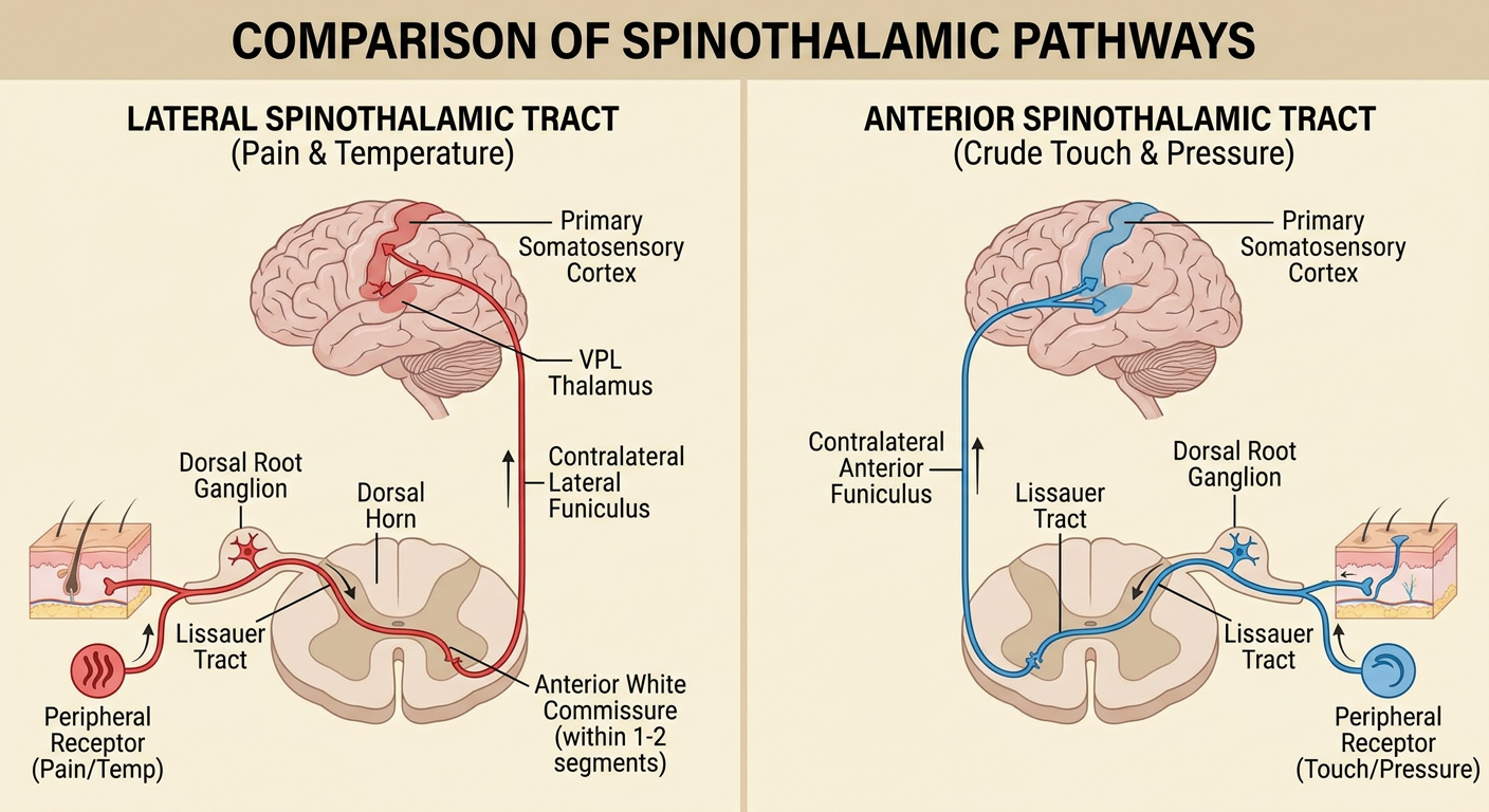
A. Lateral spinothalamic tract
Carries
- Pain
- Temperature
- Itch
- Some crude tactile sensation
Route
Pain/temperature receptor
        ↓
Dorsal root ganglion
        ↓
Enters dorsal horn of spinal cord
        ↓
May travel up/down 1-2 levels in Lissauer tract
        ↓
Synapses in dorsal horn
        ↓
Crosses in anterior white commissure
        ↓
Ascends on opposite side in lateral spinothalamic tract
        ↓
VPL thalamus
        ↓
Primary somatosensory cortex
Crossing
- Crosses in the spinal cord, through the anterior white commissure.
- Crossing usually occurs about 1-2 spinal levels above the point where the nerve entered.
Lesion
Left-sided loss of pain and temperature
Beginning 1-2 levels below the lesion
B. Anterior spinothalamic tract
Carries
- Crude touch
- Light touch
- Pressure
Route
Skin receptor
        ↓
Dorsal root ganglion
        ↓
Dorsal horn
        ↓
Cross in anterior white commissure
        ↓
Ascend contralaterally in anterior funiculus
        ↓
VPL thalamus
        ↓
Primary somatosensory cortex
Key difference
Lateral spinothalamic = pain + temperature
Anterior spinothalamic = crude touch + pressure
3. Spinocerebellar tracts
Posterior and anterior spinocerebellar pathways
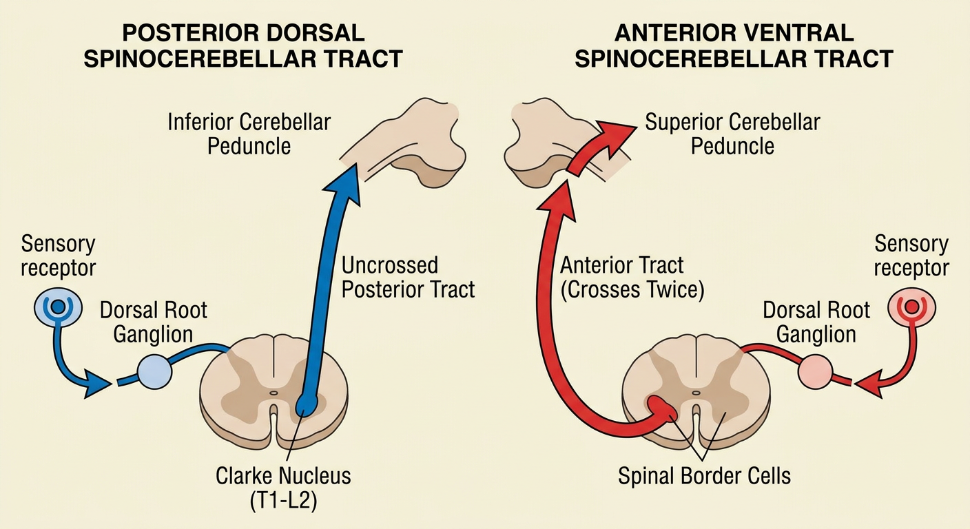
A. Posterior spinocerebellar tract
Carries
- Unconscious proprioception from lower limb and lower trunk
- Muscle spindle information
- Golgi tendon organ information
- Joint and tendon information needed for coordinated movement
Route
Muscle/joint/tendon receptor
        ↓
Dorsal root ganglion
        ↓
Clarke nucleus, mainly T1-L2
        ↓
Posterior spinocerebellar tract
        ↓
Ascends on SAME side of spinal cord
        ↓
Inferior cerebellar peduncle
        ↓
Ipsilateral cerebellar cortex
Crossing
Does not cross.
Result
Upper-limb equivalent
Upper limb receptor
        ↓
Dorsal root ganglion
        ↓
Fasciculus cuneatus
        ↓
Accessory cuneate nucleus in medulla
        ↓
Inferior cerebellar peduncle
        ↓
Ipsilateral cerebellum
B. Anterior spinocerebellar tract
Carries
- Mainly information about spinal interneuronal and motor activity
- “Feedback” about lower-limb movement
- Information important for gait and coordinated lower limb activity
Route and double crossing
Lower-limb sensory/interneuron activity
        ↓
Spinal border cells, lumbosacral spinal cord
        ↓
FIRST CROSSING:
Anterior white commissure
        ↓
Ascends on opposite side
        ↓
Superior cerebellar peduncle
        ↓
SECOND CROSSING:
In or near the cerebellum
        ↓
Ends in cerebellum on original side
Key memory rule
Posterior spinocerebellar = no crossing

Anterior spinocerebellar = crosses twice
4. Corticospinal pathways
Lateral and anterior corticospinal tracts
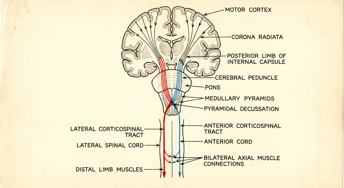
Common route before division
Primary motor cortex
        ↓
Corona radiata
        ↓
Posterior limb of internal capsule
        ↓
Cerebral peduncle, midbrain
        ↓
Pons
        ↓
Medullary pyramids
A. Lateral corticospinal tract
Function
- Voluntary movement of limbs
- Skilled movement of fingers and hand
- Fine, fractionated distal movement
- Main pathway for precise motor control
Route
Motor cortex
        ↓
Internal capsule
        ↓
Midbrain → pons → medullary pyramids
        ↓
Pyramidal decussation in caudal medulla
        ↓
Descends in lateral funiculus as LCST
        ↓
Ventral horn motor neurons
        ↓
Contralateral distal limb muscles
Crossing
- Crosses in the pyramidal decussation of the caudal medulla.
- About 85-90% of corticospinal fibers do this.
Clinical rule
Lesion above pyramidal decussation:
Contralateral upper motor neuron weakness

Lesion below pyramidal decussation, in spinal cord:
Ipsilateral upper motor neuron weakness
Upper motor neuron signs
- Spasticity
- Hyperreflexia
- Clonus
- Extensor plantar response, Babinski sign
- Weakness, especially of fine distal movement
B. Anterior corticospinal tract
Function
- Voluntary control of axial muscles
- Trunk muscles
- Proximal limb muscles
- Postural adjustments
Route
Motor cortex
        ↓
Brainstem pyramids
        ↓
Does not cross at pyramidal decussation initially
        ↓
Descends in anterior funiculus
        ↓
Many fibers cross at their spinal level of termination
        ↓
Bilateral connections to axial/proximal muscles
Crossing
- Initially uncrossed in the medulla.
- Many fibers cross near the level where they terminate in the spinal cord.
- Many terminate bilaterally.
Why unilateral cortical lesions spare the trunk relatively
5. Extrapyramidal descending pathways
Rubrospinal, vestibulospinal, and reticulospinal tracts
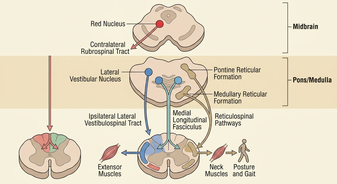
- Posture
- Balance
- Muscle tone
- Gait
- Head position
- Automatic postural reactions
- Proximal and axial movement
A. Rubrospinal tract
Origin
- Red nucleus of midbrain
Function
- Assists upper-limb flexor activity
- Contributes to distal limb control
- More important in nonhuman mammals than in humans
Route
Red nucleus, midbrain
        ↓
Crosses immediately in ventral tegmental decussation
        ↓
Descends contralaterally in lateral funiculus
        ↓
Mostly terminates at cervical spinal levels
        ↓
Influences upper-limb flexor motor neurons
Crossing
Crosses in the midbrain.
B. Lateral vestibulospinal tract
Origin
- Lateral vestibular nucleus, Deiters nucleus
Function
- Maintains balance
- Facilitates antigravity muscles
- Increases extensor tone in limb and trunk muscles
- Helps keep the body upright
Route
Vestibular apparatus
        ↓
Lateral vestibular nucleus
        ↓
Descends ipsilaterally through spinal cord
        ↓
Facilitates ipsilateral extensor muscles
Crossing
Does not cross.
Memory aid
Lateral vestibulospinal tract = lateral vestibular nucleus
                             = limb extensors
                             = balance and antigravity posture
C. Medial vestibulospinal tract
Origin
- Mainly medial vestibular nucleus
Function
- Head and neck stabilization
- Vestibulocollic reflexes
- Maintains head position during body movement
- Supports gaze stabilization with vestibulo-ocular reflex circuits
Route
Medial vestibular nucleus
        ↓
Medial longitudinal fasciculus, MLF
        ↓
Bilateral descent to cervical and upper thoracic cord
        ↓
Neck and upper trunk motor neurons
Crossing
- Has bilateral projections.
- Mainly acts at cervical and upper thoracic levels.
Memory aid
Medial vestibulospinal = MLF = neck and head control
D. Reticulospinal tracts
Origin
- Reticular formation of pons and medulla
Functions
- Posture
- Gait
- Muscle tone
- Automatic movement
- Anticipatory postural adjustments before voluntary movement
Pontine/medial reticulospinal tract
Pontine reticular formation
        ↓
Mainly ipsilateral descent
        ↓
Facilitates extensor tone
        ↓
Supports posture
Medullary/lateral reticulospinal tract
Medullary reticular formation
        ↓
Bilateral spinal influence
        ↓
Modulates or inhibits excessive extensor tone
        ↓
Contributes to gait and voluntary postural adjustment
6. Other ascending pathways
Spinotectal, spino-olivary, and spinoreticular tracts
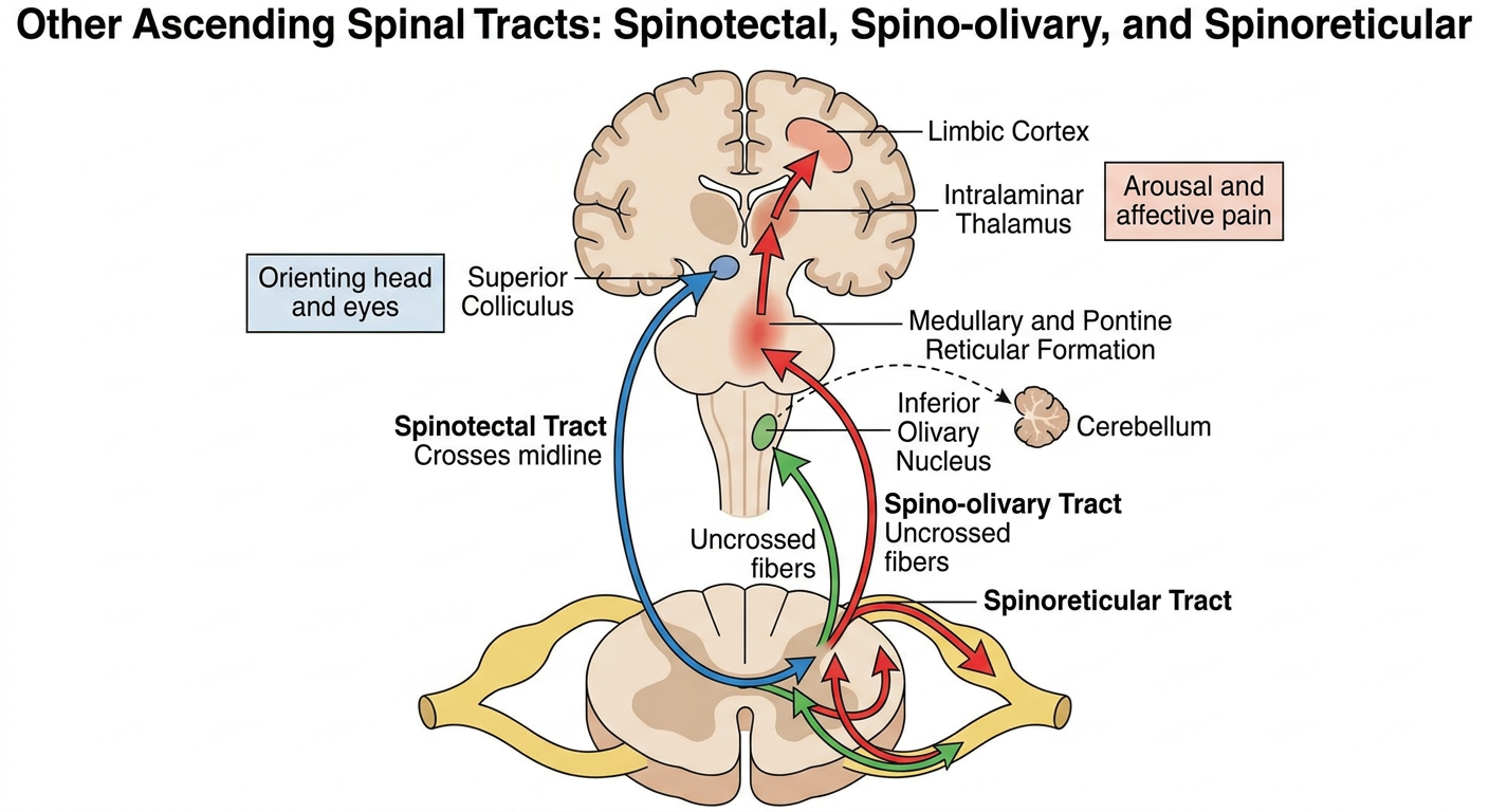
A. Spinotectal tract
Destination
- Superior colliculus in midbrain tectum
Main role
- Reflex orientation of eyes, head, and neck toward a sensory stimulus, especially a painful or unexpected one.
Route
Pain/temperature input
        ↓
Dorsal root ganglion
        ↓
Dorsal horn
        ↓
Crosses in spinal cord
        ↓
Ascends in anterolateral system
        ↓
Superior colliculus
        ↓
Orienting response of head and eyes
B. Spino-olivary tract
Destination
- Inferior olivary nucleus in medulla
- Then, indirectly, cerebellum
Main role
- Communicates sensory and proprioceptive information to cerebellar circuits.
- Believed to contribute to coordination, timing, motor learning, and error correction.
Simplified route
Peripheral sensory/proprioceptive information
        ↓
Spinal cord neurons
        ↓
Spino-olivary fibers
        ↓
Inferior olivary nucleus
        ↓
Olivocerebellar fibers
        ↓
Cerebellum
Important note
C. Spinoreticular tract
Destination
- Reticular formation in medulla and pons
- Intralaminar nuclei of thalamus
- Widespread cortical and limbic areas
Main role
- Arousal response to pain
- Emotional unpleasantness of pain
- Autonomic reaction to pain
- Diffuse, slow, persistent pain response
Route
Slow pain / visceral pain input
        ↓
Dorsal horn
        ↓
Bilateral ascent in anterolateral system
        ↓
Reticular formation
        ↓
Intralaminar thalamic nuclei
        ↓
Widespread cortex and limbic areas
Final rapid-revision chart
| Tract | What it carries/does | Crossing point | Final destination |
|---|---|---|---|
| Fasciculus gracilis | Fine touch, vibration, proprioception from lower body | Caudal medulla | Contralateral sensory cortex |
| Fasciculus cuneatus | Same sensations from upper body | Caudal medulla | Contralateral sensory cortex |
| Lateral spinothalamic | Pain and temperature | Spinal cord, anterior white commissure | Contralateral sensory cortex |
| Anterior spinothalamic | Crude touch and pressure | Spinal cord | Contralateral sensory cortex |
| Posterior spinocerebellar | Unconscious proprioception from lower body | No crossing | Ipsilateral cerebellum |
| Anterior spinocerebellar | Spinal motor/interneuron feedback | Crosses twice | Ipsilateral cerebellum |
| Spinotectal | Reflex orientation to sensory stimulus | Mainly spinal cord | Superior colliculus |
| Spino-olivary | Sensory input for cerebellar processing | Complex/bilateral | Inferior olive then cerebellum |
| Spinoreticular | Arousal and affective component of pain | Bilateral | Reticular formation, thalamus, limbic cortex |
| Lateral corticospinal | Fine voluntary distal limb movement | Pyramidal decussation | Contralateral limb motor neurons |
| Anterior corticospinal | Axial and proximal voluntary movement | Mostly segmental crossing | Bilateral axial motor neurons |
| Rubrospinal | Upper-limb flexor influence | Midbrain | Contralateral cervical cord |
| Lateral vestibulospinal | Balance and limb extensor tone | No crossing | Ipsilateral spinal cord |
| Medial vestibulospinal | Head/neck stabilization | Bilateral | Cervical and upper thoracic cord |
| Reticulospinal | Posture, gait, muscle tone | Mainly bilateral | Axial/proximal motor neurons |
Each diagram and easy to understand And easy to explain
spinal cord pathways neuroanatomy
NCBI Bookshelf neuroanatomy spinal cord ascending descending tracts decussation
A simple, clean vertical medical pathway diagram for fasciculus gracilis only. White background, very large readable labels, one thick green arrow. From lower limb receptor to dorsal root ganglion, then ipsilateral fasciculus gracilis in posterior spinal cord, nucleus gracilis in caudal medulla, crossing as internal arcuate fibers, medial lemniscus, VPL thalamus, primary somatosensory cortex. Put a prominent red label: CROSSES IN CAUDAL MEDULLA. Include a small spinal cord cross-section with gracilis highlighted medial. Minimal text, no dense details.
A simple, clean vertical medical pathway diagram for fasciculus cuneatus only. White background, very large readable labels, one thick green arrow. From upper limb receptor to dorsal root ganglion, then ipsilateral fasciculus cuneatus in posterior spinal cord, nucleus cuneatus in caudal medulla, crossing as internal arcuate fibers, medial lemniscus, VPL thalamus, primary somatosensory cortex. Put a prominent red label: CROSSES IN CAUDAL MEDULLA. Include a small spinal cord cross-section with cuneatus highlighted lateral to gracilis. Minimal text.
A simple clean vertical medical pathway diagram for the lateral spinothalamic tract only. Large readable labels, white background, thick red arrow. From pain and temperature receptor to dorsal root ganglion, dorsal horn, cross in anterior white commissure, then rise on the opposite side in lateral spinothalamic tract, VPL thalamus, primary somatosensory cortex. Show a small spinal cord cross-section and mark a large red X-shaped crossing. Prominent caption: CROSSES IN SPINAL CORD, 1 TO 2 SEGMENTS ABOVE ENTRY. Minimal text.
A simple clean vertical medical pathway diagram for anterior spinothalamic tract only. Large readable labels, white background, thick blue arrow. From crude touch and pressure receptor to dorsal root ganglion, dorsal horn, cross in anterior white commissure, then rise on opposite side in anterior spinothalamic tract, VPL thalamus, primary somatosensory cortex. Show spinal cord cross-section with anterior tract highlighted. Prominent caption: CROSSES IN SPINAL CORD. Minimal text.
A simple clean vertical medical pathway diagram for posterior dorsal spinocerebellar tract only. Large readable labels, white background, thick purple arrow. From lower limb muscle spindle and tendon receptor to dorsal root ganglion, Clarke nucleus at T1 to L2, ascends ipsilaterally in posterior spinocerebellar tract, inferior cerebellar peduncle, ipsilateral cerebellum. Include small spinal cord cross-section with tract highlighted. Prominent green label: DOES NOT CROSS. Minimal text.
A simple clean vertical medical pathway diagram for anterior ventral spinocerebellar tract only. Large readable labels, white background, thick orange arrow. From lower-limb spinal border cells to first crossing in anterior white commissure, rises on opposite side in anterior spinocerebellar tract, superior cerebellar peduncle, second crossing near cerebellum, then ends in cerebellum on original body side. Prominent labels FIRST CROSSING and SECOND CROSSING. Caption: CROSSES TWICE. Minimal text.
Error calling model 'gemini-3-pro-image' (RESOURCE_EXHAUSTED): 429 RESOURCE_EXHAUSTED. {'error': {'code': 429, 'message': 'Resource has been exhausted (e.g. check quota).', 'status': 'RESOURCE_EXHAUSTED'}}
We encountered an error while processing your request. Please try again or reach out to us for help.