I now have comprehensive material from multiple authoritative textbooks and recent PubMed evidence. Let me compile the full review:
DOWN SYNDROME (TRISOMY 21)
A Comprehensive Evidence-Based Review for MD/DNB Pediatrics Residents
Sources: Robbins & Cotran Pathologic Basis of Disease (10th ed.), Thompson & Thompson Genetics and Genomics in Medicine (9th ed.), Emery's Elements of Medical Genetics (15th ed.), Campbell's Operative Orthopaedics (15th ed., 2026), Adams & Victor's Principles of Neurology (12th ed.), Tietz Textbook of Laboratory Medicine (7th ed.), Creasy & Resnik's Maternal-Fetal Medicine, Langman's Medical Embryology, AAP Guidelines 2022, ACMG Standards, Recent PubMed Evidence (2023-2026)
SECTION 1: DEFINITION AND INTRODUCTION
Down syndrome (DS) is the most common autosomal chromosomal disorder in humans and the leading genetic cause of intellectual disability worldwide. It is defined by the presence of an extra copy (full or partial) of chromosome 21, resulting in gene dosage imbalance across hundreds of loci. The condition affects virtually every organ system and produces a recognizable constellation of dysmorphic features, intellectual disability, and multi-organ comorbidities.
High-Yield Pearl: Down syndrome = most common chromosomal disorder + most common genetic cause of intellectual disability. The three cytogenetic forms are free trisomy 21 (~95%), Robertsonian translocation (~4%), and mosaicism (~1%). - Robbins & Cotran Pathologic Basis of Disease, p. 163
SECTION 2: HISTORICAL BACKGROUND
| Year | Milestone |
|---|
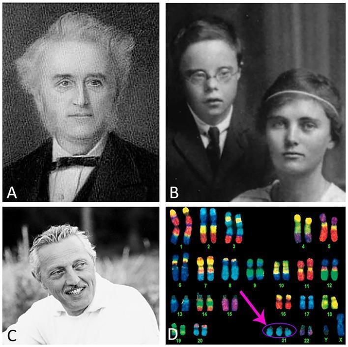
| 1866 | Dr. John Langdon Down, British physician, first described the clinical phenotype (called "Mongolism" - now considered offensive) |
| 1932 | Waardenburg first suggested a chromosomal cause |
| 1959 | Dr. Jérôme Lejeune (France) and colleagues identified trisomy 21 as the chromosomal basis - first chromosomal disorder identified in humans |
| 1961 | Term "Down syndrome" officially adopted replacing "Mongolism" |
| 1968 | Amniocentesis established for prenatal diagnosis |
| 1984 | Maternal serum AFP identified as second-trimester screening marker |
| 1997 | Cell-free fetal DNA (cffDNA) discovered in maternal plasma (Lo et al.) |
| 2000 | Long arm of chromosome 21 fully sequenced |
| 2011 | NIPT (Non-Invasive Prenatal Testing) introduced clinically |
| 2022 | AAP updates Health Supervision Guidelines for Down Syndrome |
SECTION 3: EPIDEMIOLOGY AND GLOBAL BURDEN
- Incidence: ~1 in 700 live births in the USA (most commonly cited figure)
- Prevalence: ~6 million individuals worldwide; ~400,000 in the USA
- Maternal age effect: 1 in 1,550 live births in mothers <20 years vs. 1 in 25 in mothers >45 years
- Survival: ~80% of trisomy 21 conceptuses are spontaneously aborted; only 20-25% survive to birth
- Improved longevity: Median age at death rose from 25 years (1983) to 47 years (current)
- India: Estimated prevalence ~1 in 800-1,000 live births; higher due to higher proportion of advanced maternal age pregnancies
Exam Pearl: Despite only 20% of pregnancies in the USA being in women >35 years, ~50% of Down syndrome births are in women >35. This is because the per-pregnancy risk rises steeply with age. However, the absolute number of DS births is still greater in younger mothers (larger reproductive population).
SECTION 4: GENETICS AND CYTOGENETICS
4.1 Chromosomal Types
| Type | Frequency | Mechanism | Maternal Age Effect | Recurrence Risk |
|---|
| Free Trisomy 21 (47,XX/XY,+21) | ~95% | Meiotic nondisjunction (usually meiosis I) | YES - strong | ~1% above age-related risk (empiric) |
| Robertsonian Translocation [e.g., 46,XY,der(14;21)] | ~4% | Translocation of chr 21q to chr 14, 13, 15, 21, or 22 | NO | Depends on carrier parent (see below) |
| Mosaic (47,+21/46 or similar) | ~1% | Mitotic nondisjunction post-fertilization | NO | Low (<1%) |
4.2 Free Trisomy 21 (Nondisjunction) - DETAILED MECHANISM
Meiotic nondisjunction is the failure of homologous chromosomes (meiosis I) or sister chromatids (meiosis II) to separate normally during cell division.
DIAGRAM (described): Meiotic Nondisjunction in Ovum

Normal Meiosis:           Nondisjunction (Meiosis I):
Oocyte (46)               Oocyte (46)
  |                         |
Meiosis I                 Meiosis I (failure to separate)
  |                         |
Primary oocyte + 1st       Both chr21 go to one cell
polar body (23 each)         |
  |                       Secondary oocyte (24 chr) + 1st PB (22 chr)
Meiosis II                  |
  |                       Meiosis II
Egg (23) + 2nd PB          |
                          Egg (24: n+1) + 2nd PB (22)
                          [includes TWO chr 21]
                            |
                          + Normal sperm (23, 1 chr21)
                            |
                          Zygote = 47 chromosomes (+21)
Key facts:
- In 95% of cases, the extra chromosome 21 is of maternal origin
- Maternal age effect suggests nondisjunction occurs in the ovum
- Unlike sperm (continuously produced), oocytes are arrested in prophase I from fetal life until just before ovulation - this prolonged arrest (decades in older women) predisposes to spindle defects and nondisjunction
- Recombination failure near the centromere of chromosome 21 is a predisposing factor
- No established paternal age effect
4.3 Robertsonian Translocation
- A Robertsonian translocation involves fusion of the long arms of two acrocentric chromosomes (13, 14, 15, 21, 22) at the centromere
- Most common: t(14;21) - chromosome 21 fuses to chromosome 14
- Carrier parent: karyotype 45,XX,rob(14;21) or 45,XY,rob(14;21) - phenotypically normal, carries 45 chromosomes (one being the fused chromosome)
- Child with DS: karyotype 46,der(14;21),+21 - effectively has 3 doses of chr 21 genetic material
Recurrence Risks for Translocation DS:
| Carrier Parent | Gamete Abnormality | Theoretical Risk | Empirical Risk of DS in Offspring |
|---|
| Mother is t(14;21) carrier | 1/3 gametes aneuploid | 33% theoretical | ~10-15% (empiric) |
| Father is t(14;21) carrier | Selection against aneuploid sperm | ~16% theoretical | ~2-5% (empiric) |
| Neither parent carrier | De novo translocation | Very low | ~1% above age-related |
| t(21;21) translocation carrier (either parent) | All gametes have +21 | 100% | ~100% of viable pregnancies = DS |
Critical Exam Point: If a child has DS due to translocation, BOTH parents MUST be karyotyped. If a parent carries t(21;21), ALL subsequent children will have DS. This is the only situation with 100% recurrence.
4.4 Mosaic Down Syndrome
- Results from mitotic nondisjunction after fertilization (in early embryogenesis)
- Child has two cell lines: 47,+21 and 46,normal
- Phenotype: Variable and usually milder depending on proportion of trisomic cells
- Some mosaics have near-normal intelligence and mild dysmorphic features
- Diagnosis: Standard karyotype may miss mosaicism if low-level; FISH or microarray on multiple tissues (blood + buccal cells) may be needed
- No maternal age effect; recurrence risk very low
4.5 Down Syndrome Critical Region (DSCR)
- 21q22.1-q22.3 is the "Down syndrome critical region" (DSCR)
- Key genes:
- DYRK1A (dual-specificity tyrosine-regulated kinase 1A) - overexpression implicated in cognitive deficits, craniofacial features, cardiac defects; a primary drug target
- APP (amyloid precursor protein) - three copies → excess amyloid → early Alzheimer disease
- SOD1 (superoxide dismutase 1) - premature aging phenotype
- ETS2 - cardiac defects
- COL6A1/COL6A2 - atrioventricular canal defects
- HMGN1 - leukemia predisposition
SECTION 5: ETIOLOGY AND RISK FACTORS
| Risk Factor | Details |
|---|
| Advanced maternal age | Strongest established risk factor (exponential increase after 35 years) |
| Prior child with trisomy 21 | Empiric recurrence risk ~1% (above baseline age-related risk) |
| Parental Robertsonian translocation carrier | High risk (see table above) |
| Young maternal age (<20 yr) | Low absolute risk but NOT zero (1 in 1,550) |
| Radiation exposure | Postulated, not consistently proven |
| Folate deficiency | Associated with nondisjunction in some studies (MTHFR variants) |
| Environmental factors | No well-established environmental teratogens identified |
SECTION 6: PATHOPHYSIOLOGY AND GENOTYPE-PHENOTYPE CORRELATION
Gene Dosage Effects (1.5x overexpression of chr 21 genes):
- Cognitive deficits: DYRK1A overexpression → abnormal neuronal proliferation, differentiation, synaptogenesis → intellectual disability
- Alzheimer disease: APP triplication → excess amyloid-beta → amyloid plaques + neurofibrillary tangles by age 40 in virtually all patients
- Leukemia: HMGN1 + GATA1 somatic mutations → transient myeloproliferative disorder (TMD) → acute megakaryoblastic leukemia (AML-M7)
- Cardiac defects: COL6A1/2, ETS2 overexpression → endocardial cushion defects
- Immune dysregulation: T-cell dysfunction → recurrent infections, autoimmune thyroiditis
- Premature aging: SOD1 overexpression → excessive ROS → oxidative stress
- Mitochondrial dysfunction: ~10% of overexpressed chr 21 genes regulate mitochondria → abnormal cristae, increased apoptosis
Pathophysiology Pearl: Trisomy 21 causes gene dosage imbalance at hundreds of loci, not just a single gene defect. This explains the multi-organ phenotype. The key principle is OVEREXPRESSION (1.5x normal dose) rather than loss-of-function.
SECTION 7: CHARACTERISTIC CLINICAL FEATURES
7.1 Neonatal Features (Major Diagnostic Clues)
MNEMONIC: "DOWN FEATURES"
- D = Duodenal atresia/"double bubble" sign
- O = Oblique (upslanting) palpebral fissures
- W = Wide sandal gap (1st-2nd toe)
- N = Neck loose skin / nuchal fold thickening
- F = Flat facial profile / flat nasal bridge
- E = Epicanthal folds
- A = Atlantoaxial instability (ligamentous laxity)
- T = Tongue protrusion (macroglossia/relative)
- U = Underactive thyroid (hypothyroidism)
- R = Round skull / brachycephaly
- E = Ear dysplasia (small, dysplastic pinnae)
- S = Single palmar crease (Simian crease), Short stature
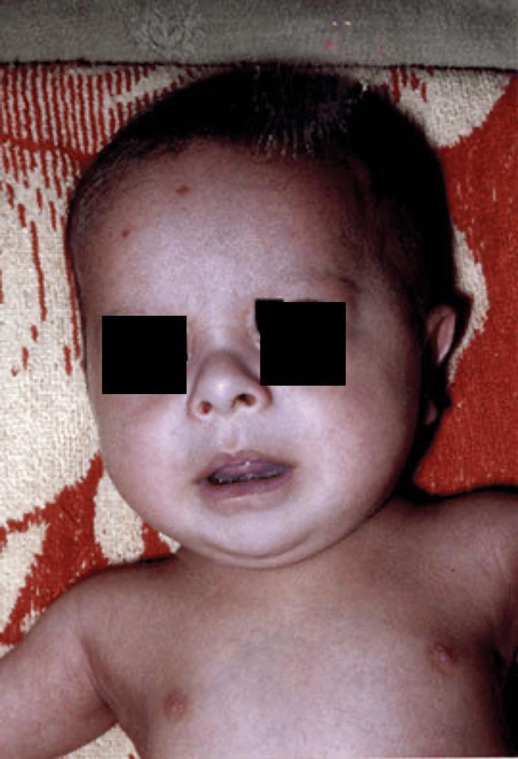
7.2 Craniofacial Features
| Feature | Description/Significance |
|---|
| Brachycephaly | Round, flat head; shortened AP diameter |
| Flat facial profile | Midface hypoplasia |
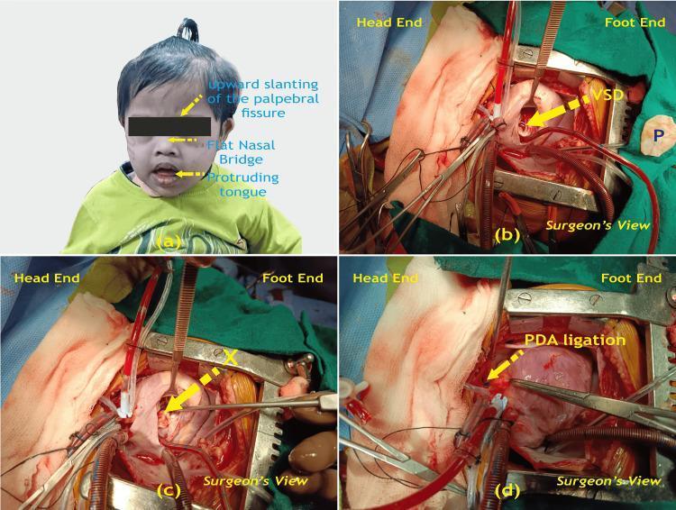
| Upslanting palpebral fissures | Lateral canthi higher than medial - CARDINAL feature |
| Epicanthal folds | Skin folds over medial canthi |
| Brushfield spots | White/gray speckles on the iris periphery (60-80% of DS; 10% of normal) |
| Flat nasal bridge | With small, upturned nose |
| Small, dysplastic ears | Often low-set with folded helices |
| Small oral cavity | Relative macroglossia (tongue appears large, protrudes) |
| Open mouth posture | Due to hypotonia + macroglossia |
| Short neck | With excess skin/nuchal fold |
| Flat occiput | Delayed/absent occipital protuberance |
7.3 Musculoskeletal Features
| Feature | Frequency | Clinical Significance |
|---|
| Hypotonia (generalized) | >99% | Neonatal feeding difficulty, delayed motor milestones |
| Ligamentous laxity | Common | Atlantoaxial instability, joint hypermobility, pes planus |
| Single transverse palmar crease (Simian line) | ~50% | Also in ~5% normals; bilateral more specific |
| Short, broad hands | Universal | "Paddle-like" appearance |
| Clinodactyly (5th finger) | ~60% | Curved inward; due to hypoplastic middle phalanx |
| Sandal gap (wide 1st-2nd toe space) | Common | Useful soft marker |
| Short stature | Universal | DS-specific growth charts essential |
| Atlantoaxial instability | 10-20% | Atlantodental interval (ADI) >5mm on lateral neck X-ray |
| Pes planus (flat feet) | Common | Requires orthotic support |
| Pectus excavatum/carinatum | Occasional | |
| Short, broad metacarpals | Universal | |
| Dysplastic pelvis | Classic radiologic finding | Flat acetabular angle, small iliac wings |
7.4 Dermatological Findings
| Finding | Description |
|---|
| Cutis marmorata | Mottled skin discoloration |
| Hyperkeratosis | Dry, rough skin in older children |
| Cheilitis | Angular cracking at mouth corners |
| Alopecia areata | Increased incidence |
| Vitiligo | Autoimmune skin depigmentation (increased) |
| Syringomas | Benign sweat gland tumors (forehead/eyelids) |
| Folliculitis | Recurrent |
| Transient neonatal pustular melanosis | Occasionally |
7.5 Neurological Findings
- Intellectual disability: IQ range 25-70; mean ~50 (moderate ID)
- Mosaic DS: IQ may be higher (60-80+)
- Approximately 80% of DS have IQ of 25-50 (moderate-severe ID)
- Hypotonia: Universal; central origin
- Seizures: 5-10% incidence; infantile spasms may occur
- Alzheimer disease: 100% neuropathological changes by age 40; clinical dementia by 50-55 years in ~50% (earlier onset than general population by 30-40 years)
- Behavioral/psychiatric: Autism spectrum disorder (ASD) in ~16-18%; depression, anxiety, ADHD
7.6 Growth Pattern
- Growth retardation begins prenatally
- Birth weight and length below normal range
- Growth velocity reduced throughout childhood
- Down syndrome-specific growth charts (Cronk 1988; updated 2015 by Zemel et al.) MUST be used - standard CDC charts are inappropriate
- Height at 18 years: Males ~155 cm; Females ~145 cm (both <3rd percentile on standard charts)
- Obesity common in older children/adults (>50%)
SECTION 8: ORGAN-SYSTEM MANIFESTATIONS
8.1 Cardiovascular (Most Life-Threatening Complication)
| Defect | Approximate Frequency in DS |
|---|
| Any congenital heart disease | ~40% of all DS |
| Atrioventricular septal defect (AVSD / endocardial cushion defect) | 45% of all DS CHD (most common in DS) |
| Ventricular septal defect (VSD) | 35% |
| Atrial septal defect (ASD) / patent foramen ovale | Common |
| Patent ductus arteriosus (PDA) | 10% |
| Tetralogy of Fallot (ToF) | 5% |
| Isolated VSD | ~30% |
Exam Pearl: AVSD (also called complete AV canal defect) is the MOST COMMON specific CHD in Down syndrome. In any child with AVSD, always suspect DS. If untreated, AVSD leads to Eisenmenger syndrome (pulmonary hypertension + right-to-left shunt reversal) more rapidly in DS than in chromosomally normal children.
- All neonates with DS require echocardiogram within first 4-6 weeks (even if no murmur, as significant defects may be "silent" early)
- Pulmonary hypertension develops faster in DS; early surgical repair strongly recommended
8.2 Respiratory
| Condition | Features |
|---|
| Obstructive sleep apnea (OSA) | 30-80% of DS children; due to midface hypoplasia, macroglossia, hypotonia, small nasopharynx, increased adenotonsillar tissue; requires overnight PSG |
| Recurrent respiratory infections | Due to immune deficiency, mucociliary dysfunction, anatomical features |
| Tracheomalacia/laryngomalacia | More common in DS |
| Subglottic stenosis | Anesthetic concern (narrow subglottis, use smaller ETT) |
| Pulmonary hypertension | See cardiac section |
8.3 Gastrointestinal
| Condition | Frequency | Notes |
|---|
| Duodenal atresia/"double bubble" sign | 2-5% (30% of all duodenal atresias = DS) | Presents as bilious vomiting in neonate |
| Hirschsprung disease (HSCR) | 2-3% | More common in DS than general population |
| Imperforate anus | Occasional | |
| Esophageal atresia | Occasional | |
| Celiac disease | 5-15% | Screen with anti-TTG IgA antibodies |
| Constipation | Very common | Rule out Hirschsprung, hypothyroidism |
| Gastroesophageal reflux | Common | |
| Feeding difficulties | Very common in neonates | Related to hypotonia, macroglossia |
Exam Pearl: "Double bubble" on antenatal ultrasound (dilated stomach + duodenum) should prompt consideration of DS. ~30% of all cases of duodenal atresia are associated with DS.
8.4 Endocrine
| Condition | Frequency | Notes |
|---|
| Hypothyroidism (congenital) | 1% (higher than general population) | Newborn screen; thyroid function annually |
| Acquired hypothyroidism (autoimmune) | 15-20% (by age 10 years) | Most common endocrine comorbidity |
| Hyperthyroidism | <1% | Graves disease possible |
| Type 1 Diabetes mellitus | Increased risk | Autoimmune mechanism |
| Hypogonadism (males) | Very common | Cryptorchidism; infertility in almost all males |
| Adrenal dysfunction | Rare | |
Exam Pearl: ALL children with DS must have annual thyroid function tests (TSH ± T4). Hypothyroidism is common, insidious, and worsens cognitive function if untreated.
8.5 Hematological
| Condition | Frequency/Notes |
|---|
| Transient Myeloproliferative Disorder (TMD/TAM) | 10% of DS neonates; leukemoid reaction (blasts, thrombocytopenia); 80% resolves spontaneously; 20% progresses to AML |
| Acute Megakaryoblastic Leukemia (AML-M7 / ML-DS) | 500-fold increased risk vs. general population |
| Acute Lymphoblastic Leukemia (B-ALL) | 20-fold increased risk |
| Polycythemia | Common in neonates |
| Thrombocytopenia | In neonates; may be part of TMD |
| Anemia | Iron deficiency common (macrocytosis also seen) |
Leukemia Pathway in DS:
DS Neonate → GATA1 somatic mutation (50% of DS neonates) →
TMD (10% clinically apparent) → [80% spontaneous resolution] →
                              → [20%] acquire additional GATA1 + other mutations →
                              → AML-M7 (ML-DS) within 1-4 years
Recent Evidence: Management of DS-Associated Leukemias (JAMA Oncol, 2023): DS-AML has BETTER prognosis than non-DS AML (70-80% cure rate with standard low-dose cytarabine protocols), but DS-ALL has WORSE outcomes than non-DS ALL. This paradox is a classic exam point.
8.6 Immunological
- Impaired T-cell function (reduced T-cell activation, abnormal thymic development)
- Reduced NK cell activity
- Impaired neutrophil chemotaxis
- Result: Recurrent serious bacterial infections (pneumonia, otitis media, sepsis)
- Increased autoimmune disease (thyroiditis, T1DM, celiac, juvenile idiopathic arthritis, alopecia areata)
- Vaccines are MORE important in DS due to immune compromise
8.7 Ophthalmological
| Condition | Frequency |
|---|
| Refractive errors (myopia, hyperopia, astigmatism) | 70-80% |
| Strabismus | 20-40% |
| Nystagmus | 10-20% |
| Congenital cataracts | 5-10% |
| Brushfield spots (iris speckling) | 60-80% |
| Blepharitis | Common |
| Nasolacrimal duct obstruction | Common in infants |
| Keratoconus | 5-8% (develop in 2nd-3rd decade) |
| Glaucoma | Rare |
Screening: Ophthalmology evaluation at 6 months, 1 year, and then annually.
8.8 ENT and Hearing
- Hearing loss: 75% by age 1 year (conductive > sensorineural); most common is conductive due to recurrent otitis media with effusion (OME)
- Narrow external auditory canals → increased OME, wax impaction
- Sensorineural hearing loss in ~20-30%
- Screening: Newborn hearing screen (AABR); audiology annually
- Enlarged tonsils and adenoids → OSA
- Recurrent sinusitis
- OSA: 30-80% prevalence; sleep study (polysomnography) at age 4 or earlier if symptomatic
8.9 Dental
- Delayed eruption of primary and permanent teeth
- Anomalous teeth (hypodontia, microdontia, malocclusion)
- Bruxism (teeth grinding): significantly more common (meta-analysis 2023 confirms high prevalence)
- Periodontal disease: aggressive form; despite better plaque control, more severe gum disease
- High rate of malocclusion, crowding
- Dental screening by 12 months; 6-monthly dental reviews
8.10 Renal
- Renal anomalies in 3-7%: hydronephrosis, duplex collecting system, renal agenesis, cystic kidney
- Renal ultrasound screening after birth if clinically indicated
- Urinary tract infections slightly increased
- Hypospadias and cryptorchidism in males (20-30%)
8.11 Orthopedic
| Condition | Notes |
|---|
| Atlantoaxial instability (AAI) | 10-20%; ADI >4.5mm pathological; symptoms: neck pain, torticollis, gait disturbance, hyperreflexia, bowel/bladder dysfunction |
| Pes planus | Universal; needs orthotic support |
| Hip subluxation/dislocation | Increased due to ligamentous laxity |
| Scoliosis | 5-8%; needs surveillance |
| Patellar instability | Increased |
| Slipped capital femoral epiphysis (SCFE) | Increased due to obesity and hypothyroidism |
AAP Current Guideline (2022): Routine screening cervical spine X-rays are NO LONGER recommended for all DS children. X-ray is indicated only for symptomatic patients OR before participation in high-impact sports/activities that stress the cervical spine. - Campbell's Operative Orthopaedics 15e, 2026
8.12 Neurological
- Intellectual disability: IQ 40-70 range most common
- Seizures: Infantile spasms, myoclonic epilepsy, tonic-clonic seizures (5-10%)
- Alzheimer disease: Neuropathological changes (amyloid plaques + neurofibrillary tangles) universally by age 40; clinical dementia by 50-55 years in 50-70%; accelerated by APP triplication
- Stroke: Small vessel cerebrovascular disease in adults
- Moyamoya disease: Rare but increased association
8.13 Psychiatric and Behavioral Disorders
| Condition | Approximate Frequency |
|---|
| Autism Spectrum Disorder (ASD) | 16-18% |
| Attention Deficit Hyperactivity Disorder (ADHD) | 25-35% |
| Anxiety disorders | 20-30% |
| Depression | 10-15% (more in adults) |
| Obsessive-compulsive behaviors | Common |
| Aggressive behavior | Occasional |
| Sleep disturbance | Very common (related to OSA) |
SECTION 9: DEVELOPMENTAL PROFILE
| Domain | Typical Milestones in DS | Comparison (Typical Development) |
|---|
| Gross motor: sits | 9-10 months | 6 months |
| Gross motor: walks | 18-24 months | 12 months |
| Language: first words | 12-18 months | 10-12 months |
| Language: two-word phrases | 3-4 years | 18-24 months |
| Social smile | 2-3 months | 6 weeks |
| Toilet training | 3-5 years | 2-3 years |
| Reading level (best case) | 2nd-3rd grade equivalent | Grade-level |
| Adaptive skills | Better than IQ suggests | - |
Key Pearl: Social skills and social awareness in DS children are DISPROPORTIONATELY STRONGER than their cognitive and language abilities. They are often described as socially engaging, affectionate, and empathetic. This strength can be leveraged in therapy.
SECTION 10: DIAGNOSIS
10.1 Clinical Diagnosis
NEONATAL DIAGNOSTIC CRITERIA (Jones' Criteria - High Sensitivity):
The clinical diagnosis is usually apparent at birth. Key features:
- Hypotonia + flat facial profile + oblique palpebral fissures → strongly suggests DS
- No single feature is pathognomonic; cluster of features is used
10.2 Differential Diagnosis
| Condition | Distinguishing Features |
|---|
| Hypothyroidism | Macroglossia, umbilical hernia, jaundice - but NO upslanting palpebral fissures, no Brushfield spots; normal karyotype; elevated TSH |
| Prader-Willi syndrome | Hypotonia, obesity, hypogonadism - almond-shaped eyes, no Brushfield spots; 15q11 deletion (paternal) |
| Noonan syndrome | Epicanthal folds, webbed neck - but cardiac = pulmonary stenosis not AVSD; Ras/MAPK pathway mutations |
| Zellweger syndrome | Hypotonia, seizures - but dysmorphic without classic DS features; peroxisomal disorder |
| Transient neonatal hypotonia | Resolves; no dysmorphic features |
| Normal neonatal variants | Individual features may overlap; karyotype confirms |
10.3 Karyotyping (Gold Standard)
- Peripheral blood lymphocyte culture + G-banding: Standard method
- Turnaround: 10-14 days
- Identifies: trisomy 21, translocation type, mosaic percentage
- Required in ALL cases of DS - clinical diagnosis alone insufficient for genetic counseling
- Resolution: ~5-10 Mb (detects large chromosomal changes)
10.4 FISH (Fluorescence In Situ Hybridization)
- Uses chr 21-specific probes → detects extra chr 21 signals
- Rapid (24-48 hours) - useful when urgent diagnosis needed
- Three red signals (chr 21) = trisomy 21
- Limitation: Does not identify translocation type; does not replace full karyotype
- Used for rapid prenatal diagnosis, NICU decision-making
10.5 Chromosomal Microarray (CMA)
- Detects copy number variants (CNVs) genome-wide
- Can detect trisomy 21 + identify additional microdeletion/duplication syndromes
- Indicated when: Clinical features not fully explained by trisomy 21 alone
- Cannot detect balanced Robertsonian translocations reliably (because total genetic material is present)
10.6 Summary of Genetic Tests
| Test | Detects | Time | First Choice? |
|---|
| G-banded karyotype | Full chromosomes: trisomy, translocation, mosaic | 10-14 days | YES (gold standard) |
| FISH (chr 21) | Extra chr 21 | 24-48h | Rapid diagnosis |
| CMA | CNVs, trisomy 21 | 3-7 days | If karyotype normal but DS suspected |
| QF-PCR | Rapid aneuploidy detection | 24-48h | Prenatal rapid |
| NGS/WES | Gene-level mutations | 2-6 weeks | Additional anomalies |
SECTION 11: PRENATAL SCREENING AND DIAGNOSIS
11.1 First Trimester Screening (11-13+6 weeks)
| Component | Method | Contribution to Risk Calculation |
|---|
| Nuchal Translucency (NT) | Ultrasound | Increased NT (≥3.5mm or ≥99th centile) strongly associated |
| Maternal Serum Free β-hCG | Biochemistry | ↑ in DS |
| Maternal Serum PAPP-A | Biochemistry | ↓ in DS |
| Combined test (NT + β-hCG + PAPP-A) | Combined | DR ~85%, FPR ~5% |
| Additional US markers | Nasal bone absence, tricuspid regurgitation, ductus venosus Pulsatility Index | Adds to detection rate |
Combined First Trimester Screen DR: ~85-90%, FPR: 5%
11.2 Second Trimester Screening (15-20 weeks)
"Quad Screen" (4 markers):
| Marker | Change in DS | Mnemonic |
|---|
| AFP | ↓ (0.75 MoM) | A=low |
| hCG (total) | ↑ (2.0 MoM) | H=high |
| Unconjugated Estriol (uE3) | ↓ (0.72 MoM) | E=low |
| Inhibin A | ↑ (1.8 MoM) | I=high |
MNEMONIC for Quad Screen in DS: "Ah Heck, Even Inhibin" (AFP↓, hCG↑, uE3↓, InhibinA↑)
Ultrasound "Soft Markers" (2nd trimester):
- Echogenic intracardiac focus (EIF)
- Short femur/humerus
- Mild renal pyelectasis
- Echogenic bowel
- Absent/hypoplastic nasal bone
- Increased nuchal fold thickness (≥6mm at 15-20 weeks)
- Atrioventricular canal defect (structural - high specificity)
- Duodenal atresia ("double bubble")
DR of Quad Screen alone: ~75-80%, FPR: 5%
11.3 Cell-Free Fetal DNA / NIPT (Non-Invasive Prenatal Testing)
- Analyzes cell-free fetal DNA (cffDNA) from maternal plasma - ~10% fetal fraction in 1st trimester
- Method: Massively parallel sequencing (shotgun) or targeted sequencing of chr 21 sequences
- Placenta-derived; highly fragmented (<200bp); cleared rapidly after delivery
- Performance:
- Detection rate for trisomy 21: >99%
- False positive rate: <0.1%
- Sensitivity/Specificity: Superior to all biochemical screening methods
| Feature | NIPT |
|---|
| Best screening test for DS | Yes |
| Diagnostic? | NO - a screening test only |
| Invasive? | No (maternal blood draw) |
| Can be done from | 10 weeks gestation |
| Fetal fraction required | ≥4% (results may be invalid if <4%) |
| Confirms translocation type? | No - karyotype still needed after positive NIPT |
| Recommended by ACMG | As primary screening for all pregnancies (2023 update) |
Critical Exam Point: A positive NIPT MUST be confirmed by invasive prenatal diagnosis (CVS or amniocentesis with karyotype/FISH) before any irreversible clinical decision. NIPT is a SCREENING test, not a DIAGNOSTIC test. - Tietz Textbook of Laboratory Medicine, 7th ed.
11.4 Invasive Prenatal Diagnosis
| Procedure | Timing | Technique | Risk of Fetal Loss | Confirms |
|---|
| Chorionic Villus Sampling (CVS) | 10-13 weeks | Transcervical or transabdominal; biopsy of chorionic villi | 0.5-1% | Karyotype, FISH, DNA |
| Amniocentesis | 15-20 weeks | Transabdominal; amniotic fluid aspiration | 0.1-0.3% | Karyotype, FISH, DNA |
| Cordocentesis (PUBS) | >18 weeks | Umbilical vein blood sampling | 1-2% | Karyotype (rapid, 48-72h) |
Indications for Invasive Testing: Positive NIPT, abnormal ultrasound markers, prior DS child, parental translocation carrier, maternal age alone (>35 years is a relative indication in India/IAP - offer NIPT first)
11.5 Interpretation of Screening Results
PRENATAL SCREENING ALGORITHM:
All Pregnant Women
        |
  First Trimester:
  Combined Test (NT + PAPP-A + β-hCG)
  OR
  NIPT (preferred per ACMG 2023)
        |
  ┌─────────────┬──────────────┐
  Low Risk       High Risk       Structural Anomaly on US
  (<1:250)       (>1:250)        (e.g., AVSD, duodenal atresia)
  Routine care   ↓               ↓
                 NIPT            Direct invasive testing
                 (if not done    (CVS or amniocentesis)
                 as primary)          |
                 ↓               Karyotype + FISH
             NIPT POSITIVE
                 ↓
         Confirm with CVS/amnio
                 ↓
         Karyotype result
         ↙              ↘
    Trisomy 21      Translocation DS
    (free)          ↓
    ↓              Parent karyotypes
    Genetic         ↓
    counseling      Recurrence risk calculation
    Multidisciplinary team referral
SECTION 12: INITIAL EVALUATION AFTER DIAGNOSIS
12.1 Neonatal Checklist (Birth to 1 Month)
| System | Investigation | Timing |
|---|
| Cardiac | Echocardiogram | Within 4-6 weeks (even without murmur) |
| Thyroid | TSH (neonatal screen) | Day 2-4 (standard newborn screen) |
| Hearing | AABR (automated ABR) newborn hearing screen | Before discharge |
| Ophthalmology | Fundoscopy/red reflex | At birth and 6 months |
| Hematology | CBC (rule out TMD, polycythemia) | At birth |
| GI | Clinical assessment; X-ray if vomiting | If symptoms (rule out duodenal atresia, EA) |
| Genetics | Karyotype | Within first few days |
| Development | Refer for early intervention | As soon as possible |
12.2 Associated Congenital Anomalies - Frequency Table
| Anomaly | Frequency in DS |
|---|
| Congenital heart disease (any) | 40-50% |
| Intellectual disability | ~100% |
| Hypotonia | ~100% |
| Short stature | ~100% |
| Hypothyroidism (any type) | 15-25% |
| Hearing loss | 75% |
| Refractive errors | 70-80% |
| Obstructive sleep apnea | 30-80% |
| Strabismus | 20-40% |
| Duodenal atresia | 2-5% |
| Hirschsprung disease | 2-3% |
| Atlantoaxial instability | 10-20% |
| Leukemia (lifetime risk) | 1-2% (20-500x increased) |
| Celiac disease | 5-15% |
| Cryptorchidism (males) | 20-30% |
| Seizures | 5-10% |
| Alzheimer disease (>40 yrs) | 100% neuropathological; 50-70% clinical |
SECTION 13: MEDICAL MANAGEMENT
13.1 Neonatal Care
- Confirm diagnosis (karyotype + parental counseling)
- ECHO within 4-6 weeks
- Newborn metabolic screen (includes thyroid)
- AABR hearing screen
- CBC (check for polycythemia, TMD)
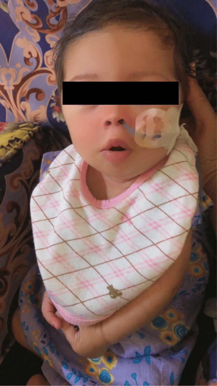
- Feeding support (often requires NGT, lactation consultant, occupational therapy)
- Inform parents compassionately; involve DS advocacy organizations early
13.2 Feeding Issues
- Due to: hypotonia, macroglossia, fatigue, CHD
- Breastfeeding encouraged but often requires support
- NGT feeds if significant feeding difficulty
- Occupational therapy and speech therapy early
- High-calorie formula if poor weight gain
- Monitor for GERD; celiac disease screen annually from age 3
13.3 Congenital Heart Disease
- Echocardiogram in ALL neonates (AVSD may be silent)
- Early surgical repair (within first 3-6 months for AVSD) is essential to prevent Eisenmenger syndrome
- Eisenmenger develops faster and at younger age in DS - this is a KEY DIFFERENCE from non-DS AVSD
- Cardiology follow-up lifelong even after repair
- Subacute bacterial endocarditis (SBE) prophylaxis as per AHA guidelines for unrepaired defects
13.4 Thyroid Disorders
- Annual TSH (and free T4 if TSH abnormal): from neonatal period, then at 6 months, then annually
- Treat hypothyroidism with levothyroxine (L-T4)
- Monitor carefully as subclinical hypothyroidism is common and debated re: treatment threshold
13.5 Sleep Apnea
- Polysomnography (PSG/sleep study) by age 4 (AAP recommends earlier if symptoms)
- Treatment: Adenotonsillectomy (T&A) is first-line (effective in 50-60% of DS, less complete than in non-DS)
- CPAP for residual or severe OSA
- Weight management to reduce OSA severity
- Nasopharyngeal airway for infants with severe obstruction
13.6 Hearing and Vision Care
- Hearing: ABR at birth, behavioral audiometry annually from 6 months; grommets (tympanostomy tubes) for recurrent OME; hearing aids if sensorineural
- Vision: Ophthalmology at 6 months, 1 year, then annually; glasses for refractive errors; treatment for strabismus (patching, surgery); monitoring for keratoconus
13.7 Hematologic Disorders
- TMD (Transient Myeloproliferative Disorder):
- Diagnose by CBC (excess blasts, thrombocytopenia), peripheral smear
- Most (80%) resolve spontaneously by 3-4 months
- 20% progress to AML; monitor with serial CBCs
- Low-dose cytarabine if symptomatic (hydrops, liver failure)
- ML-DS (DS-associated AML):
- Excellent response to chemotherapy (AML BFM protocols with cytarabine modifications)
- Overall survival 70-80% (better than non-DS AML)
- BUT increased chemotherapy toxicity (mucosal, cardiac) - dose adjustments needed
- DS-ALL:
- High-risk ALL; use intensive protocols
- Inferior outcomes to non-DS ALL (65-70% EFS)
- JAK2 mutations common in DS-ALL; emerging targeted therapies
Recent Guideline: Guideline for treating relapsed/refractory ML-DS (Pediatric Blood Cancer, 2024) - recommends second-line chemotherapy for relapsed ML-DS.
13.8 Atlantoaxial Instability (AAI)
- Clinical screening at every visit (neck pain, gait changes, hyperreflexia, bowel/bladder symptoms)
- Cervical X-rays (lateral, neutral/flexion/extension): Atlantodental interval (ADI) normal <4.5mm; pathological ≥5mm
- Current AAP guideline (2022): Routine X-ray screening NOT recommended; indicated only if symptomatic or before high-risk sport participation
- Surgical indications: Symptomatic instability, progressive neurological signs → posterior C1-C2 fusion
- Sports restriction: Avoid high-impact sports (gymnastics, diving, football, rugby) if symptomatic AAI confirmed
13.9 Obesity Prevention
- Obesity common (>50% of DS adults)
- DS-specific growth charts; regular BMI monitoring
- Low calorie density but micronutrient-dense diet
- Structured physical activity from early age
- Screen for hypothyroidism as cause of weight gain
- Treat OSA (untreated OSA promotes obesity)
13.10 Behavioral Management
- ABA (applied behavior analysis) for ASD + DS
- Positive behavior support strategies
- Sleep hygiene (address OSA first)
- Manage ADHD (methylphenidate effective but monitor cardiac effects, especially with CHD)
- Depression/anxiety: SSRIs + CBT for adults; behavioral strategies for children
SECTION 14: DEVELOPMENTAL INTERVENTION
14.1 Physiotherapy
- Starting age: 0-3 months (NICU or home)
- Goals: Strengthen trunk, improve head control, facilitate motor milestones
- Methods: NDT (neurodevelopmental therapy), hydrotherapy, sensory integration
- Specifically: Neck strengthening exercises for AAI prevention, gait training, orthotic prescription for pes planus
14.2 Occupational Therapy
- Starting age: 2-3 months
- Focus: Fine motor skills, feeding (oral motor therapy), sensory processing, self-care (ADLs)
- Equipment: Adaptive utensils, orthotics, seating aids
- School-based OT for handwriting, social participation
14.3 Speech and Language Therapy
- Starting age: 6-12 months (pre-linguistic stage)
- Focus: Augmentative and alternative communication (AAC) before speech develops, oral motor training, vocabulary, syntax
- Sign language (Makaton/baby signs) - highly effective in pre-verbal DS children
- DS children have better receptive than expressive language - leverage this
- Music therapy complements speech therapy
14.4 Early Stimulation and Educational Planning
- Early Intervention Programs: Start from birth through age 3 (Home-based in India under NRHM/RBSK/NPPCD)
- School: Inclusive education in mainstream schools with appropriate support is the goal (per RPWD Act 2016 in India; IDEA in USA)
- Individualized Education Plan (IEP)
- Reading programs (DS children can learn to read with appropriate methods - Dolch sight word approach)
- Vocational training in late adolescence
SECTION 15: IMMUNIZATION RECOMMENDATIONS
Routine Immunizations: DS children should receive ALL routine immunizations on the standard schedule (no live vaccine contraindications in DS unless on immunosuppressives).
Additional/Special Immunizations:
| Vaccine | Recommendation in DS |
|---|
| Influenza | Annual (from 6 months) - PRIORITY |
| Pneumococcal (PCV13 + PPSV23) | PCV13 per schedule; PPSV23 at 2 years + booster at 5 years |
| Hepatitis B | Standard schedule; check serology at 12 months (may need booster) |
| COVID-19 | Per current schedule; DS is high-risk for severe COVID |
| RSV prophylaxis (palivizumab/nirsevimab) | Consider if CHD + DS in first RSV season |
| Varicella | Standard 2-dose schedule |
| MMR | Standard; no contraindication in DS |
| Hepatitis A | Standard schedule |
SECTION 16: HEALTH SUPERVISION SCHEDULE (AAP 2022 - Age-Wise)
| Age | Key Screening/Assessment |
|---|
| Newborn | Karyotype, ECHO, AABR, CBC, thyroid (NBS), ophthalmology reflex, feeding assessment, genetic counseling |
| 1-6 months | Cardiology f/u, thyroid (6 months), hearing (behavior), ophthalmology (6 months), developmental assessment, OT/PT/SLT referral, feeding, celiac screen deferral |
| 6 months-2 years | Annual TSH, hearing annually, ophthalmology annually, developmental surveillance, dental from 1 year, CBC annually (year 1 for TMD monitoring) |
| 2-5 years | Annual TSH, annual hearing, annual ophthalmology, dental 6-monthly, IEP planning, PSG (sleep study by age 4), AAI clinical screen, celiac screen (TTG-IgA from age 3), thyroid antibodies |
| 5-12 years | Annual TSH, annual audiometry, annual ophthalmology, dental, annual BMI/obesity screen, AAI clinical screen, behavioral/psychiatric screen, educational review |
| 12-21 years | Annual TSH, annual audiometry, annual ophthalmology, gynecological care (for females - menstruation, contraception), diabetes screen, sleep study if OSA symptoms, cervical spine X-ray if symptomatic, behavioral health, transition to adult services |
| >21 years | Annual TSH, annual audiometry, annual ophthalmology, Alzheimer disease screening (from age 40), cardiology if CHD, dental, mental health |
SECTION 17: DOWN SYNDROME-SPECIFIC GROWTH CHARTS
- Standard charts: CDC/WHO growth charts are inappropriate for DS (ALL DS children will appear severely stunted)
- Cronk Charts (1988): First DS-specific growth charts
- Zemel et al. (2015): Updated DS-specific growth charts endorsed by AAP; available on AAP website
- Key features:
- Lower height/weight/head circumference centiles
- Obesity risk visible on DS charts (weight for height trending >85th centile on DS chart = overweight)
- BMI charts also available
- India: Use DS-specific charts (Zemel 2015 or India-validated charts if available)
SECTION 18: NUTRITIONAL MANAGEMENT
- Neonates: NGT feeds if unable to suckle; breastmilk preferred; high-calorie formula if poor weight gain
- Infants: Oral motor therapy for feeding difficulties; iron supplementation if anemic
- Children: Balanced diet; avoid excessive calories (obesity risk); adequate calcium and vitamin D (bone health)
- Celiac disease: Strict gluten-free diet if diagnosed (screen annually with TTG-IgA from age 3)
- Micronutrients: No strong evidence for megadose vitamin therapy; zinc supplementation may reduce infection risk (limited evidence); antioxidants not proven to improve cognition
- Constipation: High fiber diet, adequate hydration; rule out Hirschsprung and hypothyroidism
- Obesity management: Caloric restriction, structured exercise, behavioral strategies, treat OSA
SECTION 19: COMPLICATIONS
| Complication | Notes |
|---|
| Congenital heart disease (if untreated) | #1 cause of death in infancy (AVSD → pulmonary hypertension → Eisenmenger) |
| Recurrent infections (pneumonia, OM) | Immune deficiency + anatomical factors |
| Early onset Alzheimer disease | By age 40 (neuropathological); clinical dementia by 55 in 50-70% |
| Leukemia (AML, ALL) | 10-20x increased risk; TMD → AML pathway |
| Epilepsy | 5-10%; various types |
| Thyroid disease | Acquired hypothyroidism in 15-20% |
| Atlantoaxial instability | 10-20%; rare spinal cord compression |
| Obesity | >50% of adults; worsens OSA, Alzheimer, joint problems |
| Psychiatric comorbidities (ASD, depression) | Significant quality of life impact |
| Eisenmenger syndrome | If CHD untreated or late repair |
| Infertility | Almost universal in males; females may be fertile |
SECTION 20: PROGNOSIS AND LIFE EXPECTANCY
- Median life expectancy: ~60 years currently (major improvement from 25 years in 1983, 47 years in 2000s)
- Main causes of death:
- Infancy: Congenital heart disease, respiratory infection
- Adults: Alzheimer disease, respiratory infection, leukemia
- Functioning:
- Most DS adults can live semi-independently with support
- Supported employment, group homes, or family settings
- 10-20% can live independently with minimal support
- Quality of life is generally good; self-reported happiness is high in DS adults
- Fertility: Males almost universally infertile (azoospermia/oligospermia); females may menstruate and can conceive (risk of DS in offspring ~50% for free trisomy 21)
SECTION 21: GENETIC COUNSELING
Key Messages for Parents:
| Situation | Recurrence Risk | Counseling Points |
|---|
| Free trisomy 21, normal parents | ~1% above age-related risk | Sporadic; not heritable; low recurrence |
| Robertsonian translocation DS, mother carrier t(14;21) | ~10-15% | Mother must be tested; all pregnancies should have prenatal diagnosis |
| Father carrier t(14;21) | ~2-5% | Prenatal diagnosis offered |
| Either parent t(21;21) | ~100% (all viable pregnancies) | Donor gametes or adoption should be discussed |
| Mosaic DS | <1% | Very low recurrence |
| NIPT positive, not yet confirmed | Not applicable | Must confirm with CVS/amnio before counseling on decisions |
Counseling Principles:
- Non-directive, non-judgmental approach
- Provide balanced information (outcomes, support resources, DS adult capabilities)
- Multidisciplinary team (geneticist, pediatrician, counselor, DS advocacy)
- Address parents' emotional response first before medical information
- Connect families with Down syndrome parent support groups
- Discuss reproductive options: continuation of pregnancy, termination (legal/ethical aspects vary by country), preimplantation genetic diagnosis (PGD) for translocation carriers
SECTION 22: PREVENTION AND PRENATAL COUNSELING
- Pre-conceptional: Folic acid supplementation (though evidence for DS prevention is limited; good for NTDs)
- Universal NIPT: ACMG 2023 recommends offering NIPT as primary screening to ALL pregnant women regardless of age
- Age-based counseling: Women >35 years offered invasive testing; NIPT can reduce invasive testing rate by ~90%
- PGD (Preimplantation Genetic Diagnosis): For translocation carriers undergoing IVF - screens embryos before implantation
- No proven prevention of nondisjunction exists; avoidance of teratogens, radiation minimization, folic acid are general measures
SECTION 23: RECENT ADVANCES (2023-2026)
23.1 Gene-Silencing Research
- XIST-based chromosome silencing: Researchers (Jiang et al.) have demonstrated in induced pluripotent stem cells (iPSCs) that inserting an XIST transgene onto the extra chromosome 21 can silence its gene expression, similar to X-inactivation. This offers proof-of-concept for chromosome therapy.
- RNA interference / siRNA targeting DYRK1A: Multiple DYRK1A inhibitors in preclinical/early clinical testing
- Leucettinib-21: A DYRK1A kinase inhibitor entering Phase II clinical trials for cognitive improvement in DS + Alzheimer disease (PMID: 39422950)
23.2 Alzheimer Disease in DS - Recent Progress
- Lancet Neurology 2025 Review: DS + Alzheimer: New biomarkers (amyloid PET, tau PET, plasma p-tau217) allow early detection of amyloid accumulation before clinical dementia
- Anti-amyloid immunotherapy: Trials of lecanemab/donanemab in DS-Alzheimer populations ongoing (2025-2026)
- Calcineurin inhibition (Dohl et al., Alzheimers Dement 2025): Calcineurin pathway shown to be a therapeutic target; could prevent Alzheimer disease progression in DS (PMID: 40042516)
23.3 Targeted Therapies
- DYRK1A inhibitors: Several in clinical trials (leucettinib-21, INDY, GNF-4877) - targeting cognitive deficit
- Stem cell therapy: iPSC models of DS used to study cardiac, neuronal, and leukemia pathways; not yet in clinical use
- Growth hormone (GH) therapy: Limited evidence for height improvement; not standard of care
- Antioxidant therapy: Not proven to improve outcomes; not recommended routinely
23.4 Leukemia Research
- GATA1 mutations now used as a biomarker in neonates with DS to predict TMD → AML progression (Baruchel et al., Haematologica 2023)
- JAK inhibitors (ruxolitinib) showing promise in DS-ALL with JAK2 mutations
23.5 OSA and Sleep
- Sleep disorders in genetic syndromes with CHD - 2026 Systematic Review: Highlights complex interplay of DS + CHD + OSA; multi-level upper airway surgery may benefit those failing adenotonsillectomy
SECTION 24: HIGH-YIELD COMPARISON TABLE
Down Syndrome vs. Other Chromosomal/Genetic Syndromes
| Feature | Down Syndrome (Trisomy 21) | Edwards Syndrome (Trisomy 18) | Patau Syndrome (Trisomy 13) | Turner Syndrome (45,X) | Noonan Syndrome |
|---|
| Karyotype | 47,+21 | 47,+18 | 47,+13 | 45,X | Normal (PTPN11, SOS1, RAF1 etc. mutations) |
| Incidence | 1 in 700 | 1 in 5,000 | 1 in 10,000-20,000 | 1 in 2,000-2,500 female births | 1 in 1,000-2,500 |
| Maternal age effect | Yes | Yes | Yes | No (majority 45,X in spontaneous abortions) | No |
| Survival | Majority survive into adulthood | 90% die within 1 year | 90% die within 1 year | Normal life expectancy | Normal life expectancy |
| Intellectual disability | Moderate (IQ 40-70) | Severe | Severe | Usually normal | Usually mild/normal |
| Cardiac defect | AVSD (#1), VSD, ASD (40%) | VSD, ASD, PDA (90%) | VSD, PDA, ASD (80%) | Bicuspid aortic valve, CoA (50%) | Pulmonary stenosis, HCM (80%) |
| Facial features | Flat face, upslanting eyes, epicanthal folds, Brushfield spots | Micrognathia, low-set ears, prominent occiput | Holoprosencephaly, microcephaly, midline clefts (CLP) | Low posterior hairline, webbed neck, shield chest | Widely spaced eyes, low-set ears, webbed neck, ptosis |
| Hands | Single palmar crease, clinodactyly 5th finger | Overlapping fingers (2nd/5th over 3rd/4th), clenched fists | Postaxial polydactyly | Normal/small | Normal |
| Key feature | Flat face, AVSD, intellectual disability | Rocker bottom feet, clenched fists, micrognathia | Holoprosencephaly, CLP, polydactyly | Short stature, gonadal dysgenesis, webbed neck, CoA | Short stature, ptosis, pulmonary stenosis, HCM |
| Recurrence risk | ~1% | ~1% | ~1% | Low (<1%) | AD inheritance (50% if parent affected) |
| Dermatology | Cutis marmorata, simian crease | - | Scalp defects (aplasia cutis) | - | Lentigines (LEOPARD variant) |
| Height | Short stature | Short | Short | Short stature (#1 feature) | Short stature |
| Genetics/Etiology | Chromosome 21 trisomy | Chromosome 18 trisomy | Chromosome 13 trisomy | X monosomy | Ras/MAPK pathway AD mutations |
SECTION 25: MNEMONICS
1. DS Clinical Features: "BOWED 5S"
- Brushfield spots, Brachycephaly
- Oblique (upslanting) palpebral fissures
- Wide sandal gap
- Epicanthal folds
- Developmental delay (intellectual disability)
- Simian crease (single palmar crease)
- Short stature
- Small ears
- Sandle gap (wide 1st-2nd toe space)
- Subtle hypotonia
2. Associated Conditions: "CHILD HAS LEAKS"
- Cardiac defect (AVSD, VSD) - 40%
- Hypothyroidism
- Intestinal atresia (duodenal)
- Leukemia risk (AML, ALL)
- Deafness (hearing loss) - 75%
- Hirschsprung disease
- Atlanto-axial instability
- Sleep apnea (OSA)
- Ligamentous laxity
- Eye problems (strabismus, cataracts, keratoconus)
- Alzheimer disease (early onset)
- Kidney anomalies
- Seizures
3. Quad Screen in DS: "All High-energy Exercise Inhibits"
- AFP = LOW ↓
- HCG = HIGH ↑
- Estriol (unconjugated) = LOW ↓
- Inhibin A = HIGH ↑
4. Cardiac Defects in DS (in order of frequency): "AVSD Beats Anything"
- AV Septal Defect (#1 - 45% of CHD)
- VSD (#2 - 35%)
- ASD
- Bicuspid, other
- And PDA, ToF
SECTION 26: VIVA QUESTIONS WITH MODEL ANSWERS
Q1. What is the most common type of Down syndrome? What is its karyotype?
A: Free trisomy 21 (~95% of cases). Karyotype: 47,XX,+21 (female) or 47,XY,+21 (male). Results from meiotic nondisjunction (usually meiosis I, maternal origin in 95%).
Q2. A baby is born with DS. The parents want to know the chance of recurrence. What would you tell them?
A: First, karyotype the index child to determine the type of DS. If free trisomy 21: empiric recurrence risk is ~1% above age-related risk (i.e., approximately 1% for mothers under 35, higher for older mothers). If translocation: BOTH parents must be karyotyped urgently. If a parent carries t(14;21), risk is 10-15% (mother carrier) or 2-5% (father carrier). If t(21;21) in either parent: 100% risk - all viable pregnancies will have DS.
Q3. What is the most common cardiac defect in Down syndrome?
A: Atrioventricular septal defect (AVSD / endocardial cushion defect) - accounts for ~45% of CHD in DS. VSDs account for ~35%. Overall, CHD occurs in ~40% of DS children.
Q4. Why does Alzheimer disease occur early in Down syndrome?
A: Chromosome 21 carries the APP (amyloid precursor protein) gene. With three copies of chromosome 21, there is 1.5x overexpression of APP → excess amyloid-beta production → amyloid plaques form decades earlier than in the general population → neuropathological Alzheimer disease changes universally by age 40; clinical dementia in 50-70% by age 55.
Q5. What is Transient Myeloproliferative Disorder (TMD) in Down syndrome?
A: TMD (also called Transient Abnormal Myelopoiesis, TAM) occurs in ~10% of DS neonates. It is a clonal myeloproliferative disorder driven by GATA1 somatic mutations, presenting with leukocytosis, blasts on peripheral smear, and thrombocytopenia. ~80% resolve spontaneously within 3-4 months. ~20% progress to AML (acute megakaryoblastic leukemia/ML-DS) within 1-4 years.
Q6. What screening investigations are mandatory for a newly diagnosed DS neonate?
A: 1) Karyotype (confirm type, guide recurrence risk); 2) Echocardiogram (40% CHD, often silent); 3) Thyroid function (newborn screen TSH); 4) AABR hearing screen; 5) CBC (TMD, polycythemia); 6) Ophthalmology evaluation; 7) Genetics referral; 8) Early intervention referral.
Q7. When is routine cervical spine X-ray NOT recommended in DS?
A: The current AAP 2022 guideline does NOT recommend routine cervical spine X-ray screening in asymptomatic DS children. X-rays are indicated only in symptomatic patients (neck pain, neurological signs) or before participation in high-impact contact sports. This is because studies showed screening X-rays had poor predictive value for future instability.
Q8. What is the paradox regarding leukemia outcomes in DS?
A: DS-AML (myeloid leukemia associated with DS / ML-DS) has BETTER outcomes (70-80% cure rate) compared to non-DS AML, making it the most treatable form of childhood AML. HOWEVER, DS-ALL has WORSE outcomes compared to non-DS ALL, particularly because DS-ALL is enriched for Ph-like ALL and JAK2 mutations.
SECTION 27: IMPORTANT MCQs WITH EXPLANATIONS
MCQ 1: The most common chromosomal disorder is:
- A) Turner syndrome
- B) Klinefelter syndrome
- C) Down syndrome (Trisomy 21)
- D) Edwards syndrome
Explanation: DS (1 in 700 live births) is the most common chromosomal disorder and the most common genetic cause of intellectual disability.
MCQ 2: A baby with DS has a karyotype showing 46 chromosomes with a Robertsonian translocation. Which parental karyotype finding would result in a 100% recurrence risk?
- A) Neither parent is a carrier
- B) Mother is t(14;21) carrier
- C) Father is t(14;21) carrier
- D) Either parent is t(21;21) translocation carrier
Explanation: A parent with t(21;21) Robertsonian translocation can ONLY produce gametes with either no chr 21 (lethal/monosomy 21) or two chr 21 (always gives trisomy 21 in viable offspring). Therefore 100% of viable pregnancies will have DS.
MCQ 3: The most common cardiac defect in Down syndrome is:
- A) Atrioventricular septal defect (AVSD)
- B) Ventricular septal defect (VSD)
- C) Atrial septal defect (ASD)
- D) Patent ductus arteriosus (PDA)
Explanation: AVSD (endocardial cushion defect) occurs in ~45% of DS-CHD. It is also THE lesion most strongly associated with DS - ~50% of all AVSD cases have DS.
MCQ 4: Which gene triplication on chromosome 21 is MOST responsible for early Alzheimer disease in Down syndrome?
- A) SOD1
- B) DYRK1A
- C) APP (amyloid precursor protein)
- D) ETS2
Explanation: APP triplication → excess amyloid-beta → amyloid plaques → Alzheimer disease by age 40.
MCQ 5: The BEST and most specific prenatal screening test for Down syndrome is:
- A) Maternal serum AFP
- B) Quad screen
- C) Nuchal translucency alone
- D) Cell-free fetal DNA (NIPT)
Explanation: NIPT has DR >99% and FPR <0.1%, significantly superior to all biochemical and ultrasound screening methods. It is now recommended as primary screening by ACMG 2023.
MCQ 6: A 2-day-old DS neonate is found to have peripheral blood showing 15% blasts, thrombocytopenia (platelets 40,000), and hepatomegaly. The MOST likely diagnosis is:
- A) Acute myeloid leukemia
- B) Transient Myeloproliferative Disorder (TMD)
- C) Acute lymphoblastic leukemia
- D) Congenital leukemia
Explanation: TMD occurs in 10% of DS neonates, driven by GATA1 mutations, mimics AML but ~80% resolves spontaneously within 3-4 months. Management is watchful waiting (with low-dose cytarabine for symptomatic cases).
SECTION 28: CLINICAL CASE DISCUSSION
Case: Approach to Diagnosis and Management
Scenario: A 6-hour-old neonate is born to a 38-year-old G2P1L1 mother at 38 weeks by LSCS for fetal distress. The baby is hypotonic with Apgar scores 6/8. Examination reveals flat facial profile, upslanting palpebral fissures, bilateral epicanthal folds, small low-set ears, single transverse palmar crease bilaterally, wide sandal gap, and significant generalized hypotonia. No audible murmur. Birth weight 2.5 kg.
Step-by-Step Approach:
Step 1 - Clinical Assessment:
- Recognize DS phenotype (flat face + oblique palpebral fissures + epicanthal folds + single palmar crease + hypotonia = strong clinical DS)
- Note: No murmur does NOT rule out CHD in DS
Step 2 - Immediate Investigations:
- Karyotype (URGENT) - peripheral blood for G-banding
- FISH for rapid result if needed
- CBC: rule out TMD, polycythemia
- Blood glucose, calcium
- TSH (or ensure newborn screen done)
- ECHO within first week/month
Step 3 - Communicate with Parents:
- Sensitive, compassionate disclosure (both parents together, private room)
- Balanced information: acknowledge concerns AND strengths/potential
- Avoid prognosis statements until karyotype confirmed
- Provide written information and DS advocacy contacts
Step 4 - Referrals:
- Cardiology: Echo within 4-6 weeks
- Audiology: AABR before discharge
- Ophthalmology: 6 months
- Genetics: Counseling on recurrence risk, parental karyotypes
- Early Intervention: OT/PT/SLT from discharge
Step 5 - Karyotype Result Returns as 47,XY,+21 (Free Trisomy 21):
- Parental karyotypes: Normal (as expected for free trisomy)
- Recurrence risk counseling: ~1% above age-related risk
Step 6 - Echo Shows AVSD:
- Refer to pediatric cardiac surgery
- Plan repair at 4-6 months (before pulmonary hypertension develops)
- SBE prophylaxis education
Step 7 - Ongoing Health Supervision:
- Annual TSH, hearing, ophthalmology
- DS-specific growth charts
- Early intervention programs (state/national schemes)
- IEP planning at school entry
- Sleep study by age 4
SECTION 29: HIGH-YIELD COMPARISON OF PRENATAL TESTS
| Method | When | DR for DS | FPR | Diagnostic? | Risk |
|---|
| Maternal age alone | Any | 30% | 5% | No | None |
| Nuchal translucency alone | 11-13 wks | 70% | 5% | No | None |
| First trimester combined | 11-13 wks | 85-90% | 5% | No | None |
| Quad screen | 15-20 wks | 75-80% | 5% | No | None |
| NIPT (cffDNA) | ≥10 wks | >99% | <0.1% | NO (screening) | None |
| CVS | 10-13 wks | ~100% | None | YES | 0.5-1% fetal loss |
| Amniocentesis | 15-20 wks | ~100% | None | YES | 0.1-0.3% fetal loss |
SECTION 30: KEY CLINICAL PRACTICE GUIDELINES
| Guideline | Organization | Year | Key Points |
|---|
| Health Supervision for Children with Down Syndrome | American Academy of Pediatrics (AAP) | 2022 (current) | Comprehensive birth-to-adult screening schedule; eliminates routine AAI X-rays; emphasizes OSA screening PSG by age 4 |
| NIPT as Primary Screening | ACMG | 2023 | Recommends offering NIPT as primary screening to ALL pregnant women |
| Down Syndrome Medical Interest Group (DSMIG) | UK | 2023 | UK health guidelines for DS |
| Myeloid Leukemia in DS | International BFM consortium | 2024 | Revised protocols for ML-DS treatment; guideline for relapsed/refractory ML-DS |
| DS Alzheimer Disease | NIA/IADDRC | 2024-2025 | Biomarker-based diagnosis; emerging anti-amyloid trials |
| IAP (India) | IAP Genetics / Developmental Pediatrics | 2020-2023 | Adapted health supervision; RBSK for early identification; NIPT recommendations |
SECTION 31: CLINICAL PHOTOGRAPHS (Image References)
The following clinical images are available from the medical literature and illustrate key diagnostic features:
Composite figure: John Langdon Down (first descriptor), clinical facial features, Dr. Jérôme Lejeune (identified chromosomal basis), and spectral karyotype with arrow showing three copies of chromosome 21 (Trisomy 21).
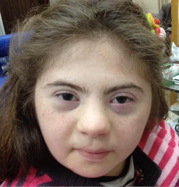
15-year-old female: flat nasal bridge, upslanting palpebral fissures, bilateral epicanthal folds, brachycephaly, midface hypoplasia - classic DS facies in an older child.
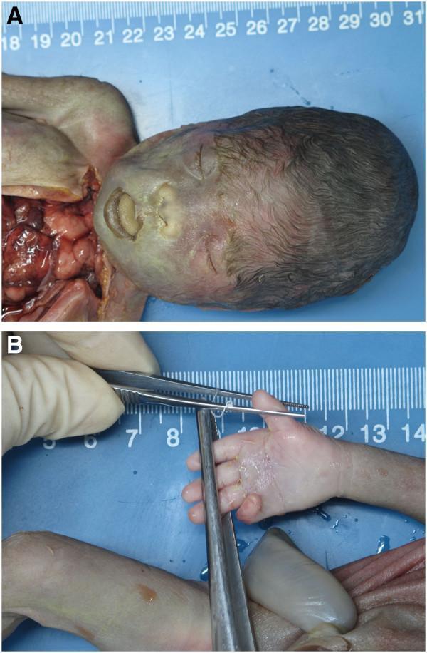
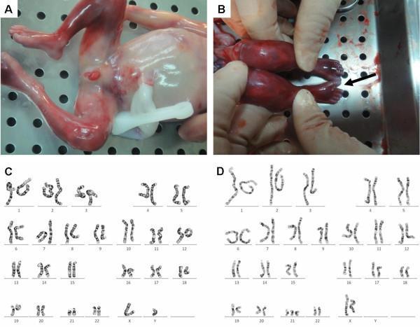
Fetal/neonatal DS: Panel A - flat profile, telecanthus, depressed nasal bridge, macroglossia/protruding tongue; Panel B - single transverse palmar crease (simian crease) on the right hand.
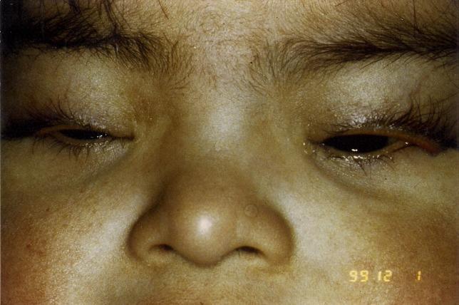
4-month-old DS infant: flat nasal bridge, depressed nasal root, upslanting palpebral fissures, protruding tongue (relative macroglossia), flat occiput, small low-set ears.
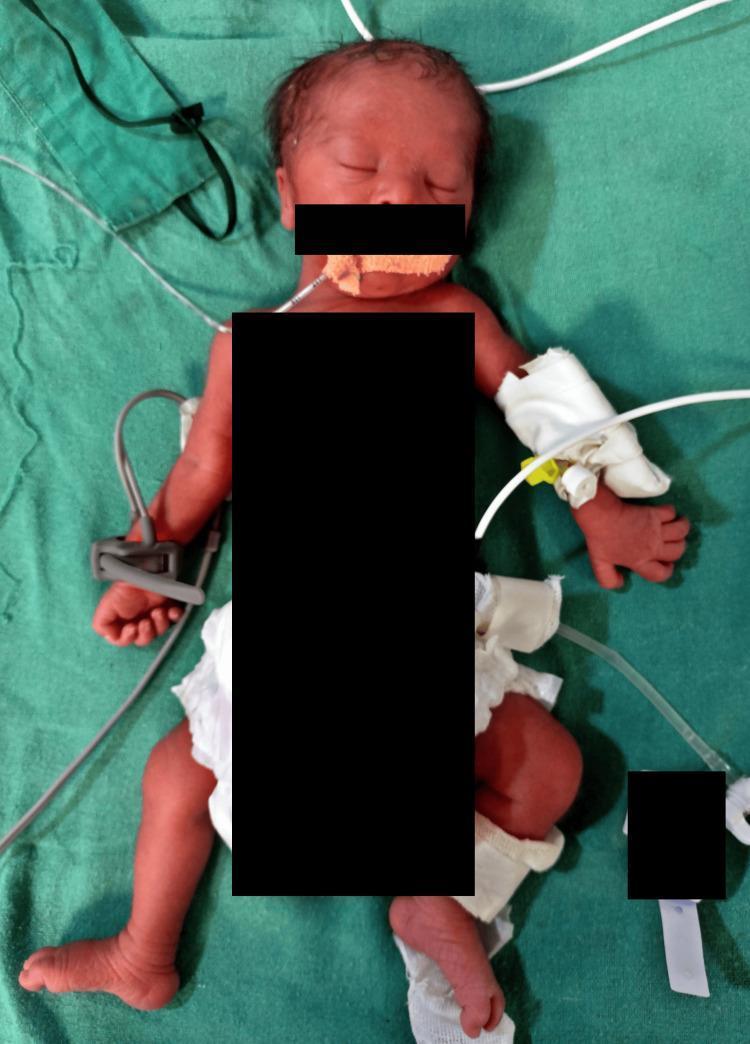
DS infant requiring nasogastric (NG) tube for nutritional support due to feeding difficulties - illustrating the common neonatal management challenge of hypotonia-related feeding problems.
RAPID REVISION SHEET (1-PAGE SUMMARY)
MUST-KNOW FACTS FOR PG EXAMINATION
| Topic | Key Fact |
|---|
| Most common chromosomal disorder | Down syndrome |
| Incidence | 1 in 700 live births |
| Most common type | Free trisomy 21 (~95%) |
| Mechanism | Maternal meiotic nondisjunction (95% maternal origin) |
| Maternal age effect | 1:1550 (<20yr) → 1:25 (>45yr); NO effect in translocation/mosaic |
| Gold standard diagnosis | G-banded karyotype |
| Best prenatal screening test | NIPT (DR >99%, FPR <0.1%) |
| Gold standard prenatal diagnosis | CVS (10-13 wks) or amniocentesis (15-20 wks) |
| Most common CHD | AVSD (endocardial cushion defect) - ~45% of DS-CHD |
| Leukemia paradox | DS-AML: BETTER outcomes; DS-ALL: WORSE outcomes |
| Quad screen pattern | AFP↓, hCG↑, uE3↓, Inhibin A↑ |
| Alzheimer disease | 100% neuropathological by age 40; gene = APP triplication |
| Atlantoaxial instability | 10-20%; routine X-ray NOT recommended (AAP 2022) |
| DYRK1A | Key drug target; DYRK1A inhibitors in clinical trials |
| TMD | 10% DS neonates; GATA1 mutation; 80% resolve spontaneously |
| Life expectancy | ~60 years (up from 25 years in 1983) |
| DS growth charts | Zemel 2015 (AAP-endorsed) - standard charts inapplicable |
| Thyroid screening | Annual TSH from birth (hypothyroidism 15-20% by age 10) |
| OSA prevalence | 30-80%; PSG by age 4 mandatory |
| Recurrence risk (free trisomy) | ~1% above age-related |
| t(21;21) carrier → recurrence | ~100% |
| Key gene for Alzheimer in DS | APP (amyloid precursor protein) on chr 21 |
| Key gene for leukemia in DS | GATA1 (somatic mutation) → HMGN1 |
| Key gene for cognition in DS | DYRK1A |
FREQUENTLY MISSED EXAM POINTS
- NIPT is a SCREENING test, NOT diagnostic - must confirm positive result with CVS/amniocentesis
- Routine cervical spine X-rays NO LONGER recommended (AAP 2022) - only for symptomatic patients
- DS-AML has BETTER prognosis than non-DS AML; DS-ALL has WORSE prognosis than non-DS ALL
- AVSD in a newborn → always suspect Down syndrome (50% of AVSD cases are DS)
- No maternal age effect in Robertsonian translocation DS or Mosaic DS
- t(21;21) carrier → 100% recurrence (not 21;21 is the crucial case - must karyotype parents)
- DS females can be fertile (unlike males who are almost universally infertile); if pregnant, ~50% chance of DS offspring
- Quad screen: AFP and uE3 both LOW (not just AFP); inhibin A HIGH (unlike in trisomy 18 where inhibin A is normal/low)
- First trimester biochemistry: PAPP-A LOW (not high); free β-hCG HIGH
- Brushfield spots are seen in 10% of NORMAL individuals too - not pathognomonic
- Celiac disease (5-15% of DS) is often asymptomatic - screen annually with TTG-IgA from age 3
- TMD vs. AML in DS neonate: TMD appears in NEONATES, self-limited; AML appears later (1-4 years); GATA1 mutation connects them
- Eisenmenger syndrome develops FASTER in DS children with AVSD compared to non-DS children - hence early surgical repair is urgent
- Subglottic stenosis in DS → use SMALLER endotracheal tube for anaesthesia
- DS-specific growth charts are ESSENTIAL - standard WHO/CDC charts make ALL DS children appear severely stunted
RECENT GUIDELINE UPDATES (2024-2026)
| Update | Source | Year |
|---|
| NIPT as primary screening for ALL pregnancies (regardless of age) | ACMG | 2023 |
| AAP Health Supervision Guidelines (birth to adulthood) | AAP | 2022 (most recent) |
| Guideline for relapsed/refractory myeloid leukemia in DS | Pediatric Blood Cancer (Int'l BFM) | 2024 |
| Leucettinib-21 (DYRK1A inhibitor) Phase II trials | J Alzheimers Dis | 2024 |
| DS + Alzheimer - biomarkers (plasma p-tau217, amyloid PET) | Lancet Neurology | 2025 |
| Sleep disorders in DS with congenital heart disease - systematic review | Sleep Medicine | 2026 |
| American Academy of Family Physicians DS management complementary guidance | AAFP | 2025 |
| Anti-amyloid therapy trials in DS-Alzheimer (lecanemab, donanemab) | Multiple ongoing | 2024-2026 |
HIGH-YIELD IMAGE-BASED DIAGNOSIS POINTS
| Image Finding | Diagnosis/Significance |
|---|
| "Double bubble" sign on prenatal US | Duodenal atresia → ~30% association with DS |
| Increased nuchal translucency (≥3.5mm) at 11-13 weeks | First trimester marker for DS (and other chromosomal abnormalities) |
| Absent nasal bone on fetal ultrasound | Soft marker for DS (specificity ~80%) |
| AVSD on echocardiogram | 50% cases = DS; confirm with karyotype |
| Three red signals on FISH (chr 21 probe) | Trisomy 21 confirmed |
| Karyotype showing 47 chromosomes with extra small chromosome in pair 21 | Free trisomy 21 (47,XX/XY,+21) |
| Flat acetabular angle + small iliac wings on pelvic X-ray | Classic DS pelvic dysplasia (Caffey sign) |
| ADI >5mm on lateral cervical spine X-ray | Atlantoaxial instability in DS |
| White speckling on iris periphery | Brushfield spots (60-80% in DS) |
| Single transverse palmar crease | Simian crease (~50% of DS) |
REFERENCES
- Robbins & Cotran Pathologic Basis of Disease, 10th Edition. Kumar, Abbas, Aster. Elsevier, 2021. Chapter 5 (Genetic Disorders), pp. 163-166.
- Robbins & Kumar Basic Pathology, 11th Edition. Chapter 4 (Genetic and Pediatric Diseases). Elsevier, 2023.
- Thompson & Thompson Genetics and Genomics in Medicine, 9th Edition. Strachan & Read. Elsevier, 2023. Chapter 6 (Down Syndrome), pp. 102-106.
- Emery's Elements of Medical Genetics and Genomics, 15th Edition. Turnpenny & Ellard. Elsevier, 2022. Chapter on Chromosomal Disorders.
- Adams & Victor's Principles of Neurology, 12th Edition. Ropper et al. McGraw-Hill, 2023. Chapter 38 (Developmental Diseases of the Nervous System).
- Tietz Textbook of Laboratory Medicine, 7th Edition. Rifai et al. Elsevier, 2023. Chapter on Prenatal Screening.
- Creasy & Resnik's Maternal-Fetal Medicine, 9th Edition. Creasy et al. Elsevier, 2022. Chapter on Aneuploidy Screening.
- Campbell's Operative Orthopaedics, 15th Edition 2026. Azar, Beaty, Canale. Elsevier. Chapter 39 (Cervical Instability in Down Syndrome).
- Bull MJ; AAP Council on Genetics. Health Supervision for Children and Adolescents with Down Syndrome. Pediatrics. 2022;149(5):e2022057010.
- Baruchel A, et al. Down syndrome and leukemia: from basic mechanisms to clinical advances. Haematologica. 2023;108(10). [PMID: 37439336]
- Verma A, et al. Management of Down Syndrome–Associated Leukemias: A Review. JAMA Oncology. 2023;9(9). [PMID: 37440251]
- Miladinovic M, et al. Guideline for treating relapsed or refractory myeloid leukemia in children with Down syndrome. Pediatric Blood & Cancer. 2024;71(9). [PMID: 38965693]
- Rafii MS, et al. Down syndrome and Alzheimer's disease: insights into biomarkers, clinical symptoms, and pathology. Lancet Neurology. 2025. [PMID: 40818475]
- Meijer L, et al. Leucettinib-21, a DYRK1A Kinase Inhibitor as Clinical Drug Candidate for Alzheimer's Disease and Down Syndrome. J Alzheimers Dis. 2024. [PMID: 39422950]
- Dohl J, et al. Calcineurin inhibition may prevent Alzheimer disease in people with Down syndrome. Alzheimers Dement. 2025. [PMID: 40042516]
- Prasad S, et al. Sleep disorders in genetic syndromes associated with congenital heart disease: A comprehensive review. Sleep Med. 2026. [PMID: 41955722]
- ACMG Statement on NIPT as Primary Screening. American College of Medical Genetics and Genomics, 2023.
- IAP Guidelines on Developmental Disabilities and Down Syndrome. Indian Academy of Pediatrics, 2020-2023.
- Zemel BS, et al. Updated Down Syndrome Growth Charts, 2015. (Endorsed by AAP)
---The comprehensive review has been compiled above. Here is a brief orientation to the document:
How to Use This Review
The document above is a complete, structured, exam-oriented review of Down Syndrome (Trisomy 21) for MD/DNB Pediatrics residents. It is organized into 31 sections covering every topic you requested. Here is a quick navigation guide:
| Section | Topic |
|---|
| 1-2 | Definition, History |
| 3 | Epidemiology |
| 4 | Genetics & Cytogenetics (with mechanism diagrams) |
| 5-6 | Etiology, Pathophysiology |
| 7 | All Clinical Features (with mnemonics) |
| 8 | All Organ-System Manifestations (tabular format) |
| 9 | Developmental Profile |
| 10 | Diagnosis (karyotype, FISH, CMA, differential diagnosis) |
| 11 | Prenatal Screening & Diagnosis (full algorithm) |
| 12 | Initial Evaluation & Anomaly Frequency Table |
| 13-14 | Medical Management & Developmental Intervention |
| 15 | Immunization Schedule (routine + additional) |
| 16 | Health Supervision Schedule (birth-to-adult, age-wise) |
| 17-18 | Growth Charts & Nutrition |
| 19-20 | Complications & Prognosis |
| 21-22 | Genetic Counseling & Prevention |
| 23 | Recent Advances (2023-2026, including DYRK1A inhibitors, Alzheimer biomarkers, GATA1 leukemia pathway) |
| 24 | Comparison Table: DS vs. Edwards / Patau / Turner / Noonan |
| 25 | All Mnemonics |
| 26 | Viva Questions with Model Answers |
| 27 | MCQs with Explanations |
| 28 | Clinical Case Discussion |
| 29 | Prenatal Test Comparison Table |
| 30 | Key Guidelines |
| 31 | Clinical Photographs |
| Rapid Revision | 1-Page Summary for Last-Night Revision |
| Frequently Missed | 15 Commonly Missed Exam Points |
| Recent Updates | 2024-2026 Guideline Table |
| Image-Based | High-Yield Imaging Diagnosis Points |
| References | 19 standard textbook + guideline references |
The content is based on: Robbins & Cotran Pathologic Basis of Disease (10th ed.), Thompson & Thompson Genetics (9th ed.), Emery's Elements of Medical Genetics (15th ed.), Campbell's Orthopaedics (15th ed., 2026), AAP Health Supervision Guidelines (2022), ACMG 2023 NIPT recommendations, and peer-reviewed PubMed literature through 2026.