
Cardiac arrest ALS cpls
ALS advanced life support cardiac arrest algorithm flowchart
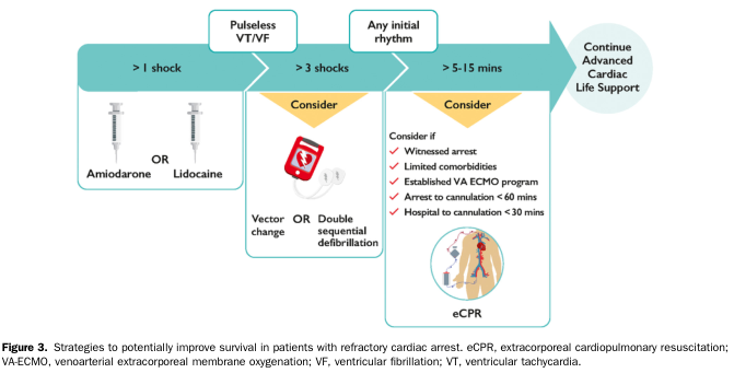
Summary : This flowchart outlines strategies to improve survival in patients with refractory cardiac arrest, detailing interventions based on the number of shocks, rhythm type, and elapsed time, and highlighting when to consider advanced therapies such as extracorporeal cardiopulmonary resuscitation (eCPR). flowchart: # Nodes : • Pulseless VT/VF (>1 shock) (rounded rectangle) • Amiodarone OR Lidocaine (rectangle with syringe icons) • Pulseless VT/VF (>3 shocks) (rounded rectangle) • Consider: Vector change OR Double sequential defibrillation (rectangle with defibrillator icon) • Any initial rhythm (>5–15 mins) (rounded rectangle) • Consider: eCPR (rectangle with eCPR illustration) – Consider if: Witnessed arrest, Limited comorbidities, Established VA ECMO program, Arrest to cannulation <60 mins, Hospital to cannulation <30 mins (checklist) • Continue Advanced Cardiac Life Support (rounded rectangle) # Connectors : • Arrows connect each node sequentially from left to right. • Decision points (e.g., “Consider”) branch to specific interventions. • Flow proceeds from initial shock management to advanced therapies and ongoing support. # Layout : • Horizontal flow from left (initial shock) to right (advanced support). • Three main stages: initial drug therapy, defibrillation strategies, and advanced resuscitation. • Decision diamonds and rectangles are used for intervention options. # Analysis : • The flowchart provides a stepwise escalation of care for refractory cardiac arrest, starting with antiarrhythmic drugs after >1 shock, considering advanced defibrillation techniques after >3 shocks, and evaluating candidacy for eCPR after 5–15 minutes of arrest based on specific criteria. The process emphasizes timely intervention and the use of specialized resources to improve survival outcomes.
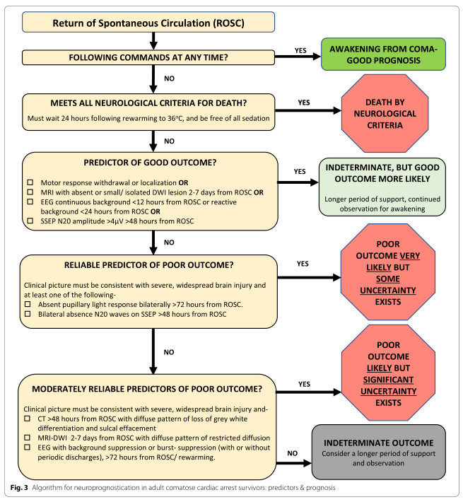
Summary : This flowchart presents an algorithm for neuroprognostication in adult comatose cardiac arrest survivors, outlining predictors and prognosis based on clinical and neurological criteria after return of spontaneous circulation (ROSC). flowchart: # Nodes : • Start (rectangle): "Return of Spontaneous Circulation (ROSC)" • Decision (rectangle): "FOLLOWING COMMANDS AT ANY TIME?" • Outcome (green rectangle): "AWAKENING FROM COMA - GOOD PROGNOSIS" • Decision (rectangle): "MEETS ALL NEUROLOGICAL CRITERIA FOR DEATH? Must wait 24 hours following rewarming to 36°C, and be free of all sedation" • Outcome (red hexagon): "DEATH BY NEUROLOGICAL CRITERIA" • Decision (rectangle): "PREDICTOR OF GOOD OUTCOME? □ Motor response withdrawal or localization OR □ MRI with absent or small/isolated DWI lesion 2-7 days from ROSC OR □ EEG continuous background <12 hours from ROSC or reactive background <24 hours from ROSC OR □ SSEP N20 amplitude >4μV >48 hours from ROSC" • Outcome (yellow rectangle): "INDETERMINATE, BUT GOOD OUTCOME MORE LIKELY Longer period of support, continued observation for awakening" • Decision (rectangle): "RELIABLE PREDICTOR OF POOR OUTCOME? Clinical picture must be consistent with severe, widespread brain injury and at least one of the following: □ Absent pupillary light response bilaterally >72 hours from ROSC. □ Bilateral absence N20 waves on SSEP >48 hours from ROSC." • Outcome (red hexagon): "POOR OUTCOME VERY LIKELY BUT SOME UNCERTAINTY EXISTS" • Decision (rectangle): "MODERATELY RELIABLE PREDICTORS OF POOR OUTCOME? Clinical picture must be consistent with severe, widespread brain injury and: □ CT >48 hours from ROSC with diffuse pattern of loss of grey white differentiation and sulcal effacement □ MRI-DWI 2-7 days from ROSC with diffuse pattern of restricted diffusion □ EEG with background suppression or burst-suppression (with or without periodic discharges), >72 hours from ROSC/rewarming." • Outcome (red hexagon): "POOR OUTCOME LIKELY BUT SIGNIFICANT UNCERTAINTY EXISTS" • Outcome (yellow rectangle): "INDETERMINATE OUTCOME Consider a longer period of support and observation" # Connectors : • Downward arrows connect each decision node to the next. • "YES" branches lead to green or red outcome nodes. • "NO" branches continue the flow to the next decision node. • The flow is strictly top-to-bottom, with each decision leading to either an outcome or further evaluation. # Layout : • Linear, vertical arrangement. • Each decision node is followed by a possible outcome (to the right) or the next decision (below). • Outcome nodes are color-coded: green for good prognosis, red for poor/death, yellow for indeterminate. • All nodes are rectangular except for outcome nodes, which are either rectangles (green/yellow) or hexagons (red). # Analysis : • The flowchart provides a stepwise approach to neuroprognostication after cardiac arrest, prioritizing early awakening as a good sign, strict neurological criteria for death, and a hierarchy of predictors for good or poor outcomes. • The algorithm emphasizes the need for multiple consistent findings before predicting poor outcome, and highlights uncertainty in many cases, recommending prolonged observation and support when prognosis is indeterminate. • Color-coding and node shapes visually distinguish between types of outcomes and decision points, aiding rapid clinical interpretation.
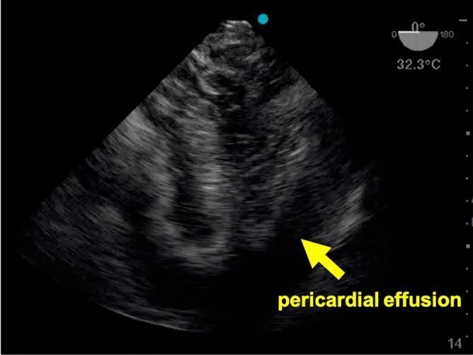
This diagnostic image is a transesophageal echocardiogram (TEE) demonstrating a mid-esophageal four-chamber view of the heart. The ultrasound captures the right and left atria and ventricles. A significant clinical finding is highlighted by a yellow arrow and text label: an anechoic (dark) space surrounding the cardiac structures, consistent with a pericardial effusion. In the clinical context of cardiopulmonary resuscitation, this finding is highly suggestive of cardiac tamponade as a potential reversible cause of cardiac arrest. Technical data on the display include a multiplane angle indicator at 0 degrees and a probe temperature reading of 32.3°C. The image serves as an educational example of using point-of-care TEE in emergency medicine and critical care to identify life-threatening obstructive pathology during advanced life support (ALS).
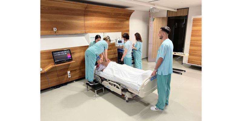
A clinical photograph depicting a high-fidelity medical simulation of an Advanced Life Support (ALS) scenario. The image shows a team of five healthcare students or professionals in light blue scrubs attending to a high-fidelity patient simulator (Apollo Patient Simulator) positioned on a standard adjustable hospital bed. One rescuer is standing on a step stool performing chest compressions on the manikin's exposed chest, while others manage the airway and monitor vital signs. The educational environment is equipped with authentic clinical technology, including a patient monitor displaying multi-parameter waveforms (ECG, SpO2) and a secondary laptop screen showing detailed cardiac rhythm data. The setting is a specialized simulation lab designed to replicate a modern hospital room, used for teaching Crisis Resource Management (CRM) skills, resuscitation algorithms, and technical medical interventions in a controlled, risk-free setting.
ALS cardiac arrest algorithm 2021 ERC CPR guidelines shockable non-shockable
Cardiac Arrest - ALS Algorithm (CPLs / Key Principles)
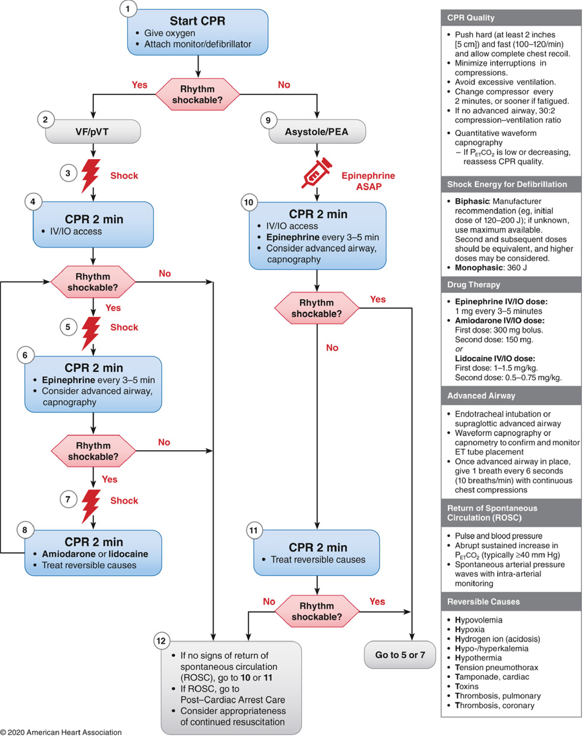
1. Start CPR Immediately
- Push hard (≥5 cm / 2 inches) and fast (100-120/min)
- Allow complete chest recoil between compressions
- Minimize interruptions - aim for CPR fraction >60%
- Give oxygen
- Attach monitor/defibrillator
- If no advanced airway: 30:2 compression-to-ventilation ratio
- Change compressor every 2 minutes to avoid fatigue
- Use quantitative waveform capnography: if PETCO₂ is low or falling, reassess CPR quality
2. Rhythm Assessment - The Fork in the Road
| Shockable | Non-Shockable |
|---|---|
| VF (Ventricular Fibrillation) | Asystole |
| pVT (Pulseless Ventricular Tachycardia) | PEA (Pulseless Electrical Activity) |
3. Shockable Arm - VF/pVT
- Shock (defibrillate) as early as possible
- Biphasic: manufacturer recommendation (typically 120-200 J); if unknown, use maximum available
- Monophasic: 360 J
- For recurrent VF/pVT: use previously successful energy level
- CPR 2 min immediately after shock - do NOT pause to check rhythm
- Establish IV/IO access
- Reassess rhythm at 2 minutes
- If still shockable: Shock again (step 5)
- CPR 2 min + start Epinephrine 1 mg IV/IO every 3-5 min + consider advanced airway
- If still shockable after 3rd shock: Shock (step 7)
- CPR 2 min + Amiodarone 300 mg IV/IO (or Lidocaine 1-1.5 mg/kg) + treat reversible causes
- Second dose amiodarone: 150 mg; Second dose lidocaine: 0.5-0.75 mg/kg
Key point: Epinephrine is given after the 3rd shock in shockable rhythms. Early defibrillation, not drugs, drives survival in VF/pVT. - Miller's Anesthesia, 10e
4. Non-Shockable Arm - Asystole/PEA
- Epinephrine 1 mg IV/IO as soon as possible (ASAP, do not delay)
- CPR 2 min + IV/IO access + repeat epinephrine every 3-5 min
- Consider advanced airway and capnography
- Treat reversible causes (see Hs and Ts below) - this is the primary focus
- Reassess rhythm every 2 minutes
- Atropine is no longer recommended for asystole/PEA
Asystole has an extremely poor prognosis. PEA requires active hunting for a treatable cause. - Miller's Anesthesia, 10e
5. The Hs and Ts - Reversible Causes
| 5 Hs | 5 Ts |
|---|---|
| Hypovolemia | Tension pneumothorax |
| Hypoxia | Tamponade (cardiac) |
| Hydrogen ion (acidosis) | Toxins (drug OD) |
| Hypo-/Hyperkalemia | Thrombosis - pulmonary (PE) |
| Hypothermia | Thrombosis - coronary (MI) |
6. Drug Summary
| Drug | Indication | Dose |
|---|---|---|
| Epinephrine (Adrenaline) | All arrests | 1 mg IV/IO every 3-5 min |
| - Non-shockable | ASAP | 1 mg |
| - Shockable | After 3rd shock | 1 mg |
| Amiodarone | Shock-refractory VF/pVT | 300 mg bolus IV/IO; 2nd dose 150 mg |
| Lidocaine (alternative) | Shock-refractory VF/pVT | 1-1.5 mg/kg IV/IO; 2nd dose 0.5-0.75 mg/kg |
7. Advanced Airway
- Endotracheal intubation or supraglottic airway (SGA/LMA)
- Confirm with waveform capnography - most reliable method
- Once placed: 1 breath every 6 seconds (10 breaths/min) with continuous chest compressions (no more 30:2)
- False negatives in ETT CO₂ detection: massive PE, very low cardiac output
- Do NOT stop CPR to intubate
8. ROSC - Signs of Return of Spontaneous Circulation
- Palpable pulse and blood pressure
- Abrupt sustained rise in PETCO₂ (typically ≥40 mmHg) - a very useful early sign
- Spontaneous arterial pressure waves on intra-arterial monitoring
9. Post-Cardiac Arrest Care (after ROSC)
- Targeted Temperature Management (TTM): prevent fever; consider 33-36°C for comatose survivors
- Coronary angiography if ST elevation or suspected ACS
- Avoid hyperoxia and hypotension
- Neuroprognostication after ≥72 hours
- Consider eCPR (extracorporeal CPR/ECMO) in refractory arrest with witnessed onset, limited comorbidities, and arrest-to-cannulation <60 min
10. CPR Quality Checklist (Remember Throughout)
- Rate: 100-120/min
- Depth: ≥5 cm (2 inches), <6 cm
- Full chest recoil each compression
- No excessive ventilation (avoids raised intrathoracic pressure)
- Rotate compressor every 2 min
- Use capnography: low PETCO₂ = poor CPR quality OR ROSC if sudden rise
- AHA 2020 ACLS Guidelines - Miller's Anesthesia, 10e, p. 12201-12205
- ERC 2025 ALS Guidelines
- ERC 2021 ALS Guidelines