
Patient with fever with inflmmed tonsils started on amoxiclav , paracetamol , rabeprazole and levocetrizine but still fever persist what is next step to proceed
tonsillitis persistent fever antibiotic treatment
persistent fever tonsillitis after amoxicillin next step management 2024
Persistent Fever with Inflamed Tonsils Despite Amoxiclav - Next Steps
Step 1: Re-evaluate the Diagnosis
| Feature | Bacterial (GABHS) | Mononucleosis (EBV) |
|---|---|---|
| Onset | Rapid | Variable |
| Fever | High | High |
| Lymphadenopathy | Large, tender cervical | Generalized + splenomegaly |
| Tonsil findings | Exudate ± petechiae | Exudate + palatal petechiae |
| Hepatomegaly | No | Present |
| Response to amoxicillin | Improves | No improvement + may develop a maculopapular rash |
Critical point: Amoxicillin/amoxiclav in EBV triggers a classic maculopapular rash in ~80% of cases. If this rash appeared, it strongly suggests EBV, not antibiotic failure.
- Monospot test (heterophile antibody) - rapid, bedside
- Complete blood count (CBC) - look for atypical lymphocytes (>10% = highly suggestive)
- EBV viral capsid antigen (VCA) IgM if monospot negative but suspicion remains high
- Liver function tests (EBV hepatitis is common)
Step 2: Investigate Cause of Treatment Failure if Still Bacterial
- Drug compliance - Is the patient actually taking it correctly and completing the course?
- Throat culture with sensitivity - Confirm GABHS and rule out resistant organisms or alternative bacteria
- Beta-lactamase producing co-pathogens - Organisms like H. influenzae or M. catarrhalis can produce beta-lactamase that inactivates penicillin-type drugs even when the primary strep is sensitive
Step 3: Investigate for Complications
- Peritonsillar abscess (quinsy) - Most common complication. Signs: unilateral tonsillar bulge, uvular deviation, muffled "hot potato" voice, trismus, drooling. Requires needle aspiration or I&D + antibiotics - does NOT resolve on oral antibiotics alone.
- Parapharyngeal abscess - Neck swelling, trismus, systemic toxicity. Needs CT neck with contrast + surgical drainage.
- Retropharyngeal abscess - Neck stiffness, drooling, stridor. Needs imaging urgently.
- Lemierre's syndrome - Fusobacterium necrophorum, septic thrombophlebitis of the internal jugular vein. Rare but life-threatening. Amoxiclav has limited coverage; needs metronidazole + ampicillin/sulbactam.
Step 4: Escalate or Switch Antibiotics (if still bacterial, no complication)
| Situation | Drug |
|---|---|
| GABHS confirmed, amoxiclav failing | Clindamycin 300-450 mg TID x 10 days (good GABHS + anaerobe coverage) |
| Suspected beta-lactamase producers | Cephalosporins - Cefdinir, Cefuroxime, or Cephalexin |
| Penicillin-allergic | Azithromycin 500 mg day 1, then 250 mg x 4 days, or Clarithromycin |
| Recurrent/culture-confirmed GABHS failure | IM Benzathine Penicillin G 1.2 million units single dose (ensures compliance) |
| Suspected anaerobes / Fusobacterium | Add metronidazole or switch to ampicillin/sulbactam |
Step 5: Consider Steroids for Severe Inflammation
Summary Algorithm
Fever persists on Amoxiclav
        │
        ├── 1. REASSESS: Is this EBV/Mono?
        │      → Monospot, CBC, LFTs
        │      → If positive: STOP antibiotics (no benefit), steroids if severe
        │
        ├── 2. EXAMINE: Any peritonsillar/deep space abscess?
        │      → Asymmetric tonsil, uvula deviation, trismus?
        │      → CT neck if yes → surgical drainage
        │
        ├── 3. CULTURE: Throat swab for C&S
        │      → Identify organism, guide antibiotic switch
        │
        └── 4. ESCALATE ANTIBIOTIC if still bacterial:
               → Clindamycin (first choice)
               → Cefuroxime / Cefdinir
               → IM Benzathine Penicillin (if compliance issue)
               → Add metronidazole if anaerobes suspected
- Cummings Otolaryngology Head and Neck Surgery, Table 201.1 (Pharyngitis Characteristics)
- Schwartz's Principles of Surgery, 11th ed., Pharyngeal and Adenotonsillar Disease, p. 2718-2720
- Harrison's Principles of Internal Medicine, 22E, Peritonsillar Abscess, p. 1388
- Medscape Tonsillitis & Peritonsillar Abscess Management
- Yoon et al. Guidelines for Antibiotic Use in Adults with Acute URTI, Infect Chemother 2017
Search for recent clinical guidelines on treatment of antibiotic-resistant pharyngotonsillitis
pharyngotonsillitis antibiotic resistant treatment guidelines
streptococcal pharyngitis antibiotic failure management
clinical guidelines antibiotic resistant pharyngotonsillitis treatment 2023 2024 2025
group A streptococcus pharyngitis antibiotic resistance penicillin
tonsillitis clindamycin cephalosporin treatment outcome
https://www.cdc.gov/group-a-strep/hcp/clinical-guidance/stre…
PMID: 38648833
PMID: 37819417
PMID: 39989986
IDSA 2025 clinical practice guidelines group A streptococcal pharyngitis update
https://pmc.ncbi.nlm.nih.gov/articles/PMC11539554
macrolide resistance streptococcus pyogenes azithromycin clindamycin tonsillitis
Recent Clinical Guidelines: Treatment of Antibiotic-Resistant Pharyngotonsillitis
1. IDSA 2025 Clinical Practice Guideline Update (October 2025)
- Use a clinical scoring system (e.g., modified Centor/McIsaac score) to stratify patients before testing. Patients with a score of 0-1 should NOT be tested or treated with antibiotics (very low probability of GAS)
- The main goal: reduce inappropriate antibiotic use, since >60% of adults with sore throat are unnecessarily prescribed antibiotics despite only 10-15% having GAS
- Penicillin and amoxicillin remain first-line - GAS has never demonstrated confirmed penicillin or cephalosporin resistance in clinical isolates (confirmed by CDC 2025)
2. Antibiotic Resistance Landscape in GAS
| Antibiotic Class | Resistance Status |
|---|---|
| Penicillin / Amoxicillin | Zero confirmed clinical resistance - MICs unchanged for 90 years |
| Cephalosporins (1st-3rd gen) | No confirmed resistance |
| Azithromycin / Clarithromycin (14/15-atom macrolides) | 10-30% resistance - varies geographically; significant in Asia, parts of Europe, US |
| Clindamycin (lincosamide) | Variable resistance emerging, particularly linked to inducible resistance in macrolide-resistant strains (erm genes) |
| Erythromycin | Resistance rates of 10-30% in many regions |
| 16-atom macrolides (josamycin, midecamycin) | Lower resistance rates than 14/15-atom macrolides |
CDC GAS Clinical Guidance (updated Nov 2025): "There has never been a report of a clinical isolate of group A strep bacteria that's resistant to penicillin or cephalosporins. However, resistance to azithromycin, clarithromycin, and clindamycin is well known and varies geographically and temporally."
3. Stepwise Antibiotic Strategy for Treatment Failure / Resistance
First-Line (Standard Treatment - No Resistance Issue)
| Drug | Adult Dose | Duration |
|---|---|---|
| Penicillin V | 500 mg twice daily | 10 days |
| Amoxicillin | 500-875 mg twice daily | 10 days |
| Benzathine Penicillin G IM | 1.2 million U single dose | Once |
When First-Line Fails (Penicillin/Amoxicillin Failure)
| Drug | Adult Dose | Duration | Rationale |
|---|---|---|---|
| Amoxicillin-clavulanate | 500/125 mg three times daily | 10 days | Covers beta-lactamase-producing co-pathogens (H. influenzae, anaerobes) |
| Clindamycin | 300 mg three times daily | 10 days | Best eradication rates for recurrent/relapsing GABHS (93-100% eradication in RCTs); covers anaerobes |
| Cefuroxime axetil | 250-500 mg twice daily | 10 days | Good for penicillin non-responders with no anaphylaxis allergy |
| Cefdinir | 300 mg twice daily or 600 mg once daily | 10 days | Broader cephalosporin option |
Penicillin-Allergic Patients
| Drug | Adult Dose | Duration | Notes |
|---|---|---|---|
| Cephalexin / Cefadroxil | 500 mg twice daily | 10 days | Use if non-anaphylactic allergy only |
| Clindamycin | 300 mg three times daily | 10 days | Preferred if beta-lactam allergy |
| Azithromycin | 500 mg day 1, then 250 mg x 4 days | 5 days | Only if local resistance rates low; check local susceptibility |
| Clarithromycin | 250 mg twice daily | 10 days | Only if local resistance rates low |
| Josamycin / 16-atom macrolides | 1 g twice daily | 10 days | Lower resistance than azithromycin where available |
Warning: Do NOT empirically use azithromycin or clarithromycin in regions with >10% macrolide resistance rates without susceptibility testing. Resistance rates in parts of the US, Asia, and Europe now reach 30%.
4. Special Scenarios
- Clindamycin 10-day course is the most effective eradication strategy (100% at 3 weeks in some studies)
- Amoxicillin-clavulanate 10 days is an equivalent alternative
- Consider IM Benzathine Penicillin to guarantee adherence
- Amoxicillin-clavulanate or ampicillin-sulbactam + metronidazole
- Penicillin alone is inadequate due to beta-lactamase production
- Treat only in selected symptomatic cases
- Same antibiotic hierarchy as GABHS applies
5. Global Guideline Comparison (Pellegrino et al., Eur J Pediatr 2023) [PMID: 37819417]
- Treat all confirmed GABHS to prevent acute rheumatic fever (ARF) - endorsed in high-ARF-risk regions (South Asia, Africa, parts of Latin America)
- Watch and wait - antibiotics only for severe/complicated cases (UK, Netherlands, Belgium)
- Risk-stratified treatment - tailor antibiotic decision to individual patient ARF risk (US, most European guidelines)
Summary: Practical Antibiotic Algorithm for Resistant/Refractory Pharyngotonsillitis
STEP 1: Is this truly GABHS failure?
  → Rule out EBV (Monospot, CBC), abscess (exam ± CT)
  → Confirm compliance with full antibiotic course

STEP 2: Still bacterial with amoxicillin/amoxiclav failure?
  → Throat C&S with sensitivity
  → Switch to CLINDAMYCIN 300 mg TID x 10 days (best eradication evidence)
  → Or CEFUROXIME/CEFDINIR if cephalosporin preferred

STEP 3: Penicillin-allergic patient?
  → Non-anaphylactic: Cephalexin or Cefadroxil x 10 days
  → Anaphylactic: Clindamycin x 10 days (first choice)
  → Only use macrolides (azithromycin) if local resistance <10%

STEP 4: Recurrent/relapsing (GABHS carrier)?
  → Clindamycin x 10 days (highest eradication rate)
  → Consider tonsillectomy if: ≥7 episodes/year, ≥5/year x 2 years,
    or ≥3/year x 3 years
Key References
| Source | Year | Evidence Type |
|---|---|---|
| IDSA GAS Pharyngitis Guideline Update | 2025 | Practice Guideline |
| CDC GAS Clinical Guidance | Updated Nov 2025 | Official Guidance |
| Hamilton & McCrea, Am Fam Physician [PMID: 38648833] | 2024 | Rapid Evidence Review |
| Pellegrino et al., Eur J Pediatr [PMID: 37819417] | 2023 | Multi-guideline Comparison Review |
| Geteneh et al., JAC Antimicrob Resist [PMID: 39989986] | 2025 | Emerging Resistance Review |
| Italian Intersociety Consensus (PMC11539554) | 2024 | Consensus Guideline |
| Spanish Adults Pharyngitis Recommendations (PMC7154615) | 2021 | Expert Consensus |
A patient came with breathless ness and contraction of both hand fingers. Spo2 drops variable started on nebulization ipratropium and budesonide
breathlessness carpopedal spasm bilateral hand fingers tetany hyperventilation management next steps
Clinical Analysis: Breathlessness + Bilateral Hand Finger Contractions + Variable SpO2
The Core Diagnostic Question: What is Causing the Hand Contractions?
Rapid/labored breathing → excess CO2 exhaled → PaCO2 falls → Respiratory Alkalosis → pH rises → ionized calcium binds to albumin → ionized hypocalcemia → neuromuscular hyperexcitability → carpopedal spasm + tetany
Differential Diagnosis to Work Through
| Diagnosis | Supporting Features | Against |
|---|---|---|
| Hyperventilation Syndrome with tetany | Breathlessness + bilateral carpopedal spasm + variable SpO2 (artifact) | Must exclude organic cause |
| Acute Asthma / Bronchospasm with secondary hyperventilation | Breathlessness, started on ipratropium + budesonide | Carpopedal spasm not explained by asthma alone |
| Hypocalcemia (true metabolic) | Bilateral hand spasm, Trousseau sign | Need labs to confirm; usually no acute breathlessness |
| Hypomagnesemia | Co-exists with hypocalcemia, same clinical picture | Requires blood test |
| Panic Disorder / Anxiety Attack | Young patient, breathlessness, spasm, no organic cause | Diagnosis of exclusion |
| COPD exacerbation with bronchospasm | Responds to nebulization | Carpopedal spasm unexplained |
Immediate Next Steps (Priority Order)
Step 1: Correct the SpO2 Measurement
- Move the pulse oximeter to the earlobe or forehead - away from the contracted fingers
- If SpO2 is actually normal (>95%) on the earlobe, this confirms the carpopedal spasm is causing the probe artifact
- If SpO2 is genuinely low, proceed with oxygen supplementation (but do not give high-flow O2 to a hyperventilating patient without cause - it can worsen respiratory alkalosis by removing the hypoxic drive)
Step 2: Immediate Bedside Assessment
Step 3: Emergency Investigations (Order All Simultaneously)
| Test | Why |
|---|---|
| ABG (Arterial Blood Gas) | Most important - will show respiratory alkalosis (low PaCO2, high pH) if hyperventilation; low PaO2 if genuine hypoxia; also shows ionized calcium |
| Serum ionized calcium | Directly measures functionally relevant calcium |
| Serum total calcium + albumin | For corrected calcium |
| Serum magnesium | Hypomagnesemia causes refractory hypocalcemia |
| Serum potassium | Hypokalemia co-exists with alkalosis |
| ECG | Prolonged QT interval in hypocalcemia; arrhythmia risk |
| Peak flow / spirometry | Assess bronchospasm severity if asthma suspected |
| Chest X-ray | Rule out pneumothorax, consolidation, effusion |
Step 4: Treat What You Find
- Do NOT paper bag rebreathing - potentially fatal in hypoxic patients
- Reassurance and controlled breathing guidance - coaching slow diaphragmatic breathing at 8-12 breaths/minute is the primary intervention
- IV Calcium Gluconate 10% - 10 mL (1g) slow IV over 5-10 minutes with continuous ECG monitoring - this directly reverses tetany
- If hypomagnesemia found: IV Magnesium Sulfate 2g over 15-20 minutes (hypoMg causes PTH resistance and refractory hypocalcemia)
- Short-acting benzodiazepine (diazepam 5 mg IV or lorazepam 1-2 mg IV) if anxiety is severe and driving the hyperventilation
- Continue and escalate bronchodilators
- Add salbutamol (albuterol) 2.5-5 mg nebulized - this is missing from the current regimen and is the most important bronchodilator in acute bronchospasm
- IV/oral corticosteroids (prednisolone 40mg oral or hydrocortisone 200mg IV)
- Consider IV magnesium sulfate 2g - also works as a bronchodilator in severe asthma (reduces smooth muscle contraction)
- Prepare for non-invasive ventilation (NIV/BiPAP) if not responding
- IV Calcium Gluconate 10%, 100 mg/kg (or 1-2 g adult dose) over 5-10 min, then calcium infusion
- Investigate underlying cause: post-thyroid surgery, hypoparathyroidism, vitamin D deficiency, chronic renal failure, malabsorption
Critical Gap in Current Management
-
Salbutamol (albuterol) is missing - it is the most rapid-acting bronchodilator and should be the first drug in any bronchospasm nebulization. Ipratropium is an anticholinergic that works as an adjunct, not as a replacement for a beta-2 agonist.
-
The carpopedal spasm has not been addressed - no calcium, no magnesium, no ABG ordered for the tetany component.
Summary Algorithm
Breathlessness + Bilateral Hand Contractions + Variable SpO2
        │
        ├── RELOCATE PULSE OX → earlobe or forehead
        │      → SpO2 normal (>95%)? Spasm was causing artifact
        │      → SpO2 still low? Genuine hypoxia
        │
        ├── BEDSIDE: Trousseau sign + Chvostek sign
        │
        ├── URGENT LABS: ABG, ionized Ca, Mg, K, ECG
        │
        ├── ABG shows LOW PaCO2 + HIGH pH (Respiratory Alkalosis)?
        │      → Hyperventilation-induced tetany
        │      → IV Calcium Gluconate 1g slow IV + reassurance + slow breathing
        │      → Add benzodiazepine if anxiety-driven
        │
        ├── ABG shows LOW PaO2 (Hypoxia)?
        │      → Add SALBUTAMOL nebulization (most important missing drug)
        │      → IV/oral corticosteroids
        │      → IV Magnesium Sulfate 2g (bronchodilator + corrects Mg)
        │      → Prepare for NIV if not improving
        │
        └── Both bronchospasm AND tetany possible:
               → Treat both concurrently
               → Magnesium sulfate 2g IV addresses BOTH bronchospasm and hypocalcemia
- Tintinalli's Emergency Medicine, Comprehensive Study Guide - Hypocalcemia section, p. 821-825
- Sabiston Textbook of Surgery - Hypocalcemia, tetany, p. 797-799
- Cleveland Clinic Journal of Medicine - Hyperventilation and Tetany
Nevus comedonicus
nevus comedonicus clinical features treatment pathology 2023 2024
Nevus Comedonicus (Acneiform Nevus / Comedo Naevus)
Clinical Photographs
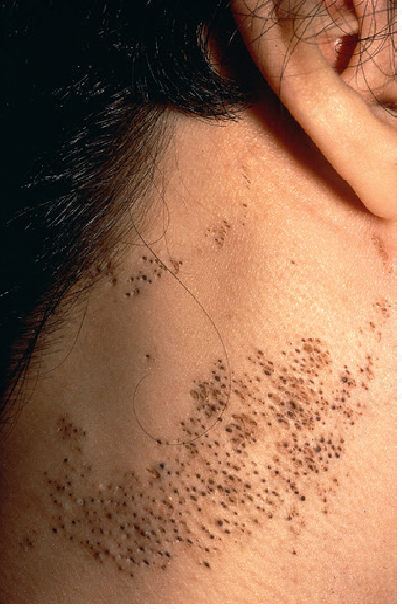
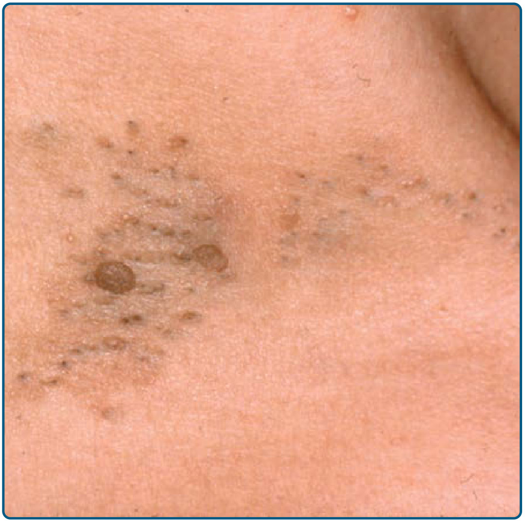
Definition
Pathogenesis
- NEK9 gene (NIMA-related kinase 9): Somatic mosaic activating mutations in NEK9 are the most frequently identified genetic cause - identified in lesional but not adjacent normal skin, confirming the mosaic nature
- FGFR2 mutations: Found in the acneiform variant (Munro acne nevus) - notably, patients with Apert syndrome (FGFR2 mutation) also develop severe cystic acne, suggesting nevus comedonicus may represent a mosaic form of Apert syndrome
- Familial cases are rare but documented; most arise sporadically
Clinical Features
- Usually unilateral, though bilateral cases occur
- Follows the lines of Blaschko (reflecting the mosaic somatic mutation pattern)
- Most common sites: face, trunk, neck, upper extremities
- Can affect hairless areas: palms, soles, glans penis (pseudocomedones on palms)
- On elbows and knees, lesions may appear verrucous
- Non-inflammatory (simple) - asymptomatic comedone plaques only
- Inflammatory - comedo lesions admixed with papules, cysts, abscesses, fistulas, and scars. Occurs in ~50% of cases. Puberty and hormonal influences worsen the inflammatory component.
Histopathology
- Dilated follicular ostia/invaginations lined by atrophic squamous epithelium
- Filled with orthokeratotic horny material (laminated keratin)
- Absent or rudimentary hair shafts - no mature terminal hairs
- Atrophic or absent sebaceous glands
- Interfollicular epidermis: papillomatous (as in typical epidermal nevi)
- Epidermolytic hyperkeratosis may sometimes be present in follicular epithelium
- On dermoscopy: dark brown to black dots (keratin-filled follicular openings) surrounded by a white halo - pathognomonic on dermoscopy
Differential Diagnosis
| Condition | Distinguishing Feature |
|---|---|
| Acne vulgaris | Diffuse, inflammatory, hormonal onset, no linear/blaschkoid pattern |
| Milia | Small white cysts, not follicular openings, no keratin plugs |
| Acne neonatorum | Transient, diffuse, no fixed pattern |
| Nevus sebaceous | Yellow-orange, alopecic plaque; sebaceous hyperplasia histologically |
| Linear Darier disease | Dyskeratosis, acantholysis on histology (corps ronds, grains) |
| Favre-Racouchot syndrome | Acquired, elderly, sun-damaged skin, periorbital |
| Chloracne | Chemical exposure history, milia + cysts |
Nevus Comedonicus Syndrome
- Ipsilateral cataracts and corneal changes
- Skeletal anomalies: hemivertebrae, scoliosis, absent 5th ray of the hand
- Neurologic defects: EEG abnormalities, cerebral anomalies
- Ocular anomalies
Treatment
Non-Inflammatory Lesions (Cosmetic)
| Modality | Evidence/Notes |
|---|---|
| Topical retinoids (tretinoin 0.1%, tazarotene) | First-line; modest improvement of keratinous plugs; does not address the structural abnormality |
| Topical calcipotriene / tacalcitol | Vitamin D analogues; some case report evidence |
| Topical 12% ammonium lactate / salicylic acid | Keratolytic; adjunctive |
| Pore strips / manual comedone extraction | Cosmetic improvement; recurrence is common |
| Ablative laser (Er:YAG, fractional CO2) | Best cosmetic outcomes; systematic review (Liu et al., J Dermatolog Treat 2023) confirms long-term efficacy of fractional CO2 |
| Full-thickness surgical excision | Most definitive option for localized lesions; prevents recurrence |
Inflammatory Lesions (Cysts, Abscesses, Scarring)
| Modality | Notes |
|---|---|
| Systemic antibiotics | Tetracycline 500 mg BD or doxycycline 100 mg BD for inflammatory flares |
| Intralesional corticosteroids | Triamcinolone 5-10 mg/cc for individual cysts/nodules |
| Oral isotretinoin (0.5 mg/kg/day minimum effective dose) | Partially suppresses cyst and nodule formation; does NOT improve the comedonal component - this is a key point. Many cases fail to respond. |
| Topical corticosteroid (e.g., mometasone furoate) | Combined with topical retinoid for anti-inflammatory effect |
| Surgical excision | For limited areas with severe cystic disease |
Emerging Therapies (investigational)
- FGFR inhibitors (targeting FGFR2 pathway)
- IL-1-alpha inhibitors
- Anti-gamma-secretase drugs
- Pinhole laser (CO2 dual-mode) - recent case reports show promise for refractory cases
Key Clinical Pearls
- Oral isotretinoin does NOT improve the comedonal lesions - it only partially suppresses inflammatory cyst formation in some patients. Families must be counseled on this limitation.
- Dermoscopy is helpful and non-invasive: dark brown-black dots in follicular openings with white halos are diagnostic.
- Puberty worsens the inflammatory variant due to hormonal stimulation of the residual pilosebaceous structures.
- Malignant transformation is exceedingly rare - SCC or keratoacanthoma within the nevus is reported but very uncommon.
- Every NC patient deserves a systemic workup for NC syndrome (eyes, skeleton, CNS) regardless of lesion size.
- Andrews' Diseases of the Skin, Clinical Dermatology - Chapter 29, p. 2022-2038
- Fitzpatrick's Dermatology, Vol 1 - Chapter 108, p. 1639-1670
- Dermatology 2-Volume Set, 5th ed. - Chapter 109, p. 1889-1920
- StatPearls - Nevus Comedonicus (updated 2023)
- DermNet - Comedo Naevus
- CosmoDerma 2024 - Dermoscopic features