
Inguinal ligament as short answer
Reading File
Now let me search the medical library for content on the inguinal ligament.
Finding Sources
Finding Sources
Reading File
Reading File
Reading File
Reading File
Finding Sources
Reading File
Reading File
Now let me fetch one of the key anatomy diagrams to include in the answer:
Loading Image
Here is a complete short-answer note on the inguinal ligament:
Inguinal Ligament
Definition / Formation
The inguinal ligament (Poupart's ligament) is the thickened, reinforced lower free border of the external oblique aponeurosis that has folded back upon itself to form a trough-like structure. It is not a true ligament in the classical sense but a condensed aponeurotic band.
Attachments
- Laterally: Anterior superior iliac spine (ASIS)
- Medially: Pubic tubercle
- Length: approximately 12-15 cm
Structure and Relations
The ligament folds inward/posteriorly on itself, forming a trough that constitutes the floor of the inguinal canal.
Several subsidiary ligaments arise from its medial end:
| Subsidiary Ligament | Description |
|---|---|
| Lacunar ligament (Gimbernat's) | Crescent-shaped extension from the medial end; passes backward to attach to the pecten pubis (pectineal line); forms the medial wall of the femoral ring |
| Pectineal (Cooper's) ligament | Further extension of lacunar ligament fibers along the pecten pubis of the pelvic brim |
| Reflected inguinal ligament | Fibers that arch medially to form the deep groove of the superficial inguinal ring |
The Gap Beneath the Inguinal Ligament
The large crescent-shaped gap between the inguinal ligament above and the anterosuperior margin of the pelvic bone below is the major route of communication between the abdomen and anteromedial thigh. Structures passing through:
- Psoas major, iliacus, and pectineus muscles (to insert on the femur)
- Femoral artery and vein
- Femoral nerve (L2-L4, lies lateral to femoral artery)
- Lymphatics of the lower limb
Role in the Inguinal Canal
| Wall | Structure |
|---|---|
| Floor | Inguinal ligament |
| Roof | Internal oblique + transversus abdominis |
| Anterior wall | External oblique aponeurosis |
| Posterior wall | Transversalis fascia + peritoneum |
Clinical Significance
- Hernia landmark: Inguinal hernia presents above the inguinal ligament and medial to the pubic tubercle; femoral hernia presents below the inguinal ligament (through the femoral ring).
- Hesselbach's (inguinal) triangle: Bounded by the inguinal ligament (base), rectus abdominis (medially), and inferior epigastric vessels (superolaterally) - the site of direct inguinal hernia.
- Femoral ring and femoral canal: The lacunar ligament forms the medial boundary of the femoral ring; femoral hernias exit here.
- Surgical repair: The inguinal ligament is a key anchoring structure in hernia repair (Bassini, Lichtenstein procedures).
- Vascular access: Femoral artery cannulation for cardiac catheterization is performed just below the inguinal ligament over the femoral head; cannulation above it risks retroperitoneal hemorrhage.
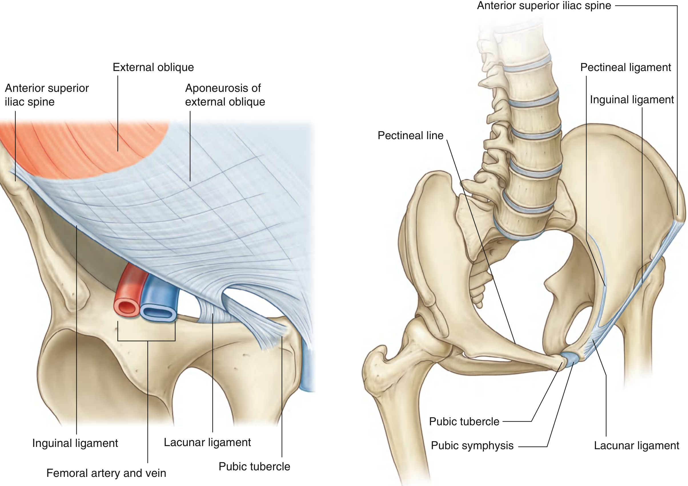
Fig. 4.28 - Ligaments Formed From the External Oblique Aponeurosis (Gray's Anatomy for Students)
Sources: Gray's Anatomy for Students, pp. 340-341 | Berek & Novak's Gynecology, p. 151 | Fischer's Mastery of Surgery, 8th ed. | THIEME Atlas of Anatomy
This is a shared conversation. Sign in to Orris to start your own chat.