
I can see this is a medical vocabulary list. Let me help you with it — but first, what would you like me to do with this content?
Asking for Preferences
What would you like me to do with this list of neurological/medical terms (items 79-100)?
femoral vein
The femoral vein is the main deep vein of the thigh, running alongside the femoral artery in the femoral triangle. It drains blood from the leg and continues upward to become the external iliac vein. It is a common site for central venous access and deep vein thrombosis (DVT).
Searching sources...
Reading File
Finding Sources
Searching the Web
femoral vein anatomy clinical significance
Reading File
Reading File
Loading Image
Femoral Vein
Anatomy
The femoral vein is the primary deep vein of the thigh. It begins at the adductor hiatus (Hunter's canal) as the proximal continuation of the popliteal vein, and ends at the inferior margin of the inguinal ligament, where it becomes the external iliac vein.
It is contained within the femoral triangle, whose boundaries are:
- Superiorly - inguinal ligament
- Laterally - sartorius muscle
- Medially - adductor longus muscle
(Mnemonic for the triangle: SAIL - Sartorius, Adductor longus, Inguinal Ligament)
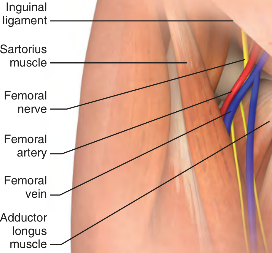
Relations - NAVEL Mnemonic
From lateral to medial within the femoral sheath:
| Letter | Structure |
|---|---|
| N | Nerve (femoral nerve) |
| A | Artery (femoral artery) |
| V | Vein (femoral vein) |
| E | Empty space |
| L | Lymphatics |
Note: As the artery and vein course distally, their relationship can rotate so the artery may lie on top of the vein - this is why ultrasound guidance helps avoid accidental arterial puncture.
Tributaries
The deep femoral vein (profunda femoris) drains into it posteriorly, about 8 cm distal to the inguinal ligament. Proximal to this junction, it is called the common femoral vein.
Clinical Importance
1. Central venous access:
The femoral vein is the most accessible central access site during critical illness - especially during cardiac arrest or trauma - because it does not require stopping CPR and avoids risks of pneumothorax.
- Cannulated 1-2 cm below the inguinal ligament, just medial to the femoral artery pulse
- In pulseless arrest, use the "V" technique: thumb on pubic tubercle, index finger on ASIS - the vein lies at the interdigital space
2. Deep Vein Thrombosis (DVT):
The femoral vein is a common site for DVT, which can embolize to cause pulmonary embolism (PE). The superficial femoral vein (despite its name) is a deep vein and DVT here carries the same embolic risk.
3. Femoral venous duplex ultrasound is the standard method for DVT diagnosis.
Sources: Roberts and Hedges' Clinical Procedures in Emergency Medicine; Tintinalli's Emergency Medicine: A Comprehensive Study Guide
This is a shared conversation. Sign in to Orris to start your own chat.