
Recombinant RDNA technology
- Core idea: Cut DNA from one organism and insert it into another (often a plasmid or viral vector), then express that gene in a host cell
- Key enzymes: Restriction endonucleases (cut DNA at specific sequences), DNA ligase (joins fragments), DNA polymerase (copies DNA)
- Applications: Insulin production, vaccines, gene therapy, transgenic organisms, forensics
- Vectors: Plasmids, bacteriophages, cosmids, BACs, YACs, viral vectors
recombinant DNA technology cloning vector plasmid diagram
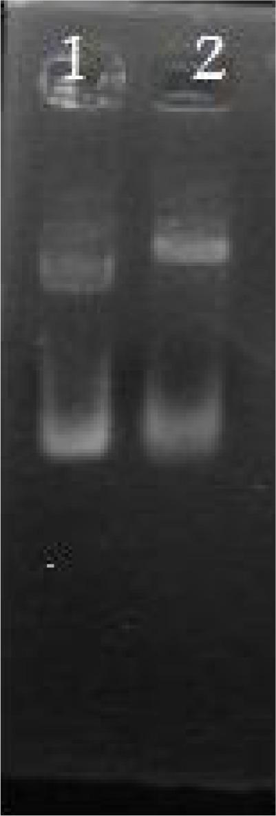
Diagnostic Image: This is an agarose gel electrophoresis image showing restriction analysis of plasmids, specifically related to the molecular cloning of recombinant SAG3 for medical research. The gel contains two numbered lanes. Lane 1 displays the migration pattern of undigested pGEMEX-1 plasmid, showing at least two distinct bright bands representing different DNA conformations (such as supercoiled and circular forms). Lane 2 shows the restriction digestion of recombinant pGEMEX43 using the PstI enzyme. A noticeable shift in band migration is visible compared to lane 1, indicating successful digestion at the PstI restriction site within the P43 gene. This diagnostic visual demonstrates the molecular biology techniques used to verify the construction of recombinant plasmids intended for the expression of proteins used in human immunological assays, such as Western blot or dot blot for infectious disease diagnostics. The image illustrates concepts of genetic engineering, restriction mapping, and plasmid vector analysis.
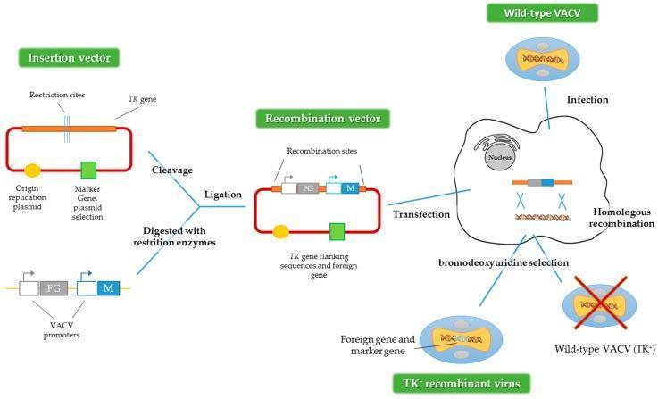
This medical pathophysiology diagram illustrates the process of generating recombinant Vaccinia virus (VACV) vectors via homologous recombination. The workflow begins with an 'Insertion vector' plasmid containing a thymidine kinase (TK) gene, an origin of replication, and a selection marker. This plasmid undergoes enzymatic cleavage and ligation with a foreign gene (FG) and a marker gene (M) driven by VACV promoters to create a 'Recombination vector.' A host cell is then dually processed: it is infected with wild-type VACV (TK+) and transfected with the recombination vector. Intracellular homologous recombination occurs between the viral genome and the vector, replacing the viral TK gene with the FG and M cassette. The final stage involves bromodeoxyuridine (BrdU) selection to isolate the resulting TK- recombinant virus while eliminating the wild-type TK+ virus. This method is a cornerstone in molecular biology for developing viral-based vaccines and gene therapy delivery systems, utilizing the TK locus as a selectable marker for identifying successful genomic integration.
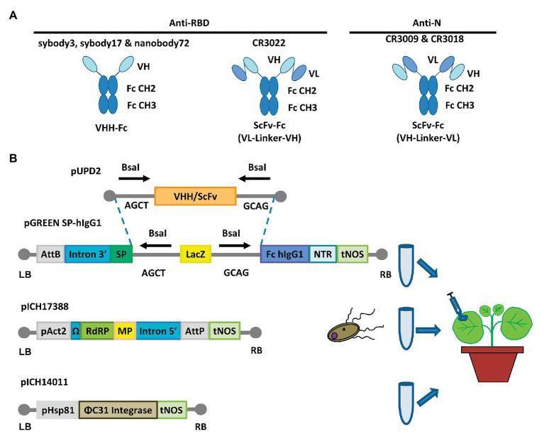
Educational diagram illustrating the recombinant production of anti-SARS-CoV-2 antibodies. Panel A shows structural schematics of engineered antibodies: VHH-Fc (single-domain nanobody fused to human IgG1 CH2-CH3) and two ScFv-Fc variants (single-chain variable fragments with VL-linker-VH or VH-linker-VL orientations). These constructs target the Receptor-Binding Domain (RBD) or Nucleocapsid (N) protein. Panel B details a plant-based transient expression system (MagnICON®). It depicts the cloning logic using BsaI restriction sites to insert VHH/ScFv sequences from a pUPD2 donor plasmid into a pGREEN SP-hIgG1 destination vector. The vector contains a signal peptide (SP), human Fc region, and regulatory elements (AttB, Intron 3', tNOS). The process demonstrates co-infiltration of Agrobacterium containing three plasmids (pGREEN, pICH17388 with RNA-dependent RNA polymerase and movement protein, and pICH14011 with ΦC31 integrase) into Nicotiana benthamiana leaves for in planta viral reconstruction and recombinant protein synthesis.
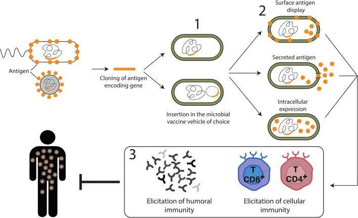
Educational diagram illustrating the development and mechanism of action of microbial-vectored vaccines. The process is divided into three sequential steps: (1) Antigen cloning and insertion, where a gene encoding a specific pathogen antigen (orange segment) is cloned and inserted into a microbial vaccine vehicle, such as a bacterium (green-outlined oval) or viral vector. (2) Antigen expression strategies, showcasing three modalities: surface antigen display (antigens anchored to the microbial membrane), secreted antigen (antigens released into the extracellular environment), and intracellular expression (antigens retained within the microbial cytoplasm). (3) Host immunization and immune response elicitation. This section demonstrates the induction of both humoral immunity, represented by Y-shaped antibodies, and cellular immunity, represented by CD8+ cytotoxic T cells and CD4+ helper T cells. The final stage illustrates how these elicited immune responses protect a human host from the target pathogen. The diagram highlights key concepts in recombinant vaccine technology and immunology, suitable for medical education regarding immunization strategies against viruses and intracellular pathogens.
Recombinant DNA (rDNA) Technology
1. Definition & Essence
2. Key Tools & Reagents
A. Restriction Enzymes (Restriction Endonucleases)
- Named after the organism of origin: EcoRI (from Escherichia coli strain R), BamHI (from Bacillus amyloliquefaciens)
- Nomenclature: First letter of genus + first two of species + strain + Roman numeral of discovery order
- They recognize palindromic sequences, typically 4-8 bp long
- Cutting frequency: A 4-cutter cuts on average every 4⁴ = 256 bp; a 6-cutter every 4⁶ = 4,096 bp
- Cuts produce two types of ends:
- Blunt ends (e.g., HpaI) - straight cut across both strands
- Sticky/cohesive ends (e.g., BamHI) - staggered cut leaving single-stranded overhangs that are particularly useful for constructing hybrid DNA molecules
B. DNA Ligase
C. Reverse Transcriptase
D. DNA Polymerase
3. Vectors
| Vector | Insert Size | Notes |
|---|---|---|
| Plasmids | 1-10 kb | Most common; carry antibiotic resistance for selection |
| Bacteriophage (λ phage) | 10-20 kb | Replaces non-essential phage genes with insert |
| Cosmids | 35-45 kb | Plasmid + phage cos sites; packaged as phage |
| BACs (Bacterial Artificial Chromosomes) | 100-300 kb | Used in Human Genome Project |
| YACs (Yeast Artificial Chromosomes) | 100-1000+ kb | Largest inserts; contain centromere, telomeres |
| Viral vectors | Variable | Retroviruses, adenoviruses, AAV - used for gene therapy |
- An origin of replication (ori)
- A selectable marker (antibiotic resistance gene)
- One or more unique restriction sites in a multiple cloning site (MCS)
4. Steps in Gene Cloning
- Obtain the gene of interest - by restriction digestion of genomic DNA, PCR amplification, or synthesis of cDNA from mRNA
- Cut both the insert and the vector with the same restriction enzyme(s) to produce compatible ends
- Ligate - DNA ligase joins insert into the vector, creating a recombinant plasmid
- Transform the recombinant plasmid into a host (usually E. coli) - bacteria take up the plasmid
- Select transformants - grow on antibiotic plates; only bacteria carrying the vector survive
- Screen colonies for the correct insert (blue-white selection, colony hybridization, PCR)
- Express the cloned gene and harvest the protein product
5. Libraries
Genomic Library
- Made by partially digesting the entire genome with restriction enzymes and cloning all fragments into vectors
- Contains all coding and non-coding sequences (introns, regulatory regions, etc.)
- Used when studying gene structure, regulation, or chromosomal context
cDNA Library
- Made by reverse-transcribing mRNA from a specific tissue at a specific time into cDNA
- Represents only expressed genes (no introns)
- Preferred for protein expression in bacteria and studying transcriptomes
6. The Polymerase Chain Reaction (PCR)
- Denaturation (~94°C) - separate double-stranded DNA
- Annealing (~55-65°C) - short oligonucleotide primers bind to complementary flanking sequences
- Extension (~72°C) - heat-stable Taq polymerase (from Thermus aquaticus) synthesizes new strands
- Diagnosis of infections (HIV, TB, COVID-19, Ebola)
- Prenatal genetic testing
- Forensic DNA fingerprinting (from a single cell)
- Quantitative gene expression (RT-PCR / qPCR)
- Site-directed mutagenesis
7. Detection & Analysis Techniques
Blotting Methods
| Technique | Target | Use |
|---|---|---|
| Southern blot (Edward Southern) | DNA | Gene copy number, deletions, mutations, RFLP |
| Northern blot | RNA | mRNA size, tissue expression levels |
| Western blot | Protein | Protein size, expression, antibody detection |
| Southwestern blot | Protein-DNA interaction | Transcription factor binding studies |
DNA Sequencing
- Sanger sequencing (chain termination) was the gold standard for decades
- Next-Generation Sequencing (NGS) / High-Throughput Sequencing (HTS) has reduced the cost of sequencing an entire human genome (3 × 10⁹ bp) from ~$350 million to less than $1,000 - a >99.97% cost reduction
8. Practical Applications
Pharmaceutical / Therapeutic Proteins
- Insulin (first rDNA drug, 1982) - replaced animal insulin for diabetes
- Human growth hormone - replaced pituitary-derived HGH
- Erythropoietin - for anemia
- Tissue plasminogen activator (tPA) - thrombolysis
- Interferons - antiviral and anticancer
- Clotting factors VIII and IX - for hemophilia
Vaccines
- Hepatitis B vaccine - first recombinant vaccine; HBsAg produced in yeast
- COVID-19 mRNA vaccines (Pfizer/Moderna) - encode spike protein
- Live-attenuated recombinant vaccines - safer versions using rDNA to delete virulence genes
- Recombinant viral vectors (e.g., Vaccinia, Adenovirus) can express foreign antigens for immunization
Diagnostics
- ELISA using recombinant antigens (HIV, hepatitis C)
- PCR-based detection of virtually any pathogen
- RFLP and DNA fingerprinting in forensic medicine
Gene Therapy
- Replacement of defective genes - applicable to single-gene disorders:
- Sickle cell disease, thalassemias
- Adenosine deaminase (ADA) deficiency (first human gene therapy trial, 1990)
- Cystic fibrosis, hemophilia
- Bone marrow precursor cells are introduced with corrected gene → resettle in marrow → produce functional protein
- iPSCs (Induced Pluripotent Stem Cells): adult somatic cells reprogrammed by transfecting cDNAs encoding transcription factors (Oct4, Sox2, Klf4, c-Myc) → can differentiate into any cell type
Agriculture & Other Applications
- Herbicide-resistant, drought-tolerant, nitrogen-fixing transgenic crops
- Crops with complete essential amino acid profiles (rice, wheat, corn)
- Forensic DNA analysis from single cells
9. Sensitivity & Precision of rDNA Technology
- Isolating a specific ~1,000 bp gene from the human genome requires discriminating 1 part in 1,000,000
- Identifying a regulatory region of ~10 bp requires sensitivity of 1 in 300,000,000
- Detecting the single-base mutation causing sickle cell anemia means identifying 1 wrong nucleotide in 3,000,000,000
10. Impact on Medicine
| Field | Impact |
|---|---|
| Hematology | Recombinant clotting factors, EPO, gene therapy for hemoglobinopathies |
| Endocrinology | Recombinant insulin, growth hormone, glucagon |
| Oncology | Monoclonal antibodies, tumor suppressor gene analysis |
| Infectious disease | Recombinant vaccines, rapid molecular diagnostics |
| Personalized medicine | Genomic sequencing guides drug choice and dosing |
| Forensics | DNA fingerprinting from single cells |
- Harper's Illustrated Biochemistry, 32nd Ed - Chapter 39 (Recombinant DNA Technology)
- Emery's Elements of Medical Genetics and Genomics - Therapeutic Applications
- Janeway's Immunobiology 10e - Recombinant vaccines
- Roitt's Essential Immunology - Attenuation by rDNA technology
4th ventricle of brain essay
fourth ventricle anatomy diagram rhomboid fossa
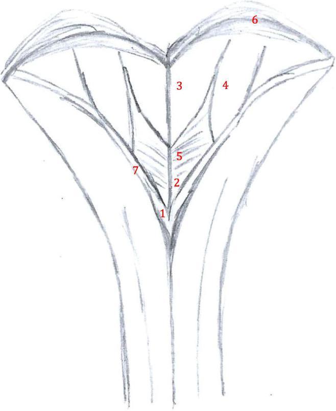
Anatomical diagram depicting the surgical anatomy of the rhomboid fossa, specifically the inferior aspect of the floor of the fourth ventricle. The illustration uses a schematic sketch format to identify key neuroanatomical landmarks. At the most inferior point of the ventricular floor is the obex (1), marking the transition to the central canal. Just superior to the obex lies the area postrema (2). The ventricular floor displays distinct trigones: the trigonum nervi hypoglossi (3) located medially, and the trigonum nervi vagi (5) situated laterally and slightly inferiorly. Further lateral is the area acustica (4). The superior boundary of this region is demarcated by the striae medullares (6), which run transversely across the floor. The lateral borders of the inferior ventricle are formed by the taenia (7). This diagram serves as an educational resource for identifying critical brainstem structures during sub-occipital surgical approaches, emphasizing the spatial relationships within the medulla oblongata's posterior surface.
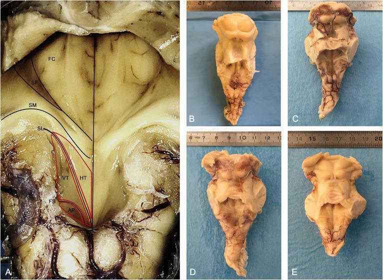
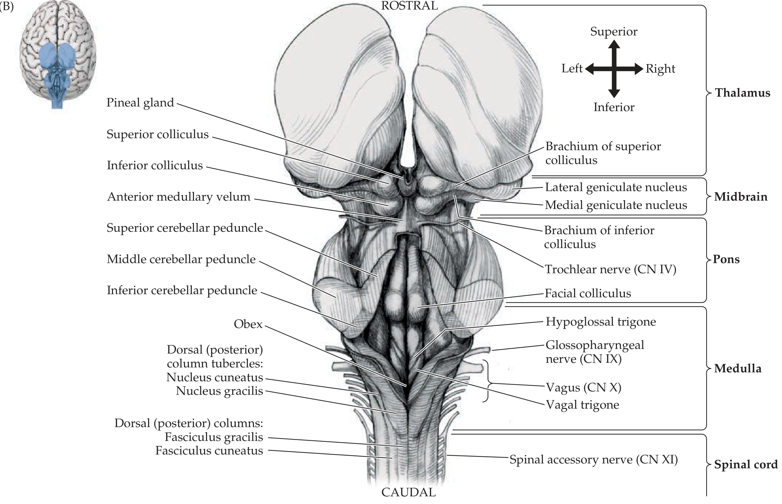
Anatomical anatomical photographs showing the posterior view of the human brainstem and the morphology of the rhomboid fossa (floor of the fourth ventricle). Image A provides a detailed, labeled view of the left half of the rhomboid fossa. Key neuroanatomical landmarks are identified: the Facial Colliculus (FC) and Superior Fovea (SF) in the pontine part; the Striae Medullares (SM) traversing horizontally; and the medullary part containing the Hypoglossal Trigone (HT), Vagal Trigone (VT or Ala Cinerea), and Area Postrema (AP). The Sulcus Limitans (SL) is marked as the longitudinal groove separating medial motor nuclei areas from lateral sensory areas, while the Funiculus Separans (FS) demarcates the Vagal Trigone from the Area Postrema. Images B, C, D, and E display four separate fixed human brainstem specimens sectioned at the mesencephalic level cranially and at the cerebellar peduncles laterally. These macro-anatomical views highlight the natural variation in the triangular shape of the rhomboid fossa and the prominence of surface landmarks like the superior and inferior colliculi. The collection serves as an educational reference for brainstem surface anatomy and the topographical arrangement of cranial nerve nuclei.
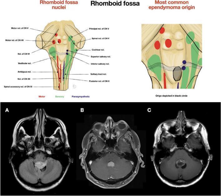
Educational medical composite image focusing on the neuroanatomy of the rhomboid fossa and its relationship to ependymoma. The upper section contains two anatomical diagrams: the left depicts the cranial nerve nuclei of the rhomboid fossa, color-coded by function (Motor in red, Sensory in green, Parasympathetic in blue). Key structures include motor nuclei of CN V, VI, VII, and XII; sensory nuclei like the vestibular and solitary tract; and parasympathetic salivary nuclei. The right diagram highlights the 'Most common ependymoma origin' located in the inferior portion of the fourth ventricle, medially near the obex. The bottom section displays three axial MRI brain images (A, B, C) showing variations in ependymoma presentation within the fourth ventricle. Image (A) shows an asymmetrical tumor extending laterally into the left foramen of Luschka. Images (B) and (C) demonstrate midline-restricted tumors. This clinical illustration serves to teach neurosurgical planning, showing how tumor location relative to the rhomboid fossa and lateral foramina dictates craniotomy approach and surgical navigation.
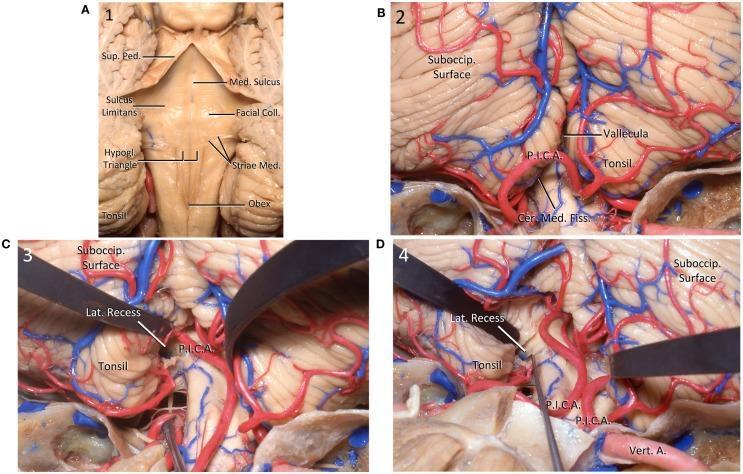
This anatomical illustration consists of four cadaveric dissection photographs (A-D) detailing the neuroanatomy of the posterior fossa, specifically the fourth ventricle and cerebellum. Figure A shows the posterior floor of the fourth ventricle (rhomboid fossa), highlighting the median sulcus, superior peduncle, facial colliculus, hypoglossal triangle, striae medullares, and the obex. Figure B depicts the posterior cerebellum, illustrating the suboccipital surface, cerebellar tonsils, and the course of the posterior inferior cerebellar artery (PICA) within the cerebellomedullary fissure. Figures C and D demonstrate a surgical approach via tonsillar retraction. In Figure C, the left tonsil is retracted superolaterally to expose the medulla and the lateral recess of the fourth ventricle. Figure D shows a dissector placed within the lateral recess, highlighting its spatial relationship to the PICA and the vertebral artery. These images serve as educational resources for neurosurgical planning and posterior fossa anatomy, emphasizing critical vascular and neural landmarks required for safe microsurgical navigation.
The Fourth Ventricle of the Brain - A Comprehensive Essay
1. Introduction
2. Embryological Development
- The cerebellum develops from thickenings of the dorsal parts of the alar plates. The cerebellar swellings initially project into the fourth ventricle. As they enlarge and fuse in the median plane, they overgrow the rostral half of the fourth ventricle and come to overlap the pons and medulla (The Developing Human)
- The choroid plexus of the fourth ventricle forms from the tela choroidea - the thin ependymal roof of the fourth ventricle covered externally by pia mater derived from hindbrain mesenchyme. Because of active proliferation of the pia, the tela choroidea invaginates the ventricle and differentiates into the choroid plexus (infoldings of choroidal arteries of the pia)
- The thin roof of the fourth ventricle evaginates at three locations; these outpouchings rupture to form the apertures: one median (foramen of Magendie) and two lateral (foramina of Luschka), allowing CSF to enter the subarachnoid space
- The cerebral aqueduct forms as the neural canal of the developing midbrain narrows, connecting the third and fourth ventricles
3. Gross Anatomy
Position and Shape
- Anteriorly (ventrally): the pons and the rostral medulla oblongata (forming its floor)
- Posteriorly (dorsally): the cerebellum (forming its roof)
Boundaries
| Boundary | Structure |
|---|---|
| Anterior wall (floor) | Pons (rostrally) + medulla oblongata (caudally) |
| Posterior wall (roof) | Cerebellum (superior/anterior medullary velum rostrally; inferior medullary velum + tela choroidea caudally) |
| Lateral walls | Superior, middle, and inferior cerebellar peduncles |
| Apex (tip of roof) | Fastigium |
| Inferior angle | Obex |
Communications
| Communication | Structure |
|---|---|
| Superiorly | Cerebral aqueduct (of Sylvius) - connects to 3rd ventricle through the midbrain |
| Inferiorly | Central canal of the spinal cord (closed/obliterated in most adults) |
| Into subarachnoid space | Foramen of Magendie (median aperture - single, midline in roof) |
| Into subarachnoid space | Foramina of Luschka (lateral apertures - two, one on each side) |
4. The Floor of the Fourth Ventricle (Rhomboid Fossa)
Key Surface Landmarks (from rostral to caudal)
- Medial motor areas (from basal plate)
- Lateral sensory areas (from alar plate)
- Facial colliculus: A prominent bulge on each side, formed by the abducens nucleus (CN VI) and the fibers of the facial nerve (CN VII) looping around it. This is an important landmark - damage here affects both CN VI (lateral gaze) and CN VII (facial movement)
- Superior fovea: A small depression in the sulcus limitans at the level of the facial colliculus
- Striae medullares (acoustic striae): Horizontal white fiber bundles that cross the floor transversely, marking the junction between pontine and medullary parts
- Hypoglossal trigone: An eminence overlying the hypoglossal nucleus (CN XII), responsible for tongue movements
- Vagal trigone (ala cinerea): Lateral to the hypoglossal trigone, overlying the dorsal motor nucleus of the vagus (CN X) - important in parasympathetic control of thoracic and abdominal viscera
- Area acustica: An area lateral to the sulcus limitans overlying the vestibular nuclei
- Area postrema: A small, darkly pigmented region at the caudal end, near the obex. It contains the chemoreceptor trigger zone (CTZ) for vomiting and is a circumventricular organ (lacks the blood-brain barrier)
- Obex: A small tongue-like fold of tissue at the caudal extreme of the fourth ventricle, marking the point where the fourth ventricle narrows into the central canal of the spinal cord
5. The Roof of the Fourth Ventricle
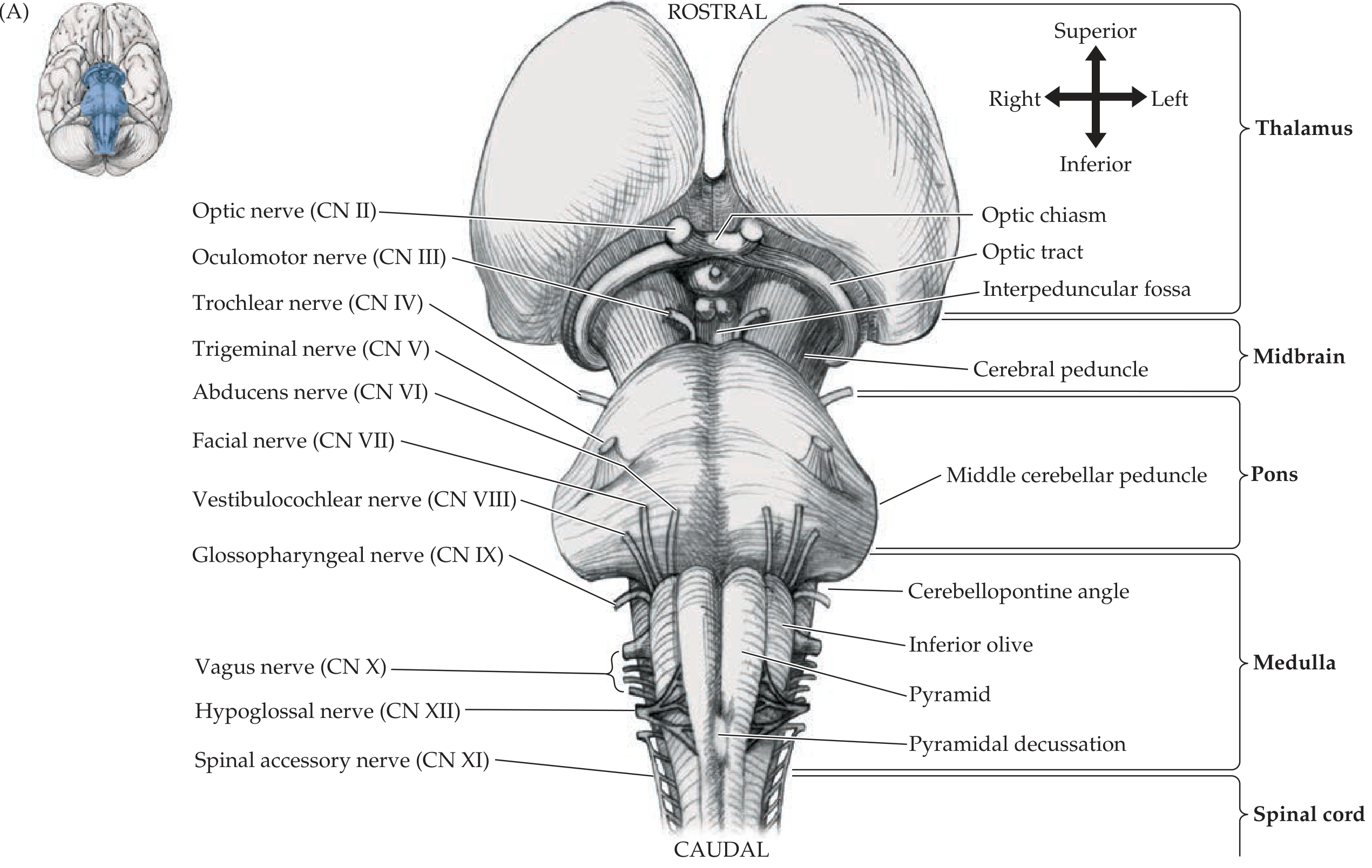
- Superior (anterior) medullary velum: A thin sheet of white matter stretching between the two superior cerebellar peduncles rostrally. The trochlear nerve (CN IV) - the only cranial nerve to exit dorsally - decussates within and emerges from it
- Inferior (posterior) medullary velum: Thin, paired sheets of cerebellar white matter caudally
- Tela choroidea: A two-layered membrane (ependyma + pia) forming the lower part of the roof, from which the choroid plexus is derived
- The fastigium is the tent-like apex of the roof, deepest into the cerebellum
6. Lateral Recesses
7. Choroid Plexus and CSF Production
- Lies along a portion of the tent-shaped roof (tela choroidea)
- Extends laterally into both foramina of Luschka, often projecting into the subarachnoid space (tufts of choroid plexus visible on imaging and can calcify with age)
- Blood supply: Branches of the posterior inferior cerebellar artery (PICA), anterior inferior cerebellar artery (AICA), and the superior cerebellar artery (SCA)
8. CSF Circulation
- Lateral ventricles (choroid plexus produces CSF)
- → Foramina of Monro (interventricular foramina) → 3rd ventricle
- → Cerebral aqueduct of Sylvius (through midbrain) → 4th ventricle
- → Foramen of Magendie (midline) and Foramina of Luschka (bilateral) → Subarachnoid space
- → Circulates around brain and spinal cord
- → Reabsorption: primarily via cranial nerve sheaths, cribriform plate into lymphatics, spinal epidural space, and paraarterial/perivenous spaces (the glymphatic system). The role of arachnoid granulations in CSF reabsorption remains debated in current literature (Grainger & Allison's Diagnostic Radiology)
9. Relations and Applied Anatomy
Cerebellar Peduncles (Lateral Relations)
| Peduncle | Connection | Main tracts |
|---|---|---|
| Superior cerebellar peduncle (brachium conjunctivum) | Cerebellum → Midbrain | Dentatorubrothalamic tract (main output) |
| Middle cerebellar peduncle (brachium pontis) | Pons → Cerebellum | Pontocerebellar fibers (largest peduncle) |
| Inferior cerebellar peduncle (restiform + juxtarestiform body) | Medulla → Cerebellum | Spinocerebellar tracts, olivocerebellar fibers |
Cranial Nerves of the 4th Ventricle Region
10. Imaging Anatomy
- Sagittal T2: Shows the fastigium (tented apex), the foramen of Magendie, and the CSF flow void in the aqueduct
- Coronal T2: Demonstrates the rhomboid shape and lateral recesses funneling into the foramina of Luschka
- Axial MRI: Shows both the foramen of Magendie (midline) and foramina of Luschka (bilateral)
11. Applied Pathology
Hydrocephalus
- Aqueductal stenosis (narrows cerebral aqueduct → dilates 4th, 3rd, and lateral ventricles)
- Obstruction of the foramina of Magendie and Luschka
Dandy-Walker Malformation
- Agenesis or hypoplasia of the cerebellar vermis
- Cystic dilation of the fourth ventricle
- Enlarged posterior fossa Results in hydrocephalus and cerebellar dysfunction.
Tumours of the Fourth Ventricle
| Tumour | Origin | Key Features |
|---|---|---|
| Medulloblastoma | Primitive neuroectodermal (vermis) | Most common malignant childhood brain tumor; hyperdense midline mass on CT; abutting the roof of the 4th ventricle; does NOT tend to extrude through foramina |
| Ependymoma | Ependymal cells lining the floor | Arises from the floor (near obex); characteristically extrudes ("squeezes") through foramina of Luschka and Magendie into the subarachnoid space ("toothpaste" sign); may invade the medulla |
| Choroid plexus papilloma | Choroid plexus epithelium | 40% arise in the 4th ventricle; papillary growths that overproduce CSF, causing hydrocephalus |
Wernicke's Encephalopathy
Multiple Sclerosis (MS) Plaques
Ependymitis / Ventriculitis
12. Surgical Significance
Suboccipital Craniotomy
Lateral Recess / Foramen of Luschka
- Auditory brainstem implant (ABI) placement - the cochlear nucleus is found in the floor of the lateral recess, entered through the foramen of Luschka
- Drainage of fourth ventricular hematomas
Fourth Ventriculostomy / Shunting
13. Summary
- Anatomically: a rhomboid CSF cistern bounded by pons/medulla anteriorly and cerebellum posteriorly, with its tented roof, lateral recesses, and three apertures
- Physiologically: a critical link in CSF circulation; its choroid plexus contributes significantly to CSF production
- Developmentally: derived from the rhombencephalon; its roof gives rise to the choroid plexus and forms the foramina of Magendie and Luschka
- Clinically: obstruction causes hydrocephalus; the floor contains life-critical cranial nerve nuclei; the region is home to important posterior fossa tumors (medulloblastoma, ependymoma), demyelinating plaques, and metabolic lesions
- Neuroanatomy through Clinical Cases, 3rd Edition - Chapters 5 and 12
- Grainger & Allison's Diagnostic Radiology - Chapter 53: Ventricular System and Subarachnoid Space
- The Developing Human: Clinically Oriented Embryology - Chapter 17: Metencephalon and Choroid Plexus
- Medical Physiology (Boron & Boulpaep) - Chapter 11: CSF and Ventricular System
- Adams and Victor's Principles of Neurology, 12th Ed - Fourth ventricular tumors
- Bradley and Daroff's Neurology in Clinical Practice - Posterior fossa tumors
Short note on fornix
fornix brain anatomy hippocampus mammillary bodies diagram
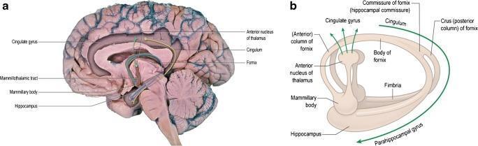
Educational neuroanatomical visual containing a clinical photograph and a schematic diagram (a and b) illustrating the Papez circuit and limbic system connectivity within the human brain. Image (a) presents a medial view of the right cerebral hemisphere with color-coded fiber tracts superimposed. It depicts the fornix (blue) originating from the hippocampal fimbria and terminating at the mammillary bodies, and the mammillothalamic tract of Vicq d’Azyr (red) connecting the mammillary bodies to the anterior nucleus of the thalamus. The cingulate gyrus and cingulum (green) are highlighted as key components projecting back to the hippocampal formation. Diagram (b) provides a simplified schematic of these relationships, labeling the crus, body, and anterior columns of the fornix, as well as the hippocampal commissure. The visual emphasizes the closed-loop functional anatomy of the Papez circuit, which is critical for memory processing and emotional regulation. Key structures shown include the hippocampus, parahippocampal gyrus, thalamus, and mammillary bodies. This content is suitable for intermediate to advanced medical education regarding neuroanatomy and limbic system pathways.
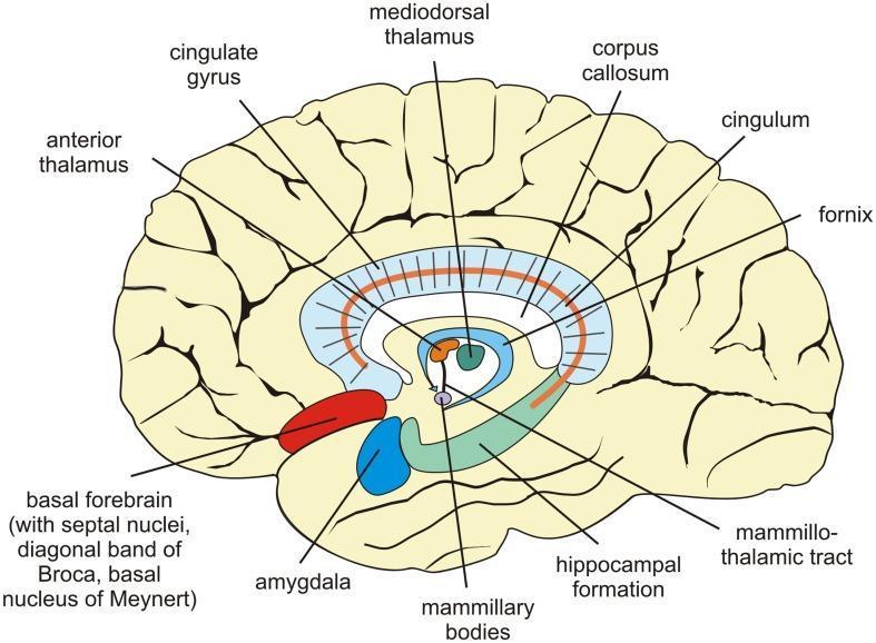
This anatomical diagram presents a mid-sagittal section of the human brain, specifically highlighting the structures of the limbic system and diencephalon involved in memory processing. Key anatomical landmarks include the C-shaped corpus callosum and the overlying cingulate gyrus. Below these, the fornix and cingulum are illustrated as major white matter tracts. Central structures are color-coded for clarity: the anterior and mediodorsal nuclei of the thalamus are depicted in orange and green respectively, while the mammillary bodies are shown at the terminus of the mammillothalamic tract. In the temporal region, the amygdala (red) and the hippocampal formation (teal) are highlighted to demonstrate their spatial orientation, with the amygdala positioned anterior and superior to the hippocampus. Other indicated regions include the basal forebrain, encompassing the septal nuclei and basal nucleus of Meynert. This visual is designed for medical education to illustrate the neuroanatomical circuits essential for episodic-autobiographical memory encoding and long-term storage.
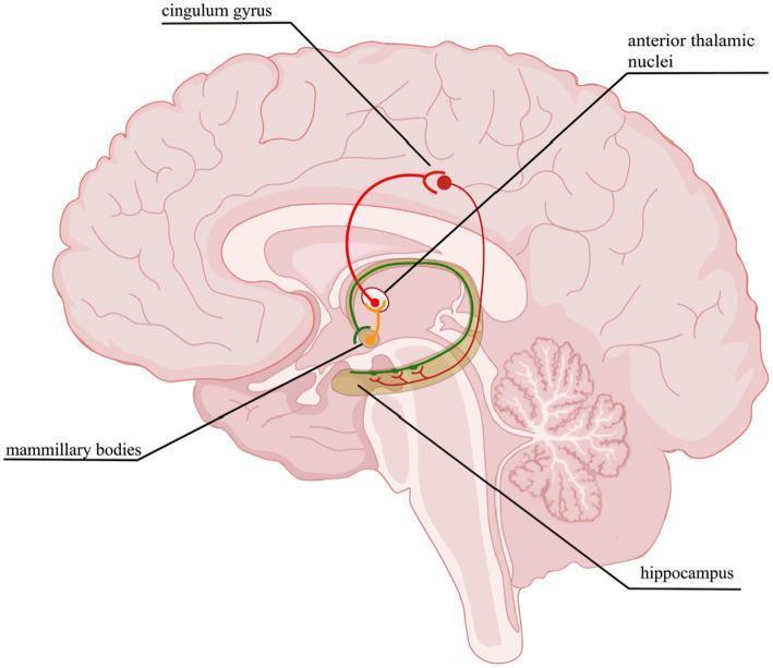
This medical anatomical diagram illustrates the Papez circuit, a fundamental neural pathway of the limbic system involved in emotional expression and memory consolidation. Depicted on a sagittal section of the human brain, the diagram highlights the sequential loop connecting key subcortical and cortical structures. The circuit begins at the hippocampus, which projects to the mammillary bodies via the fornix. From the mammillary bodies, the mammillothalamic tract reaches the anterior thalamic nuclei. The loop continues as the thalamocingulate tract extends to the cingulum gyrus (cingulate cortex). Finally, the circuit completes by projecting from the cingulate cortex back to the hippocampus via the parahippocampal gyrus. Key labeled anatomical landmarks include the cingulum gyrus, anterior thalamic nuclei, mammillary bodies, and hippocampus. This illustration serves as an educational tool for neuroanatomy and clinical neurology, demonstrating the functional connectivity disrupted in conditions such as hypoxic-ischemic encephalopathy and various forms of amnesia.
Short Note on the Fornix
Definition and Etymology
Gross Anatomy and Course
1. Alveus
- The fibers of the hippocampal pyramidal neurons converge on the ventricular surface of the hippocampus as a thin white sheet called the alveus
- These fibers sweep medially to form the next segment
2. Fimbria
- As the alveus fibers gather medially, they form a discrete bundle along the medial edge of the hippocampus called the fimbria of the fornix
- The fimbria is continuous with the crus of the fornix posteriorly
3. Crus (Crura) - "legs"
- The two crura leave the hippocampal formation and curve upward and forward beneath the corpus callosum
- As the crura approach each other in the midline, they are connected by the hippocampal commissure (commissure of the fornix), which allows fibers from one hippocampus to reach the contralateral side
- The crura then converge to form the body
4. Body
- The body of the fornix runs forward as a single midline structure on the undersurface of the corpus callosum, just above the third ventricle
- It is visible on sagittal MRI between the corpus callosum above and the thalamus below
- The body then divides anteriorly into two columns
5. Columns (Anterior Pillars)
- The two columns of the fornix curve downward in front of the interventricular foramina of Monro, pass through the hypothalamus, and divide at the anterior commissure into:
- Precommissural fibers (pass anterior to the anterior commissure)
- Postcommissural fibers (pass posterior to the anterior commissure)
Fiber Composition and Targets
| Division | Fiber Origin | Destination | Function |
|---|---|---|---|
| Postcommissural fornix (majority) | Subiculum | Medial and lateral mammillary nuclei of hypothalamus | Core Papez circuit for episodic memory |
| Precommissural fornix | Subiculum + hippocampus | Lateral septal nucleus | Limbic-autonomic integration |
| Direct thalamic fibers | Subiculum | Anterior thalamic nucleus | Direct contribution to Papez circuit |
The Papez Circuit
Relations
- Superiorly: Corpus callosum (body of fornix runs along its undersurface)
- Inferiorly: Thalamus and third ventricle (body of fornix forms part of the roof of the third ventricle)
- Anteriorly: Anterior commissure (columns divide here into pre- and postcommissural)
- Laterally: Lateral ventricles (fornix follows their curve)
- The septum pellucidum is a thin membrane stretching between the fornix and the corpus callosum, forming the medial walls of the frontal horns of the lateral ventricles
Applied / Clinical Significance
1. Amnesia - Korsakoff Syndrome
2. Fornix Transection / Surgical Injury
3. Alzheimer's Disease
4. Deep Brain Stimulation (DBS) of the Fornix
5. Imaging
Summary
| Feature | Detail |
|---|---|
| Type | White matter tract (limbic system) |
| Origin | Hippocampal formation (alveus → fimbria) |
| Segments | Alveus → Fimbria → Crus → Body → Columns |
| Main target | Mammillary bodies (postcommissural) |
| Other targets | Septal nuclei, anterior thalamus |
| Key commissure | Hippocampal commissure (between crura) |
| Key circuit | Papez circuit for memory |
| Clinical importance | Korsakoff amnesia, Alzheimer's, fornix DBS |
- Neuroanatomy through Clinical Cases, 3rd Edition - Chapter 18: The Fornix and Medial Diencephalic Memory Pathways
- Neuroscience: Exploring the Brain, 5th Edition - Forebrain Structures
- Ganong's Review of Medical Physiology, 26th Edition - Hippocampus and Medial Temporal Lobe
- Kaplan & Sadock's Comprehensive Textbook of Psychiatry - Deep Brain Stimulation and Memory
Neural crest cells and it’s derivatives
neural crest cells migration derivatives diagram embryology
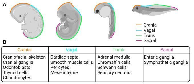
This educational infographic illustrates the comparative embryology and physiological derivatives of the neural crest (NC). Panel A features anatomical diagrams of four animal models—human, mouse, zebrafish, and frog—during various stages of embryogenesis. Color-coded lines map the longitudinal distribution of specific NC populations: Cranial (orange), Vagal (light blue), Trunk (green), and Sacral (purple). Panel B provides a structured comparison chart detailing the differentiated adult tissue derivatives of each population. The Cranial NC is linked to the craniofacial skeleton, cranial ganglia, odontoblasts, thyroid cells, and chondrocytes. The Vagal NC contributes to cardiac septa, smooth muscle cells, pericytes, and mesenchyme. The Trunk NC gives rise to the adrenal medulla, chromaffin cells, Schwann cells, and sensory neurons. The Sacral NC develops into enteric and sympathetic ganglia. This visual serves as a summary of developmental biology, emphasizing the evolutionary conservation of neural crest migration and its diverse contributions to vertebrate anatomy across species.
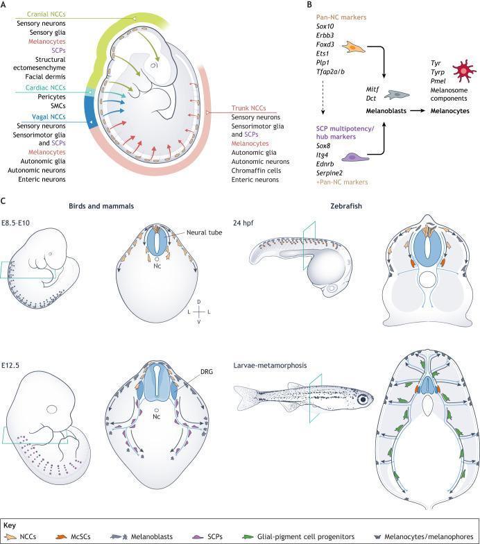
This composite educational diagram illustrates the embryonic origins and differentiation pathways of melanocytes from neural crest cells (NCCs). Section A displays a sagittal view of an embryo, mapping the regional fates of cranial, cardiac, vagal, and trunk NCCs. Cranial NCCs differentiate into sensory neurons, glia, melanocytes, and facial ectomesenchyme. Vagal and trunk NCCs contribute to the enteric and autonomic nervous systems and melanocyte lineages. Section B outlines the molecular progression from Pan-NC markers (Sox10, Foxd3) to Schwann cell precursors (SCPs) and melanoblasts, triggered by the Mitf transcription factor to produce mature melanocytes. Section C provides comparative embryology across species. In birds and mammals, it shows melanoblast migration from the neural tube at E8.5-E10 toward the dorsal root ganglion (DRG) and epidermis by E12.5. In zebrafish, it highlights the transition from 24 hours post-fertilization (hpf) to metamorphosis, showing the emergence of melanophores from glial-pigment cell progenitors along peripheral nerves. Key anatomical landmarks include the neural tube, notochord (Nc), and DRG, emphasizing the dual origin of melanocytes from direct NCC migration and nerve-associated progenitors.
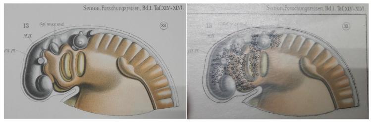
This comparative anatomical diagram illustrates the embryological development of the pharyngeal arches, using a stage 33 lungfish embryo as a model for vertebrate morphogenesis. The image is presented in two panels: the left displays a shaded specimen with key landmarks labeled, including 'Gl. Pl.' (Glandular Plate), 'M.H.' (Midbrain/Mittelhirn), and 'Ggl. max. md.' (Maxillomandibular ganglion). The right panel utilizes a historic transparent overlay method to demonstrate the migration of neural crest cells, represented by small black ovals. These cells are concentrated along the cephalic region and the dorsal-ventral axis of the pharyngeal arches, which appear as segmented bulges. The illustration highlights the pathways of migratory cells from the neural tube into the pharyngeal apparatus, which eventually contribute to the development of the aorticopulmonary septum, cardiac outflow tract, and craniofacial structures. This comparison is an educational resource for developmental biology and embryology, focusing on neural crest cell distribution and the evolution of vertebrate circulatory systems.
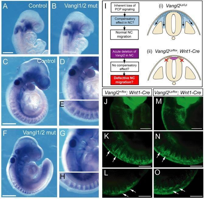
Educational panel of mouse embryology demonstrating neural crest (NC) cell migration patterns under different genetic conditions. Panels A-H show whole-mount in situ hybridization (WISH) for Erbb3 at E8.5 (A, B) and E9.5 (C-H) stages. Control embryos (A, C, D, E) are compared with Vangl1/2 double mutants (B, F, G, H). Despite the open neural tube in double mutants, Erbb3-positive NC cell distribution in the cranial and trunk regions appears normal and comparable to controls. Panel I provides a conceptual flowchart and anatomical diagrams of the neural tube cross-section, illustrating hypotheses regarding Planar Cell Polarity (PCP) signaling and potential compensatory mechanisms. Panels J-O utilize YFP lineage labeling regulated by Wnt1-Cre to visualize NC migration at E9.5. Comparison between control (J-L) and acute Vangl2 downregulation (M-O) shows identical streams of YFP-positive NC cells migrating from the trunk neural tube (white arrows). The figure demonstrates that Vangl1 and Vangl2 functions are not essential for the specification or migration of mouse neural crest cells in vivo.
Neural Crest Cells and Their Derivatives
1. Introduction
2. Origin and Formation
Induction (Week 3 of Embryogenesis)
- The notochord induces the overlying ectoderm to form the neural plate (neuroectoderm)
- BMP (Bone Morphogenetic Protein) concentration gradients are the key inductive signal:
- Very high BMP → epidermis
- Intermediate BMP (at the neural plate border) → neural crest
- Very low BMP → neural ectoderm (CNS)
- BMP inhibitors Noggin and Chordin fine-tune these gradients
- Intermediate BMP levels, combined with FGF and WNT signals, activate transcription factors PAX3 and others that "specify" the neural plate border
Transcription Factors Governing NCC Specification
| Signal | Transcription Factor | Role |
|---|---|---|
| BMP gradient + FGF + WNT | PAX3 | Specifies neural plate border |
| PAX3 downstream | SNAIL, FOXD3 | Specifies cells as neural crest |
| FOXD3 | SLUG | Promotes delamination and EMT |
| Later NCC | MITF | Melanocyte lineage specification |
| Pan-NCC marker | SOX10 | Maintains multipotency |
Epithelial-to-Mesenchymal Transition (EMT)
3. Timing of Departure
- Cranial NCCs: depart from neural folds before neural tube closure in the head region - they are highly migratory and contribute to the craniofacial skeleton
- Trunk NCCs: depart after neural tube closure; migration is delayed until tube closure is complete
4. Migration Pathways
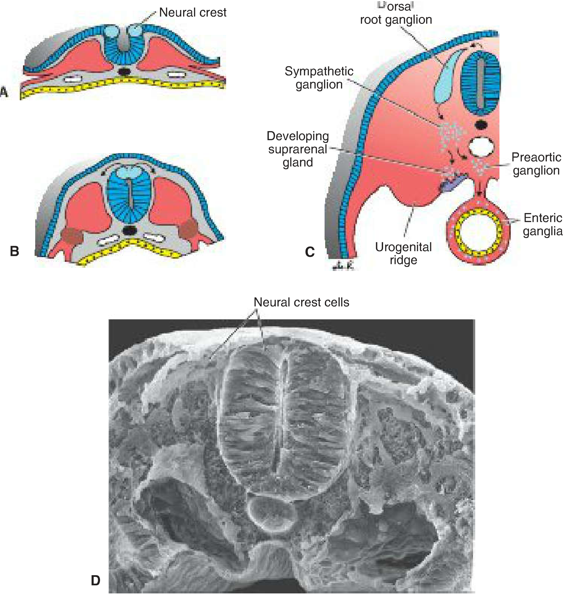
Pathway 1: Dorsolateral (Dorsal) Pathway
- NCCs migrate between the ectoderm and the dermomyotome
- Cells become melanoblasts → differentiate into melanocytes in skin and hair follicles
- Enter the ectoderm through holes in the basal lamina
Pathway 2: Ventrolateral Pathway
- NCCs migrate through the anterior (rostral) half of each somite
- Form sensory neurons of the dorsal root ganglia (DRG / spinal ganglia)
- The restriction to the anterior half of somites creates the segmental pattern of the peripheral nervous system
Pathway 3: Ventral Pathway
- NCCs migrate ventrally past the notochord and aorta
- Become:
- Sympathetic chain ganglia (paravertebral)
- Preaortic ganglia (prevertebral)
- Chromaffin cells of the adrenal medulla
- Enteric ganglia of the gut wall
5. Derivatives of Neural Crest Cells
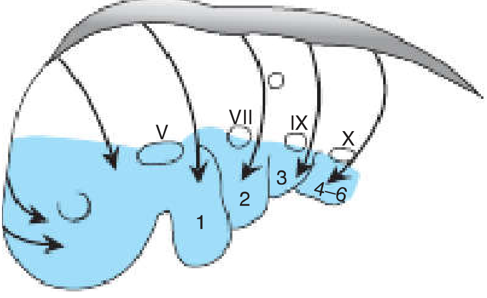
A. Cranial Neural Crest (Cephalic)
| Derivative | Structure Formed |
|---|---|
| Craniofacial skeleton | Bones of the face and skull (frontal, nasal, lacrimal, zygomatic, maxilla, mandible, vomer) |
| Cartilages | Meckel's cartilage, thyroid cartilage (parts), cricoid, arytenoids |
| Hyoid bone | Greater cornu and body of hyoid |
| Dermis | Dermis of the face and neck |
| Teeth | Odontoblasts (form dentin of teeth) |
| Glands | Stroma of salivary, lacrimal, thymus, thyroid, and pituitary glands |
| Endocrine | C cells (parafollicular cells) of the thyroid gland - produce calcitonin |
| Cranial ganglia | Ganglia of CN V (trigeminal), VII (geniculate), IX (superior), X (jugular) |
| Connective tissue | Stroma of thymus; connective tissue of pharyngeal arches |
B. Cardiac Neural Crest (Vagal)
- NCCs from the vagal region (somites 1-7) migrate into the conotruncal endocardial cushions of the heart
- Form the aorticopulmonary septum that divides the truncus arteriosus into the aorta and pulmonary trunk
- Contribute smooth muscle cells and pericytes to the walls of the great vessels
C. Trunk Neural Crest
| Derivative | Structure |
|---|---|
| Sensory neurons | Dorsal root ganglia (spinal ganglia) - unipolar neurons giving dorsal roots of spinal nerves |
| Autonomic - sympathetic | Sympathetic chain ganglia (paravertebral) and preaortic ganglia |
| Autonomic - parasympathetic | Postganglionic parasympathetic neurons of thoracic and abdominal viscera |
| Adrenal medulla | Chromaffin cells (modified sympathetic neurons) - secrete epinephrine and norepinephrine |
| Schwann cells | Myelinate peripheral axons; form the neurolemma |
| Satellite cells | Surround cell bodies in peripheral ganglia |
| Melanocytes | Pigment cells of the skin, hair follicles, inner ear (stria vascularis), uveal tract of the eye |
D. Sacral Neural Crest
- Contributes to pelvic ganglia and the most distal portion of the enteric nervous system (hindgut ganglia)
- Joins the vagal NCCs to complete the enteric nervous system
E. Special Derivatives
| Derivative | Notes |
|---|---|
| Meninges (forebrain) | Pia mater and arachnoid (leptomeninges) of the cranial region derived from cranial NCC; spinal meninges from NCC + mesenchyme |
| Smooth muscle | Tunica media of arteries supplying the face and forebrain |
| Corneal stroma and endothelium | Cranial NCC migrate into the anterior segment of the eye |
| Ciliary ganglion | Parasympathetic ganglion in the orbit |
6. Complete List of Neural Crest Derivatives (Table 6.1, Langman's)
- Connective tissue and bones of the face and skull
- Cranial nerve ganglia
- C cells (parafollicular cells) of the thyroid gland
- Conotruncal septum in the heart
- Odontoblasts
- Dermis of face and neck
- Spinal (dorsal root) ganglia
- Sympathetic chain and preaortic ganglia
- Parasympathetic ganglia of the GI tract (enteric nervous system)
- Adrenal medulla (chromaffin cells)
- Schwann cells
- Glial cells (satellite cells)
- Meninges (forebrain)
- Melanocytes
- Smooth muscle cells of blood vessels of the face and forebrain
7. Clinical Significance - Neurocristopathies
| NCC Derivative Affected | Disease/Condition |
|---|---|
| Enteric nervous system (ganglia absent from colon) | Hirschsprung disease (aganglionic colon) - colonic obstruction in neonates |
| Melanocytes | Waardenburg syndrome - white forelock, heterochromia iridis, deafness (stria vascularis melanocyte loss) |
| Melanocytes | Malignant melanoma - most lethal skin cancer |
| Melanocytes | Albinism - failure of melanin synthesis |
| Adrenal medulla chromaffin cells | Pheochromocytoma - catecholamine-secreting tumour |
| Sympathetic neuroblasts | Neuroblastoma - most common solid malignancy of infancy |
| Schwann cells / glial cells | Neurofibromatosis type 1 (von Recklinghausen disease) - NF1 mutation |
| C cells of thyroid | Medullary thyroid carcinoma - calcitonin-secreting; part of MEN2 |
| Cranial NCC (conotruncal heart) | DiGeorge syndrome (22q11.2 deletion) - conotruncal defects, thymic aplasia, parathyroid aplasia |
| Cranial NCC (first arch) | Treacher Collins syndrome (TCOF1 mutation) - mandibular/zygomatic hypoplasia, cleft palate, ear defects |
| Cranial NCC (mandibular arch) | Robin sequence - micrognathia, cleft palate, glossoptosis |
| Enteric NCC + Schwann cells | Multiple endocrine neoplasia type 2B (MEN2B) - mucosal neuromas |
| Cranial NCC | Alcohol embryopathy / retinoic acid teratogenesis - NCCs are particularly sensitive to alcohol and excess retinoic acid |
8. Evolutionary Significance
Summary Diagram Concept
Neural Plate Border (BMP intermediate gradient)
       ↓
NCC Specification (PAX3 → SNAIL/FOXD3/SLUG)
       ↓
Epithelial-to-Mesenchymal Transition (EMT)
       ↓
MIGRATION along three pathways:
  ├─ Dorsolateral → MELANOCYTES
  ├─ Ventrolateral → DORSAL ROOT GANGLIA (sensory neurons)
  └─ Ventral → SYMPATHETIC GANGLIA / ADRENAL MEDULLA / ENTERIC NS

CRANIAL NCCs → FACE, SKULL, TEETH, CRANIAL GANGLIA, C-CELLS
CARDIAC NCCs → AORTICOPULMONARY SEPTUM
SACRAL NCCs  → HINDGUT ENTERIC GANGLIA
- Langman's Medical Embryology - Chapter 6: Neural Crest Cells (Table 6.1 - Neural Crest Derivatives)
- The Developing Human: Clinically Oriented Embryology - Chapter 17: Spinal Ganglia and Meninges
- THIEME Atlas of Anatomy: General Anatomy and Musculoskeletal System - Neural Crest Derivatives and Diseases
Cadaver is our first torcher explain
cadaver is our first teacher meaning medical education anatomy dissection significance
https://indianmentalhealth.com/pdf/2018/new-issue/1-4-2018/1…
"The Cadaver Is Our First Teacher" - A Medical Education Essay
"Hic locus est ubi mors gaudet succurrere vitae" "This is the place where death rejoices to help those who live"
- Latin inscription found above many anatomy dissection halls worldwide
1. Understanding the Phrase
2. Historical Background of Cadaveric Dissection
| Era | Development |
|---|---|
| ~300 BC | Herophilus and Erasistratus of Alexandria - first systematic human dissections |
| Renaissance (1543) | Andreas Vesalius - De Humani Corporis Fabrica - revolutionized anatomy using cadaveric dissection; corrected centuries of Galenic error |
| Medieval period | Dissection was banned by the Church as offensive and sacrilegious |
| 18th-19th century | Body snatching ("resurrection men") became rampant due to shortage of legal cadavers |
| 1832 | Anatomy Act (UK) - legalized use of unclaimed bodies for dissection |
| Modern era | Voluntary body donation programs replaced involuntary sources; ethical frameworks developed |
3. What the Cadaver Teaches - A Multi-Dimensional Education
A. Anatomical Knowledge (The Core Lesson)
- Three-dimensional spatial relationships between structures - how the femoral nerve, artery, and vein relate to each other in the femoral triangle; how the brachial plexus winds around the subclavian artery
- Normal anatomical variation - textbooks show the "standard" anatomy, but every cadaver reveals that real human bodies vary considerably. One person's median nerve may bifurcate early; another's appendix may be retrocaecal. This prepares students for the variability they will encounter in operating theatres and clinics
- Tissue texture and feel - the density of fascia, the slipperiness of a serous membrane, the consistency of a healthy liver versus a cirrhotic one cannot be taught from pictures
- Depth perception - understanding how deep the ureter lies, how thin the tympanic membrane is, how close the facial nerve is to the parotid gland surface - all require hands-on experience
- Surgical planes - recognizing the avascular tissue planes that surgeons use for safe dissection is only learned by actually finding them in tissue
B. Clinical Correlation
- Dissecting the axilla teaches them why axillary lymph node clearance in breast surgery risks injuring the long thoracic nerve, causing winged scapula
- Exposing the femoral triangle teaches them where to compress the femoral artery in bleeding trauma
- Identifying the recurrent laryngeal nerve in the tracheoesophageal groove teaches them why thyroid surgeons must protect it to prevent hoarseness
C. Professional Attitudes and Values
- Respect for human dignity - the body is treated with care and reverence even in death
- Empathy - realizing that this was once a living person, with a family, a history, a name
- Detachment - learning to perform a necessary technical task without being overwhelmed by emotion; a balance every physician needs
- Teamwork - dissection is done in groups, requiring communication, shared work, and collaborative learning
- Responsibility - the body is entrusted to the students; carelessness or disrespect is unacceptable
D. Confronting Death
- It allows students to process their own emotional responses to death in a structured environment
- It begins the desensitization necessary for clinical practice - not a cold, unfeeling desensitization, but the professional ability to function clearly in the presence of death or severe injury
- Students often report that this experience fundamentally changed their understanding of mortality and the preciousness of life
4. The Body Donor - A Selfless Teacher
- The "teacher" made an active, voluntary, ethical choice to teach
- Many medical colleges around the world now hold annual memorial services for body donors (e.g., Mayo Medical College, Rochester, USA) where students, faculty, and the families of donors gather to express gratitude
- In India, at some colleges (e.g., St. John's Medical College, Bangalore), anatomy dissection classes begin with a prayer thanking the donor
- At other colleges, a cadaveric oath is administered to first-year students, in which they pledge to honor the dignity and integrity of the human remains they will work on
5. The Dissection Hall as a Transformative Space
- The Latin proverb above its doors reminds students of their purpose
- The smell of formalin becomes inseparably linked to the memory of learning
- Nervousness and fear give way, over weeks and months, to confidence and curiosity
- The human body, once overwhelming, becomes a map that a future physician knows intimately
6. Modern Relevance
- No technology fully replaces the tactile, sensory, three-dimensional experience of real dissection
- Cadaveric dissection uniquely combines anatomy, clinical application, and professional attitude formation in one setting
- Surgical simulation on fresh/perfused cadavers (non-embalmed) has actually increased in importance for training surgeons in robotic and minimally invasive techniques
7. Summary
| Dimension | What the Cadaver Teaches |
|---|---|
| Cognitive | 3D anatomy, variation, spatial relationships, surgical planes |
| Clinical | Surface anatomy, procedural landmarks, tissue feel |
| Attitudinal | Respect, empathy, professional detachment |
| Psychological | First encounter with death, emotional resilience |
| Ethical | Gratitude to donors, dignity of the human body |
| Social | Teamwork, communication, shared responsibility |
"Let conversation cease. Let laughter flee. This is the place where death delights to help the living."
- Inscription in anatomy dissection halls worldwide