
Search some dermatology images
Asking for Preferences
What type of dermatology images are you looking for?
Searching for melanoma, basal cell carcinoma, and squamous cell carcinoma images now.
Searching Images
melanoma skin cancer clinical appearance
Searching Images
basal cell carcinoma BCC skin lesion
Searching Images
squamous cell carcinoma SCC skin
The image search service appears to be unavailable right now (returning 404). Let me try fetching some dermatology images from a reliable web source instead.
Searching the Web
melanoma basal cell carcinoma squamous cell carcinoma clinical images dermatology
Here are clinical images of the three main skin cancers - Melanoma, Basal Cell Carcinoma (BCC), and Squamous Cell Carcinoma (SCC):
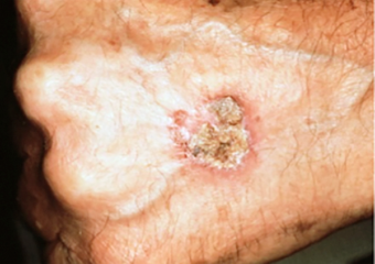
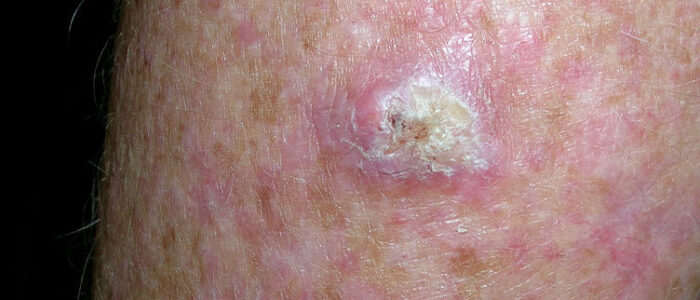
Squamous Cell Carcinoma (SCC)
Scaly patch on back of hand - a classic early SCC presentation:
Large crusty SCC - advanced, ulcerated nodule:
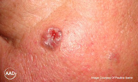
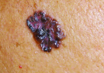
Basal Cell Carcinoma (BCC)
BCC on lighter skin - pearly, flesh-colored nodule:
Pigmented BCC on darker skin (Fitzpatrick type IV):
Key Visual Distinguishing Features
| Cancer | Classic Appearance |
|---|---|
| Melanoma | Asymmetric pigmented lesion; irregular borders; multiple colors (brown, black, red, white); may bleed |
| BCC | Pearly/translucent nodule with rolled borders; telangiectasias; may ulcerate ("rodent ulcer"); rarely pigmented |
| SCC | Rough, scaly, raised red plaque or nodule; may form a cutaneous horn; ulcerates; arises from actinic keratosis |
For a full gallery with early and late-stage examples across all skin tones, the Skin Cancer Foundation's image library and the AAD clinical image collection are excellent resources. DermNet NZ also has one of the most comprehensive free dermatology image databases available.
Note: The internal medical image library search service is temporarily unavailable (404 error). The images above are sourced from the Skin Cancer Foundation, a peer-reviewed clinical resource.
This is a shared conversation. Sign in to Orris to start your own chat.