
hip joint anatomy
Reading File
The hip joint is a ball-and-socket synovial joint formed by the femoral head and the acetabulum. Let me pull detailed content from the medical library.
Searching sources...
Finding Sources
Finding Sources
Good results. Now let me read the detailed content from the THIEME Atlas (primary anatomy source) and the imaging anatomy atlas, and also fetch the images.
Reading File
Reading File
Reading File
Good content. Let me now read more THIEME Atlas content covering the joint surfaces, capsule, ligaments, and muscles, and fetch a few key images in parallel.
Reading File
Reading File
Loading Image
Loading Image
Good. Now let me read the dedicated hip joint section from the THIEME Atlas (section 1.10 starts at line 6639) and get more ligament/capsule content, plus fetch more key images.
Reading File
Reading File
Loading Image
Excellent content. Let me now fetch the ligament diagram and the hip joint anterior/posterior overview images, and also get the muscles section from THIEME.
Reading File
Loading Image
Loading Image
Excellent - I now have comprehensive content and images. Let me compile the full detailed answer.
Hip Joint Anatomy
The hip joint (articulatio coxae) is a ball-and-socket (spheroidal) synovial joint formed by the spherical head of the femur articulating within the cup-shaped acetabulum of the pelvis. It is the most stable joint in the body, designed to bear weight while allowing a wide range of movement.
1. Articulating Bones
The Femoral Head
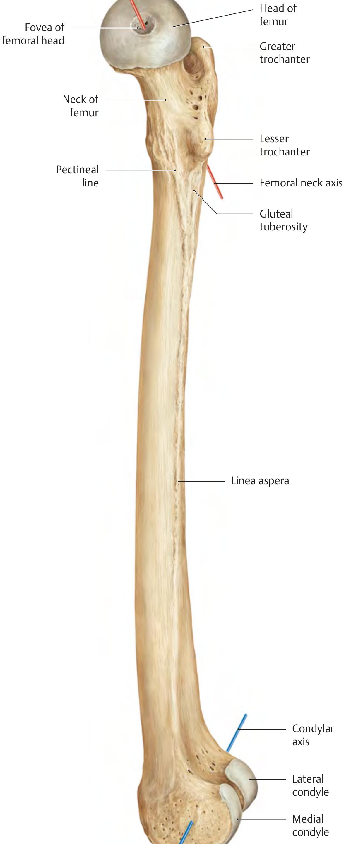
The femoral head forms approximately two-thirds of a sphere with an average radius of curvature of ~2.5 cm. It is largely enclosed within the acetabulum. At its center is the fovea capitis, a small pit that anchors the ligamentum teres (ligament of the femoral head). The head is connected to the femoral shaft via the femoral neck, which projects superiorly and medially at a neck-shaft angle (angle of inclination) of ~126° in adults (coxa vara if <120°, coxa valga if >135°).
The femoral neck also has an anteversion angle of ~12° in adults (the neck axis projects anteriorly relative to the condylar axis). At birth this angle is 30-40° and decreases through the 2nd decade. Increased or decreased torsion leads to in-toeing or out-toeing gaits.
Key femoral landmarks:
- Greater trochanter - attachment of gluteal muscles; palpable laterally
- Lesser trochanter - attachment of iliopsoas; posteromedial
- Intertrochanteric line (anterior) - where the joint capsule and iliofemoral ligament attach
- Intertrochanteric crest (posterior)
- Linea aspera - posterior longitudinal ridge for muscle attachment
The Acetabulum
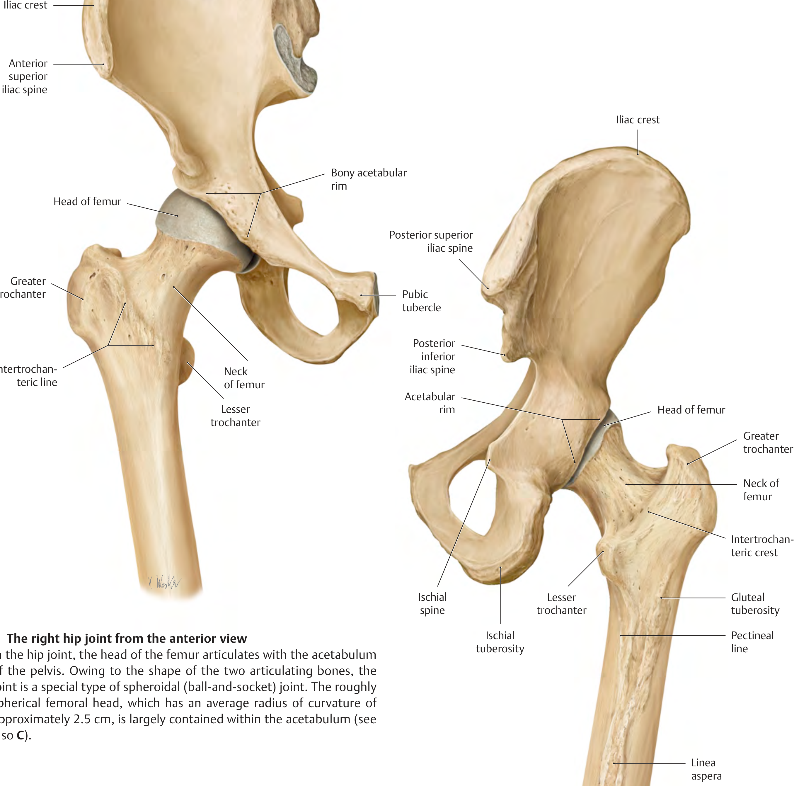
The acetabulum is formed by the junction of three bones: the ilium (superiorly, ~40%), ischium (posteroinferiorly, ~40%), and pubis (anteroinferiorly, ~20%), which fuse at the triradiate cartilage, typically completing fusion by age 16-18.
The acetabulum forms approximately two-thirds of a sphere (incomplete inferiorly), with an acetabular notch at the inferior aspect bridged by the transverse acetabular ligament. The articular surface is horseshoe-shaped (lunate surface), lined with hyaline cartilage, and surrounds the central non-articular acetabular fossa (filled with fat and the ligamentum teres).
Key acetabular angles:
- Transverse (inclination) angle: ~40° in adults (51° at birth, decreases with growth). Determines lateral coverage of the femoral head.
- Sagittal (anteversion) angle: ~17° in adults (7° at birth). The acetabulum faces anteroinferiorly.
The acetabulum is subdivided into:
- Anterior and posterior walls (portions covering the femoral head laterally)
- Anterior and posterior columns (structural pillars not covering the femoral head)
- Acetabular roof (superior rim, bears most of the load)
- Medial wall (thin portion between columns)
This column/wall architecture forms the basis of the Judet-Letournel classification of acetabular fractures.
2. Articular Cartilage and Labrum
- The lunate surface of the acetabulum and the femoral head are both lined by hyaline cartilage
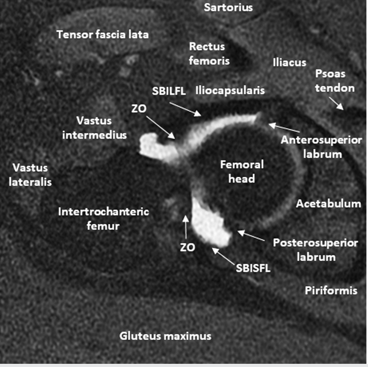
- The acetabular labrum is a fibrocartilaginous rim attached around the entire bony acetabular rim and bridging the acetabular notch as the transverse acetabular ligament. It deepens the socket by ~22%, increasing coverage of the femoral head from ~55% to ~72%. It also creates a negative intra-articular pressure (suction) that resists distraction, plays a role in joint lubrication, and contains nerve endings that contribute to proprioception
3. Joint Capsule
The fibrous capsule extends from the margins of the acetabular rim and labrum to:
- Anteriorly: intertrochanteric line (full femoral neck intracapsular)
- Posteriorly: ~1.5 cm proximal to the intertrochanteric crest (posterior femoral neck is partially extracapsular)
This is clinically important: femoral neck fractures within the capsule (intracapsular - subcapital, transcervical) jeopardize the blood supply to the femoral head, risking avascular necrosis.
The capsule has two layers:
- Outer fibrous layer - reinforced by the three main extracapsular ligaments
- Inner synovial layer - lines the intracapsular surfaces and produces synovial fluid
The zona orbicularis (annular ligament) is a circular thickening of the deep capsule that encircles the femoral neck like a buttonhole, contributing to joint stability.
4. Ligaments
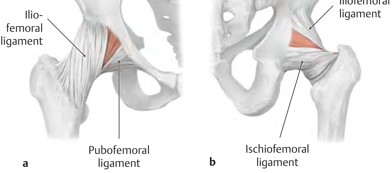
Three main extracapsular ligaments reinforce the capsule. During hip extension, all three twist upon themselves, pressing the femoral head into the acetabulum (stabilizing function). During flexion, they relax, allowing greater mobility.
Iliofemoral Ligament (Y-ligament of Bigelow)
- Origin: Anterior inferior iliac spine (AIIS) and acetabular rim
- Insertion: Intertrochanteric line (two bands - medial and lateral)
- Form: Inverted Y-shape
- Function: Strongest ligament in the human body (tensile strength >350 N). Limits extension, adduction, and external rotation. Crucially, it prevents posterior pelvic tilt in upright stance without muscular effort, and stabilizes the pelvis on the stance side during gait
Pubofemoral Ligament
- Origin: Superior pubic ramus and obturator crest
- Insertion: Blends with the inferior part of the iliofemoral ligament and joint capsule
- Function: Limits abduction and extension; particularly active at the end range of abduction
Ischiofemoral Ligament
- Origin: Posterior acetabular rim (ischial portion)
- Insertion: Greater trochanter (medial surface), blending with zona orbicularis
- Function: Limits internal rotation and extension; the weakest of the three
Ligamentum Teres (Ligament of the Femoral Head)
- Intracapsular, intra-articular
- Runs from the fovea capitis of the femoral head to the transverse acetabular ligament and margins of the acetabular notch
- No significant mechanical function in adults
- Transmits the medial epiphyseal artery (branch of obturator artery), which supplies a small portion of the femoral head in adults (more significant in children)
Weak spots: Gaps exist between the ligaments both anteriorly (between the iliofemoral and pubofemoral) and posteriorly (below the ischiofemoral). Traumatic dislocations occur at these sites - posterior dislocation is far more common (~90%) due to a thinner posterior capsule.
5. Blood Supply to the Femoral Head
This is critically important clinically:
- Medial femoral circumflex artery (MFCA) - the dominant supply; retinacular vessels run along the femoral neck beneath the synovial membrane and enter the femoral head superiorly. Most vulnerable in intracapsular fractures.
- Lateral femoral circumflex artery (LFCA) - minor contribution
- Obturator artery (via ligamentum teres) - negligible in adults; more important in children
Disruption of the MFCA retinacular branches in femoral neck fractures causes avascular necrosis (AVN) of the femoral head.
6. Nerve Supply
The hip joint receives its nerve supply from branches of the femoral, obturator, sciatic, and superior gluteal nerves (following Hilton's law - the joint is supplied by nerves that cross it).
- Anterior: Femoral nerve and obturator nerve
- Posterior: Nerve to quadratus femoris (branch of sacral plexus) and superior gluteal nerve
The obturator nerve supply explains referred pain from hip pathology to the medial thigh and knee.
7. Surrounding Muscles and Movements
The hip is a multiaxial joint capable of:
| Movement | Primary Muscles |
|---|---|
| Flexion | Iliopsoas (primary), rectus femoris, sartorius, tensor fascia lata |
| Extension | Gluteus maximus (primary), hamstrings (biceps femoris, semitendinosus, semimembranosus) |
| Abduction | Gluteus medius (primary), gluteus minimus, tensor fascia lata |
| Adduction | Adductors (longus, brevis, magnus), gracilis, pectineus |
| Internal rotation | Gluteus medius/minimus (anterior fibers), TFL |
| External rotation | Piriformis, obturator internus/externus, gemelli (superior and inferior), quadratus femoris ("short external rotators") |
The short external rotators (piriformis, obturator internus, superior and inferior gemelli, quadratus femoris) pass posterior to the hip and are analogous to the rotator cuff of the shoulder.
Axial MRI cross-section showing surrounding structures:
8. Stability Mechanisms Summary
The hip's exceptional stability (compared to, e.g., the shoulder) comes from:
- Deep bony socket - acetabulum covers ~55% of the femoral head
- Labrum - deepens socket to ~72% coverage, creates suction seal
- Capsular ligaments - extremely strong, especially the iliofemoral
- Zona orbicularis - grips the femoral neck
- Negative intra-articular pressure - opposes distraction
- Surrounding musculature - 17 muscles cross the joint
9. Clinical Correlations
- Intracapsular femoral neck fractures: Risk of AVN due to disruption of retinacular blood supply; treated with hemiarthroplasty or total hip replacement in the elderly
- Hip dysplasia: Shallow acetabulum (increased transverse angle, reduced center-edge angle of Wiberg <25°) leads to poor femoral head coverage and secondary OA
- Femoral acetabular impingement (FAI): Developmental incongruity of femoral head and acetabulum; cam type (non-spherical head), pincer type (overcoverage), or mixed; causes labral tears and early OA
- AVN of femoral head: Disrupted blood supply (trauma, steroids, alcohol, sickle cell); progresses to femoral head collapse
- Trendelenburg sign: Weakness of ipsilateral gluteus medius causes the contralateral pelvis to drop during single-leg stance (disruption of the hip abductor - iliofemoral ligament stabilization mechanism)
Sources:
- General Anatomy and Musculoskeletal System, THIEME Atlas of Anatomy, Sections 1.10-1.14
- Imaging Anatomy: Text and Atlas, Volume 3 - Bones, Joints, Vessels and Nerves, Chapter 20
- Rockwood and Green's Fractures in Adults (10th ed., 2025), Ligamentous Anatomy and Biomechanics
prostate gland anatomy related to 1st year mbbs for university exams 10 marks
Reading File
This is a broad anatomy topic (Visual + Broad category). Searching the library now.
Searching sources...
Finding Sources
Finding Sources
Reading File
Finding Sources
Finding Sources
Good content so far. Let me read more from this section and also search for the THIEME Atlas content on prostate, and fetch the key diagrams.
Reading File
Finding Sources
Loading Image
Excellent. Let me now read more of the prostate section (blood supply, lymphatics, nerve supply, zones) and also search for THIEME Atlas prostate content.
Reading File
Finding Sources
Gray's Anatomy for Students is perfect for 1st year MBBS. Let me read that section and the Smith's Urology blood supply section simultaneously, plus fetch the prostate cross-section diagram.
Reading File
Reading File
Loading Image
Now let me fetch the zonal anatomy diagram from Gray's Anatomy for Students, and the zones diagram from Smith's Urology.
Loading Image
Loading Image
I now have comprehensive, well-illustrated content for a complete 10-mark MBBS answer. Let me compose it.
Prostate Gland - Anatomy (10 Marks - 1st Year MBBS)
1. Introduction
The prostate gland is an unpaired, fibromuscular and glandular accessory sex organ of the male reproductive system. It is the largest accessory gland of the male reproductive system and has no counterpart in females. Its secretion forms ~30% of seminal fluid volume and contributes to sperm motility and viability.
2. Position
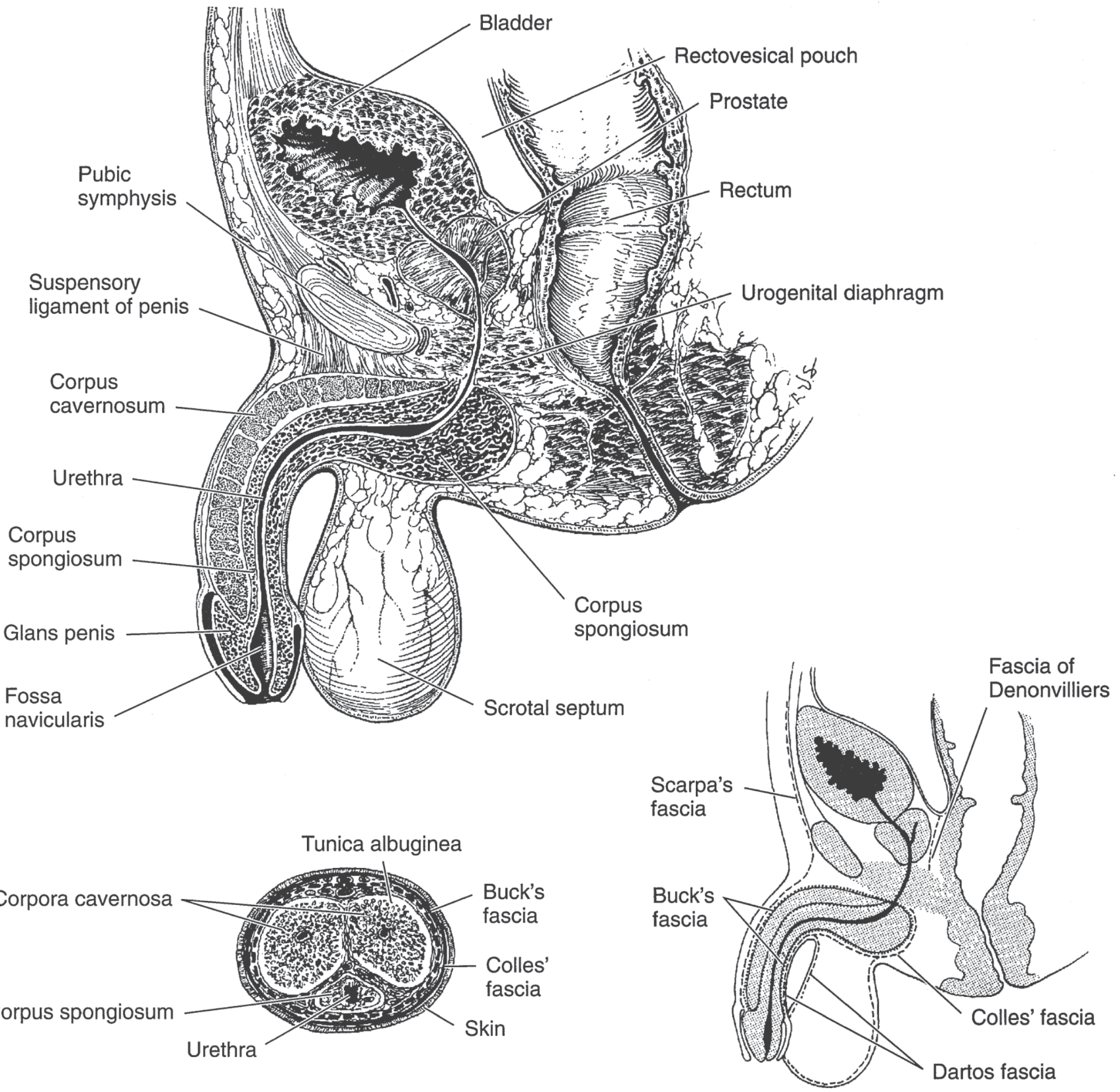
The prostate lies in the pelvic cavity:
- Inferior to the urinary bladder
- Posterior to the pubic symphysis
- Anterior to the rectum
- Superior to the urogenital diaphragm (perineal membrane)
It surrounds the prostatic urethra, which traverses it vertically.
3. Size, Shape and Weight
- Shape: Inverted rounded cone (like a chestnut)
- Weight: ~20 g in adult males (normal range 15-25 g)
- Dimensions: ~3 cm long, 4 cm wide, 2 cm anteroposteriorly
- Has a larger base (continuous with the neck of the bladder above) and a narrower apex (rests on the urogenital diaphragm below)
4. Relations
| Surface/Aspect | Relation |
|---|---|
| Superior (base) | Neck of urinary bladder; continuous with it |
| Inferior (apex) | Urogenital diaphragm (deep perineal pouch) |
| Anterior | Pubic symphysis; separated by retropubic (cave of Retzius) space; puboprostatic ligaments attach here |
| Posterior | Rectum (separated by Denonvilliers' fascia - two layers of peritoneal remnant); this is why prostate is palpable on digital rectal examination (DRE) |
| Inferolateral | Levator ani muscles (pubococcygeus part) - cradle the prostate on each side |
| Posterosuperior | Seminal vesicles and vas deferens (ampullae) |
The posterior surface is separated from the rectum by Denonvilliers' fascia, making it accessible to DRE. A normal prostate feels rubbery/firm on DRE; carcinoma feels "rock hard."
5. Capsule and Fascia
The prostate has two coverings:
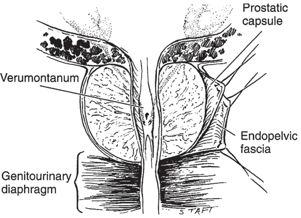
- True (prostatic) capsule - a thin fibrous capsule under which lie circularly oriented smooth-muscle fibres and collagenous tissue surrounding the urethra. This smooth muscle forms the involuntary smooth muscle sphincter of the posterior urethra.
- False capsule (prostatic sheath) - derived from the endopelvic (pelvic) fascia; a condensation of pelvic fascia surrounding the true capsule and containing the prostatic venous plexus between the two layers.
6. Lobar Classification (Cystourethroscopic/Clinical)
Five lobes (used clinically during cystourethroscopy and DRE):
| Lobe | Location | Clinical Significance |
|---|---|---|
| Anterior lobe | Anterior to urethra | Little glandular tissue; rarely involved in disease |
| Posterior lobe | Posterior to urethra, below ejaculatory ducts | Site of carcinoma (palpable on DRE) |
| Median (Middle) lobe | Between urethra and ejaculatory ducts | Prone to BPH; may obstruct bladder neck |
| Right lateral lobe | Lateral to urethra | Enlarges in BPH |
| Left lateral lobe | Lateral to urethra | Enlarges in BPH |
7. Zonal Classification (McNeal, 1981 - Pathological)
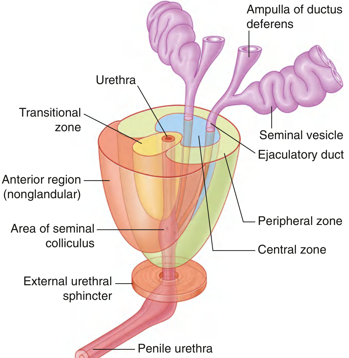
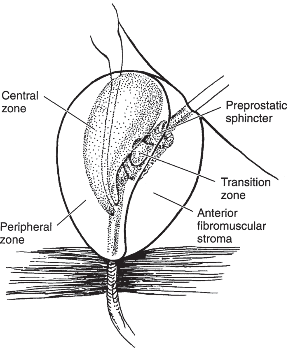
McNeal divided the prostate into four zones based on histology and disease patterns:
| Zone | % of glandular tissue | Disease |
|---|---|---|
| Peripheral zone | ~70% | Site of prostate carcinoma (70-80% of cancers) |
| Central zone | ~25% | Surrounds ejaculatory ducts; resistant to disease |
| Transitional zone | ~5% (enlarges in BPH) | Surrounds prostatic urethra; site of BPH |
| Anterior fibromuscular stroma | Non-glandular | No glands; purely muscle and fibrous tissue |
8. Prostatic Urethra and Structures Within
The prostatic urethra (~2.5 cm long) passes through the prostate. On its posterior wall is a longitudinal ridge called the urethral crest (crista urethralis). In the midpoint of this crest is a rounded eminence called the verumontanum (seminal colliculus), on which open:
- The prostatic utricle (a blind-ended pouch in the midline - Mullerian duct remnant)
- The two ejaculatory ducts (one on each side of the utricle)
On either side of the urethral crest are the prostatic sinuses, into which the ~25 prostatic ducts drain.
The ejaculatory ducts pierce the posterior aspect of the prostate and pass obliquely anteroinferiorly to open at the verumontanum.
9. Histology
- Outer layer: Thin fibrous capsule with smooth muscle fibres
- Stroma: Fibromuscular tissue embedded with 30-50 compound tubuloalveolar exocrine glands arranged in three concentric layers:
- Mucosal glands (innermost, periurethral) - drain directly into urethra
- Submucosal glands (middle layer)
- Main (peripheral) glands (outermost) - constitute bulk of prostate
- Epithelium: Pseudostratified columnar (secretory) with basal cells; produces prostatic secretion (citric acid, acid phosphatase, PSA, zinc, spermine)
- Corpora amylacea: Calcified concretions in prostatic ducts - increase with age, clinically insignificant
10. Blood Supply
Arterial
- Inferior vesical artery (main supply, branch of internal iliac)
- Internal pudendal artery
- Middle rectal artery
Venous
- Drain into the periprostatic (prostatic) venous plexus (of Santorini)
- This plexus communicates with:
- Deep dorsal vein of penis (anteriorly)
- Internal iliac (hypogastric) veins (laterally)
- Vertebral venous plexus (of Batson) - explains metastasis of prostate cancer to lumbar vertebrae and pelvis (retrograde venous spread)
11. Lymphatic Drainage
Lymphatics drain to:
- Internal iliac (hypogastric) nodes (primary)
- External iliac nodes
- Sacral nodes
- Vesical nodes
- Common iliac nodes
12. Nerve Supply
- Sympathetic (T10-L2 via hypogastric plexus): causes smooth muscle contraction during ejaculation, closing the bladder neck (prevents retrograde ejaculation)
- Parasympathetic (S2-S4 via pelvic splanchnic nerves / nervi erigentes): stimulates glandular secretion
- Both reach the gland via the inferior hypogastric (pelvic) plexus
The neurovascular bundles of Walsh run posterolaterally to the prostate and are critical to preserve during prostatectomy to maintain erectile function.
13. Development (Embryology)
The prostate develops from the urogenital sinus (endoderm) under the influence of androgens (testosterone/DHT) from the fetal testis. About 30-40 individual glands bud from the urethral epithelium into the surrounding mesenchyme at weeks 10-12 of gestation.
14. Function
The prostate secretes ~30% of the seminal fluid. Key secretions include:
- Prostate-specific antigen (PSA) - liquefies the seminal coagulum formed by seminal vesicle secretions
- Citric acid - major energy source for sperm
- Zinc - bactericidal properties
- Acid phosphatase - marker of prostate tissue (forensic and clinical use)
15. Clinical Correlations (Important for Exams)
| Condition | Zone/Anatomy Involved | Key Point |
|---|---|---|
| BPH (Benign Prostatic Hyperplasia) | Transitional zone + median lobe | Compresses urethra → obstructive LUTS; treated by TURP |
| Carcinoma Prostate | Peripheral zone (posterior lobe) | Palpable on DRE; raised PSA; metastasizes to lumbar vertebrae via Batson's plexus |
| Prostatitis | Peripheral zone | Tenderness on DRE; perineal pain |
| Retrograde Ejaculation | Bladder neck / sympathetic | Failure of sympathetic-mediated bladder neck closure |
| DRE Examination | Posterior surface through rectum | Separated by Denonvilliers' fascia; only posterior lobe palpable |
Sources:
- Smith and Tanagho's General Urology, 19th Edition, pp. 22-26
- Gray's Anatomy for Students, p. 549-550
- Guyton and Hall Textbook of Medical Physiology - Function of the Prostate Gland
This is a shared conversation. Sign in to Orris to start your own chat.