
Detail explain of ra vs os vs reactive arthritis vs gout vs pseudo gout
Reading File
Now I'll run parallel searches across all 5 conditions simultaneously.
Finding Sources
Finding Sources
Finding Sources
Finding Sources
Finding Sources
Now let me read the most comprehensive sources in parallel:
Reading File
Reading File
Reading File
Reading File
Reading File
Good. Now let me get more RA details and the reactive arthritis full section, plus gout treatment:
Reading File
Reading File
Reading File
Searching Images
rheumatoid arthritis hand joints X-ray
Searching Images
gout tophus urate crystals podagra
I now have comprehensive data from textbooks. Let me also get the RA treatment/extraarticular section and the reactive arthritis treatment from Goldman-Cecil, plus search for comparison images:
Reading File
Searching Images
pseudogout CPPD calcium pyrophosphate crystals knee chondrocalcinosis
Searching Images
reactive arthritis keratoderma blenorrhagica sausage digit dactylitis
I have comprehensive data from all sources. Now I'll compile the full detailed comparison.
Arthritis Comparison: RA vs OA vs Reactive Arthritis vs Gout vs Pseudogout
1. Rheumatoid Arthritis (RA)
Pathophysiology
RA is a chronic systemic autoimmune inflammatory disease in which cellular and humoral immune mechanisms cause destruction of synovial tissue. The key steps:
- Genetic predisposition via HLA-DR (particularly HLA-DR4 "shared epitope") triggers an immune response against citrullinated self-proteins (e.g., citrullinated vimentin, fibrinogen)
- Activated T-cells, B-cells, and macrophages infiltrate the synovium → synovial lining cells and subsynovial vessels proliferate → forming pannus (invasive granulation tissue)
- Pannus secretes matrix metalloproteinases (MMPs) and cytokines (TNF-α, IL-1, IL-6) → cartilage and bone erosion
- Rheumatoid factor (RF) = IgM autoantibody against Fc portion of IgG; Anti-CCP antibodies = highly specific (95%) and appear years before symptoms
- TNF-α plays a central role → basis for biologic therapy
Epidemiology
- Prevalence: 1–2% of adults; peak onset 20–50 years
- Women:Men = 3:1 (estrogen effect); 70% have insidious onset
- Higher concordance in monozygotic twins (MHC class II gene influence)
Clinical Features
| Feature | Detail |
|---|---|
| Pattern | Symmetric polyarthritis |
| Joints affected | PIP, MCP, wrists (sparing DIP) → "MCP/wrist" hallmark |
| Morning stiffness | >1 hour (hallmark) |
| Onset | Insidious, weeks to months |
| Deformities | Swan-neck, boutonnière, ulnar deviation, Z-thumb |
| Extra-articular | Rheumatoid nodules (20–30%), scleritis, pleuritis, pericarditis, Felty syndrome, vasculitis, interstitial lung disease |
ACR 1987 Diagnostic Criteria (need ≥4 of 7; symptoms ≥6 weeks)
- Morning stiffness ≥1 hour
- Arthritis of ≥3 joint areas
- Arthritis of hand joints (PIP, MCP, wrist)
- Symmetric arthritis
- Rheumatoid nodules
- Positive RF
- Radiographic changes (erosions, juxta-articular osteopenia on X-ray)
Investigations
- RF positive: ~70–80% (not specific — can be positive in SLE, Sjögren, infections, elderly)
- Anti-CCP (anti-citrullinated peptide): highly specific (~95%), positive even in seronegative RF
- ESR/CRP: elevated (disease activity markers)
- Synovial fluid: WBC >2,000/mm³ (inflammatory), no crystals
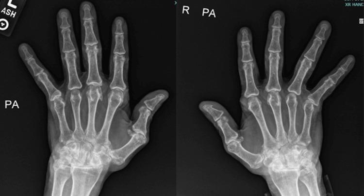
- X-ray: juxta-articular osteopenia → joint space narrowing → erosions → deformities
Treatment
Goal: Treat to target (T2T); early aggressive DMARD therapy
- NSAIDs: symptomatic only, no disease modification
- Conventional DMARDs (csDMARDs):
- Methotrexate (MTX) — anchor drug, 15–25 mg/week; monitor LFTs, CBC
- Hydroxychloroquine (mild disease): 200 mg BID, ophthalmology follow-up
- Sulfasalazine: 2–3 g/day in divided doses
- Leflunomide
- Biologic DMARDs:
- Anti-TNF: etanercept, adalimumab, infliximab
- IL-6 inhibitors: tocilizumab
- Anti-CD20: rituximab
- Co-stimulation blocker: abatacept
- Glucocorticoids: bridge therapy or for flares (not long-term due to side effects)
- Surgery: joint replacement for end-stage disease
2. Osteoarthritis (OA)
Pathophysiology
OA is a degenerative (non-inflammatory) joint disease — the most common joint disease. Matrix breakdown exceeds synthesis:
- Mechanical stress + aging → chondrocyte dysfunction → reduced proteoglycan/collagen II production
- Subchondral bone remodeling → sclerosis + osteophyte formation
- Mild inflammation (synovitis) is secondary, driven by locally produced cytokines (IL-1β, IL-6), but NOT the primary pathology
- Risk factors: age, obesity, prior joint injury, repetitive use, female sex (post-menopause)
Clinical Features
| Feature | Detail |
|---|---|
| Pattern | Asymmetric or bilateral (weight-bearing joints + DIP/PIP of hands) |
| Joints affected | DIP > PIP (hands), 1st CMC, knees, hips, spine (sparing MCP/wrist) |
| Stiffness | <30 min (post-rest stiffness/"gelling"), worsens with use |
| Bony nodules | Heberden's nodes (DIP) and Bouchard's nodes (PIP) |
| No systemic features | No extra-articular manifestations |
| Crepitus | Joint crepitus on movement |
Investigations
- Labs: ESR, CRP, RF — all NORMAL (distinguishes from RA)
- Synovial fluid: non-inflammatory WBC < 2,000/mm³, clear/yellow
- X-ray (4 classic signs):
- Joint space narrowing (asymmetric)
- Osteophytes (bony spurs)
- Subchondral sclerosis
- Subchondral cysts (Mnemonic: "JOSS")
Treatment
- Non-pharmacological: weight loss, physiotherapy, exercise, orthotics
- Topical NSAIDs: first-line for knee/hand OA
- Oral NSAIDs: use cautiously (GI/renal risk)
- Intra-articular corticosteroids: short-term relief
- Intra-articular hyaluronic acid: modest benefit
- Duloxetine: for chronic pain component
- Surgery: osteotomy, joint replacement (TKR/THR)
- Note: No DMARDs — OA is not autoimmune; NSAIDs/physiotherapy remain cornerstone
3. Reactive Arthritis (ReA)
Definition & Pathophysiology
Reactive arthritis = aseptic arthritis triggered by an extra-articular infection (GI or GU tract), occurring 1–3 weeks after the infection. The joint itself is sterile.
- GI pathogens: Salmonella typhimurium, Yersinia enterocolitica, Shigella flexneri, Campylobacter jejuni
- GU pathogen: Chlamydia trachomatis (most common in developed countries)
- Mechanism: Bacterial antigens (fragments or even viable Chlamydia) traffic to the synovium → activate immune response → synovitis. PCR studies have found viable Chlamydia in joints in a metabolically altered state
- HLA-B27 strongly associated (found in 60–80% of cases) — B27 confers risk for onset, axial involvement, and chronicity
- Part of the seronegative spondyloarthropathy spectrum (RF negative)
- Affects 2–7% of those with triggering GI infection; up to 20% in B27-positive individuals
Clinical Features
Classic triad (Reiter's syndrome): "Can't see, can't pee, can't bend the knee"
- Arthritis: asymmetric oligoarthritis, lower extremity predominance (knee, ankle, MTP)
- Urethritis/cervicitis: dysuria, urethral discharge (cervicitis may be asymptomatic in women — leads to underdiagnosis)
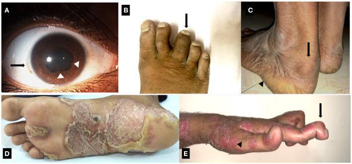
- Conjunctivitis/uveitis: bilateral conjunctivitis (painful); acute anterior uveitis (unilateral, less painful)
Additional features:
- Dactylitis ("sausage digit"): diffuse swelling of an entire digit — pathognomonic of spondyloarthropathy
- Enthesitis: Achilles tendinitis, plantar fasciitis
- Keratoderma blennorrhagicum: painless hyperkeratotic papules/plaques on palms and soles — clinically identical to pustular psoriasis
- Circinate balanitis: painless shallow ulcerations on glans penis
- Oral painless ulcers
- Sacroiliitis: asymmetrical (vs. bilateral in AS)
- Nail dystrophy (pitting, onycholysis)
Investigations
- RF and ANA: negative (seronegative)
- HLA-B27: positive in majority
- ESR/CRP/WBC: elevated during acute phase
- Synovial fluid: inflammatory WBC; no crystals; culture negative (aseptic)
- STI screen: urethral/cervical swab for Chlamydia; stool cultures for GI pathogens
- X-ray: periostitis, new bone formation at entheses; asymmetric sacroiliitis (vs. symmetric in AS); "fluffy" periosteal reaction at calcaneus; bulky non-marginal syndesmophytes (vs. marginal in AS)
Treatment
- NSAIDs: first-line for arthritis symptoms (indomethacin preferred)
- Antibiotics: for active Chlamydia infection (doxycycline/azithromycin); little effect once arthritis established; prolonged antibiotics (3-month doxycycline/azithromycin) may benefit post-Chlamydia chronic ReA
- Intra-articular corticosteroids: for persistent monoarticular disease
- Sulfasalazine or MTX: for chronic/refractory cases (>6 months)
- Anti-TNF biologics: refractory/severe chronic disease
- Prognosis: Most resolve in 3–12 months; ~15% develop chronic arthritis; B27-positive patients more likely to develop ankylosing spondylitis
4. Gout
Pathophysiology
Gout = monosodium urate (MSU) crystal deposition disease due to hyperuricemia
- Uric acid is the end product of purine catabolism (humans lack uricase)
- 90% of patients: reduced renal excretion of uric acid
- 10%: overproduction of uric acid
- Triggers for acute attacks: dietary purines (red meat, shellfish), alcohol (especially beer — ethanol blocks renal urate excretion), dehydration, surgery, diuretics (thiazides, loop), aspirin, trauma
- MSU crystal formation occurs in cooler, more acidic environments → explains predilection for 1st MTP, heel, ankle
- Crystals → phagocytosed by neutrophils → activate NLRP3 inflammasome → IL-1β release → acute inflammatory cascade (neutrophil recruitment, prostaglandins, bradykinin)
Clinical Stages
| Stage | Features |
|---|---|
| Asymptomatic hyperuricemia | Elevated uric acid (>7 mg/dL men; >6 mg/dL women) with no symptoms; usually takes ≥20 years before first attack; treatment NOT recommended |
| Acute gouty arthritis | Sudden severe monoarthritis, peak within 24 hrs, resolves in days–weeks even untreated |
| Intercritical gout | Symptom-free periods between attacks; crystals still present in joints |
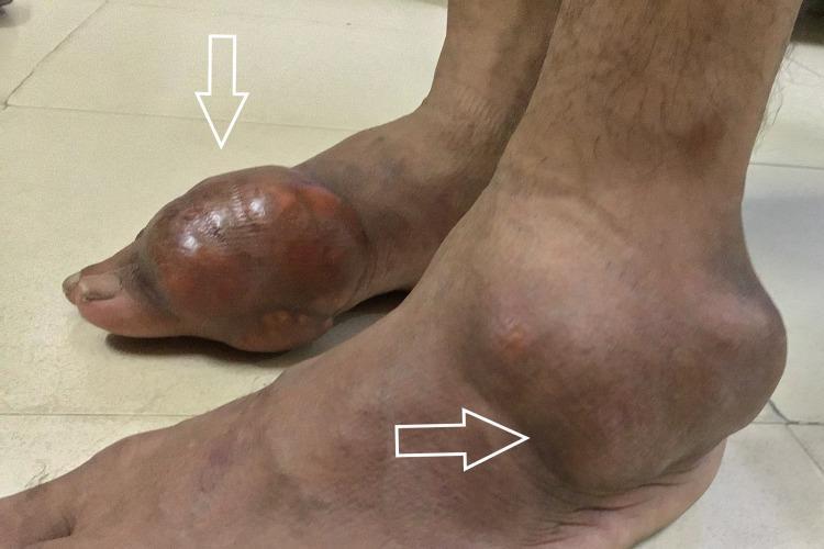
| Chronic tophaceous gout | Persistent tophi (chalky urate deposits) in joints, earlobes, Achilles tendon; can mimic RA |
Clinical Features
- 1st MTP joint (podagra) in 50% of initial attacks; eventually involved in 75–90%
- Also: heel, ankle, knee, midtarsal joints, olecranon bursa
- Severe pain, swelling, erythema, warmth — patient can't tolerate even bedsheet touching the joint
- Attacks often begin at night (↓ temperature, dehydration during sleep)
- Low-grade fever, leukocytosis may accompany severe attacks (can mimic septic arthritis)
- Tophi: chalky-white subcutaneous nodules; common sites = 1st MTP, olecranon bursa, ear helix, Achilles tendon
- Gout in women and elderly: often polyarticular, can mimic RA; tophi mistaken for rheumatoid nodules
Investigations
- Serum uric acid: usually >8 mg/dL during attack (can be normal during acute flare as uric acid redistributes)
- Synovial fluid (GOLD STANDARD): needle-shaped crystals with negative birefringence (yellow when parallel to compensator axis, blue when perpendicular) under polarized light; WBC 10,000–100,000/mm³; culture negative
- 24-hr urine uric acid: distinguishes underexcretion vs. overproduction
- X-ray (chronic gout): "rat-bite" erosions with overhanging edge (punched-out erosions with sclerotic margins), soft tissue tophi; periarticular calcification uncommon (contrast with pseudogout)
Treatment
Acute attack:
- NSAIDs (e.g., indomethacin 50 mg TID): first-line if no contraindications
- Colchicine: 1.2 mg then 0.6 mg 1 hr later (first 36 hours most effective); inhibits microtubule polymerization → inhibits neutrophil migration
- Corticosteroids: (oral, IM, or intra-articular) if NSAIDs/colchicine contraindicated; prednisone 0.5 mg/kg/day tapering over 7–10 days
- IL-1 inhibitors (anakinra, canakinumab): refractory/polyarticular attack
Urate-lowering therapy (ULT) — start when attack fully resolved:
- Indications: ≥2 attacks/year, tophi, uric acid nephropathy/stones, radiographic erosions
- Allopurinol (xanthine oxidase inhibitor): start low (100 mg/day), titrate; paradoxically can trigger attacks if started during active gout
- Febuxostat: alternative XOI, more potent; cardiovascular concerns in some guidelines
- Probenecid: uricosuric; second-line (avoid if GFR <50 or uric acid stones)
- Pegloticase (IV recombinant uricase): refractory tophaceous gout
- Target: serum uric acid <6 mg/dL (<5 mg/dL with tophi)
- Prophylaxis during ULT initiation: low-dose colchicine or NSAID for 3–6 months (mobilization flares)
5. Pseudogout (CPPD — Calcium Pyrophosphate Crystal Deposition Disease)
Pathophysiology
Pseudogout = calcium pyrophosphate dihydrate (CPPD) crystal deposition in hyaline cartilage and fibrocartilage:
- Degradation of articular cartilage proteoglycans (which normally inhibit mineralization) allows CPP crystallization around chondrocytes
- Crystals form in cartilage matrix → rupture → seed the joint → NLRP3 inflammasome activation → IL-1β → acute inflammation (like gout)
- Even without inflammation, crystals disrupt cartilage structure/function
- ANKH gene mutation: encodes inorganic pyrophosphate transporter — linked to familial CPPD
- Sporadic (idiopathic) vs. hereditary vs. secondary
Secondary CPPD causes (associated metabolic disorders):
- Hyperparathyroidism (↑ Ca²⁺)
- Hemochromatosis (↑ Fe²⁺ inhibits pyrophosphatase)
- Hypomagnesemia (Mg²⁺ normally inhibits CPP crystallization)
- Hypothyroidism
- Ochronosis (alkaptonuria)
- Diabetes mellitus
- Previous joint damage/trauma
Epidemiology
- Age: predominantly >50 years; prevalence rises sharply with age — up to 60% of those ≥85 years
- Equal sex distribution (vs. gout's male predominance)
- Often asymptomatic (incidental chondrocalcinosis on X-ray)
Clinical Features
| Presentation | Description |
|---|---|
| Acute pseudogout | Sudden monoarthritis — most commonly knee (then wrist, elbow, shoulder, ankle); mimics septic arthritis; lasts weeks to months (vs. days in gout) |
| Chronic CPPD | Persistent joint pain with superimposed flares — can mimic OA or RA |
| Chondrocalcinosis | Asymptomatic X-ray finding of cartilage calcification |
| Crowned dens syndrome | CPPD deposition around odontoid process → acute neck pain, fever, meningism |
- Unlike gout, CPPD commonly affects the knee (gout prefers 1st MTP)
- Large joint predominance: knee > wrist > shoulder > ankle
- Attacks may be triggered by: illness, surgery, trauma, IV contrast, bisphosphonates
Crystal Characteristics
| Feature | Gout (MSU) | Pseudogout (CPPD) |
|---|---|---|
| Shape | Needle-shaped | Rhomboid (0.5–5 μm) |
| Birefringence | Negatively birefringent | Positively birefringent (weakly) |
| Color under compensator | Yellow when parallel | Blue when parallel |
| Site of crystal formation | Periarticular (synovial fluid) | In hyaline cartilage / fibrocartilage |
| X-ray | "Rat-bite" erosions | Chondrocalcinosis (cartilage calcification) |
Investigations
- Synovial fluid: rhomboid crystals with weak positive birefringence (blue when parallel to axis under compensated polarized light); WBC 10,000–50,000/mm³
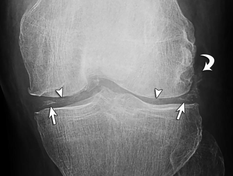
- X-ray: chondrocalcinosis — linear/punctate radiopaque calcifications in hyaline cartilage or fibrocartilage (knee menisci, wrist TFCC, symphysis pubis) — pathognomonic
- Ultrasound: hyperechoic deposits within the substance of hyaline cartilage (vs. gout's "double contour" sign on cartilage surface)
- Dual-energy CT (DECT): can color-code CPPD from MSU crystals
- Metabolic screen: calcium, PTH, magnesium, iron/ferritin, TFTs, blood glucose (for secondary causes)
Treatment
Acute attack:
- NSAIDs: high-dose (indomethacin, naproxen)
- Colchicine: effective (same mechanism as gout)
- Intra-articular corticosteroids: excellent if septic arthritis ruled out and ≤2 joints involved; aspiration alone also provides symptomatic relief
- Systemic corticosteroids: polyarticular or contraindications to above
- IL-1 inhibitors: refractory cases
Chronic/prophylaxis:
- Low-dose colchicine (0.5–1 mg/day): reduces frequency of recurrent attacks
- Hydroxychloroquine or MTX: for chronic inflammatory CPPD mimicking RA
- No equivalent of ULT for CPPD: no treatment currently prevents or reverses crystal formation
Comprehensive Comparison Table
| Feature | RA | OA | Reactive Arthritis | Gout | Pseudogout |
|---|---|---|---|---|---|
| Mechanism | Autoimmune (anti-CCP, RF) | Mechanical degeneration | Post-infectious (sterile) | MSU crystal deposition | CPP crystal deposition |
| Age/Sex | 20–50 yrs, F>M (3:1) | >50 yrs, F>M | 20–40 yrs, M>F | 40–60 yrs M, post-menopausal F | >50 yrs, equal |
| Onset | Insidious, weeks | Gradual, years | Acute, 1–3 wks post-infection | Sudden (overnight), acute | Sudden, acute |
| Pattern | Symmetric polyarthritis | Asymmetric, weight-bearing | Asymmetric oligoarthritis LL | Mono → polyarthritis | Mono/oligoarthritis |
| Joints | PIP, MCP, wrist (sparing DIP) | DIP, PIP, knee, hip (sparing MCP) | Knee, ankle, MTP (LL predominance) | 1st MTP (podagra), ankle, knee | Knee > wrist, shoulder |
| Stiffness | Morning stiffness >1 hr | Morning stiffness <30 min (gelling) | Variable | Not a feature | Not a feature |
| Key exam | Pannus, subluxation | Heberden's/Bouchard's nodes, crepitus | Dactylitis, keratoderma blenorrhagica | Tophi, podagra | No tophi; chondrocalcinosis on X-ray |
| RF / ANA | RF+, anti-CCP+ | Negative | Seronegative | Negative | Negative |
| HLA assoc. | HLA-DR4 | None | HLA-B27 (60–80%) | None | ANKH gene |
| Serum marker | ↑ESR, CRP, RF, anti-CCP | Normal | ↑ESR/CRP; HLA-B27+ | ↑Uric acid (>8 mg/dL) | Normal uric acid; ↑Ca²⁺ if hyperparathyroid |
| Synovial fluid | Inflammatory (WBC >2000); no crystals | Non-inflammatory (WBC <2000); no crystals | Inflammatory; no crystals; culture negative | Needle crystals, negative birefringence | Rhomboid crystals, positive birefringence |
| X-ray | Erosions, juxta-articular osteopenia, joint space narrowing | Osteophytes, subchondral sclerosis/cysts, JSN | Periostitis, asymmetric sacroiliitis | "Rat-bite" punched-out erosions with overhanging edge | Chondrocalcinosis (cartilage calcification) |
| Key pathogen | None (autoimmune) | None | Chlamydia, Salmonella, Shigella, Campylobacter, Yersinia | None (metabolic) | None (metabolic/metabolic disease) |
| Systemic features | Nodules, scleritis, ILD, vasculitis | None | Urethritis, conjunctivitis, uveitis, mucocutaneous | Renal stones, nephropathy | Usually none (except secondary metabolic cause) |
| 1st-line Rx | MTX (DMARD) | NSAIDs, PT, weight loss | NSAIDs ± antibiotics (Chlamydia) | NSAIDs / colchicine (acute); allopurinol (ULT) | NSAIDs / colchicine / intra-articular steroids |
| Disease-specific Rx | DMARDs, biologics (anti-TNF) | Joint replacement | Sulfasalazine/MTX if chronic | Allopurinol, febuxostat | Low-dose colchicine prophylaxis; no ULT |
Key Distinguishing Pearls
- Morning stiffness >1 hr → think RA (or other inflammatory arthritis); <30 min → think OA
- DIP involvement → OA (Heberden's) or psoriatic arthritis; MCP/wrist sparing DIP → RA
- 1st MTP sudden overnight pain → GOUT until proven otherwise
- Knee calcification (chondrocalcinosis) on X-ray → PSEUDOGOUT
- Urethritis + conjunctivitis + arthritis + recent GI/GU infection → REACTIVE ARTHRITIS
- Sausage digit (dactylitis) → reactive arthritis, psoriatic arthritis (spondyloarthropathy)
- Negatively birefringent needle crystals → GOUT; positively birefringent rhomboid crystals → PSEUDOGOUT
- Tophi on ear helix → chronic gout; subcutaneous nodules on extensor surface → RA nodules
- Seronegative + HLA-B27 + sacroiliitis → spondyloarthropathy (ReA, AS, psoriatic)
- Metabolic screen for pseudogout: always check hyperparathyroidism, hemochromatosis, hypomagnesemia, hypothyroidism
Sources: Textbook of Family Medicine 9e — Goldman-Cecil Medicine International Edition — Robbins & Kumar Basic Pathology — Robbins, Cotran & Kumar Pathologic Basis of Disease — Miller's Review of Orthopaedics 9e
This is a shared conversation. Sign in to Orris to start your own chat.