

446265ad-17b0-4293-b815-3baf8490c538.jpg
Explain me this based on pbl format detailed
Reading File
Searching sources...
Finding Sources
Finding Sources
Finding Sources
Reading File
Reading File
Reading File
Reading File
Finding Sources
Finding Sources
Reading File
Reading File
Now I have all the information needed to build a thorough PBL response. Let me compile it:
PBL Analysis - Day 1 (Trigger 1)
STEP 1: SCENARIO SUMMARY
Patient: 25-year-old married woman
Presenting to: Emergency Department
Complaints:
- Lower abdominal pain (crampy, intermittent, suprapubic) - 1 day
- Vaginal bleeding - started as spotting, gradually increasing
- Dizziness + generalized weakness
- No fever, no rash, no medications
Relevant History:
- Irregular menstrual cycles
- LMP: ~2-3 months ago (uncertain)
- Obstetric history: G2P1 (1 living child, age 3, normal delivery; no prior miscarriages)
STEP 2: CUES (What stands out?)
| Cue | Significance |
|---|---|
| Married woman of reproductive age | Pregnancy-related pathology likely |
| LMP ~2-3 months ago | Possible pregnancy (~8-12 weeks) |
| Crampy lower abdominal pain | Uterine/tubal involvement |
| Vaginal bleeding - increasing | Threatened abortion or ectopic |
| Dizziness + weakness | Possible hemodynamic compromise/anaemia |
| No fever | Infectious cause less likely |
| Irregular cycles | Makes dating difficult; may have underlying hormonal issue |
| Para 1, No miscarriages | Baseline reproductive health context |
STEP 3: PROBLEM IDENTIFICATION
Main Clinical Problems:
- Bleeding in early pregnancy (amenorrhoea ~2-3 months + vaginal bleeding)
- Acute lower abdominal/pelvic pain
- Dizziness and weakness - suggesting haemodynamic instability or anaemia
STEP 4: HYPOTHESES / DIFFERENTIAL DIAGNOSES
Primary Hypothesis: Ectopic Pregnancy
The classic symptom triad of ectopic pregnancy is pain, amenorrhoea, and vaginal bleeding - present in approximately 50% of patients. This presentation fits well.
"Ectopic pregnancy accounts for up to 20% of diagnoses among women presenting with vaginal bleeding or abdominal pain in the first trimester of pregnancy."
- Rosen's Emergency Medicine
Differential Diagnoses (ranked by likelihood):
| Diagnosis | Supporting Features | Against |
|---|---|---|
| Ectopic pregnancy | Amenorrhoea, pain, bleeding, dizziness | No history of IUD/PID/previous ectopic mentioned |
| Threatened abortion | Bleeding + cramping + amenorrhoea | Usually cervix closed, less pain |
| Incomplete abortion | Cramping + increasing bleeding | Usually more severe pain |
| Inevitable abortion | Progressive bleeding + pain | Cervical os would be open |
| Corpus luteum cyst/haemorrhage | Pelvic pain, can mimic ectopic | Pregnancy test negative usually |
| Appendicitis | Abdominal pain | No fever, no nausea/vomiting mentioned |
| Ovarian cyst accident | Pelvic pain | Irregular but not amenorrhoeic pattern |
Most Dangerous Diagnosis to Rule Out FIRST: Ectopic Pregnancy (Ruptured)
- Dizziness and weakness suggest haemodynamic compromise
- This is a life-threatening emergency
"Ectopic pregnancy is the most common obstetric cause of maternal death in the first trimester."
- Roberts & Hedges' Clinical Procedures in Emergency Medicine
STEP 5: LEARNING OBJECTIVES (What do we need to know?)
- What is ectopic pregnancy? Where does it implant?
- What are the risk factors?
- What is the pathophysiology of ectopic pregnancy?
- How do you diagnose ectopic pregnancy?
- What investigations are needed?
- How do you manage ectopic pregnancy?
- How do we classify spontaneous abortions?
STEP 6: APPLYING KNOWLEDGE
A. What is Ectopic Pregnancy?
An ectopic pregnancy is a pregnancy that implants outside the uterine cavity. The most common site is the fallopian tube (95%), specifically the ampullary portion. Other sites include the ovary, cervix, interstitial/cornual region, and abdominal cavity.
B. Risk Factors
| Risk Factor | Mechanism |
|---|---|
| Previous salpingitis/PID | Tubal damage, impaired cilia motility |
| IUD use | Alters tubal transport |
| Previous tubal ligation/surgery | Scarring |
| Previous ectopic pregnancy | Tubal damage |
| Assisted reproduction (IVF) | Embryo displacement |
| Smoking | Impairs ciliary function |
(This patient has none of these mentioned - but ectopic can occur without risk factors.)
C. Pathophysiology
The fertilised ovum is unable to travel to the uterine cavity due to mechanical obstruction or impaired tubal motility. It implants in the tube, where trophoblastic tissue invades the tubal wall. As the pregnancy grows:
- The tube stretches and eventually ruptures
- Bleeding into the peritoneal cavity (haemoperitoneum) occurs
- This causes peritoneal irritation, pain, and haemodynamic shock
Dizziness + weakness in this patient may represent early haemodynamic compromise from a leaking/ruptured ectopic.
D. Classic Triad (Ectopic Pregnancy)
Pain + Amenorrhoea + Vaginal Bleeding
- Present in ~50% of patients
- Pain can be unilateral, bilateral, sharp, dull, or crampy
- Bleeding is usually scanty/dark (unlike normal menstruation)
- Shoulder-tip pain = diaphragmatic irritation from haemoperitoneum
E. Spontaneous Abortion Classification (differential)
| Type | Cervical Os | Tissue Passed | Bleeding | Pain |
|---|---|---|---|---|
| Threatened | Closed | No | Present | Mild cramping |
| Inevitable | Open/dilated | No | Present | Present |
| Incomplete | Open | Partial | Heavy | Severe cramping |
| Complete | Closed/closing | Yes (all) | Minimal | Reducing |
| Missed | Closed | No | Absent | Absent |
STEP 7: INVESTIGATIONS NEEDED
Immediate (Emergency)
| Investigation | Rationale |
|---|---|
| Urine/Serum β-hCG | Confirms pregnancy (urine detects ≥20 mIU/mL; serum detects >5 mIU/mL) |
| Transvaginal Ultrasound (TVUS) | Locates gestation sac (intrauterine vs extrauterine) |
| FBC (Full Blood Count) | Check for anaemia, haematocrit (blood loss) |
| Blood group + Rh factor | Critical - if Rh negative, RhoGAM needed |
| Serum Progesterone | Low levels (<5 ng/mL) suggest non-viable pregnancy |
Interpretation of β-hCG
- Single β-hCG is of limited use alone (overlap between normal and abnormal pregnancies)
- Serial β-hCG (48 hours apart): A rise of ≥63% suggests viable IUP; <53% rise or a fall suggests ectopic or failing pregnancy
- Discriminatory zone (~1,500-2,000 mIU/mL): At this level, an IUP should be visible on TVUS; absence of IUP = suspect ectopic
Ultrasound Findings Suggesting Ectopic
- Absence of intrauterine gestational sac with positive β-hCG
- Adnexal mass (may or may not contain a gestational sac)
- Free fluid in the Pouch of Douglas = haemoperitoneum (ruptured ectopic)
- A "tubal ring" sign on TVUS
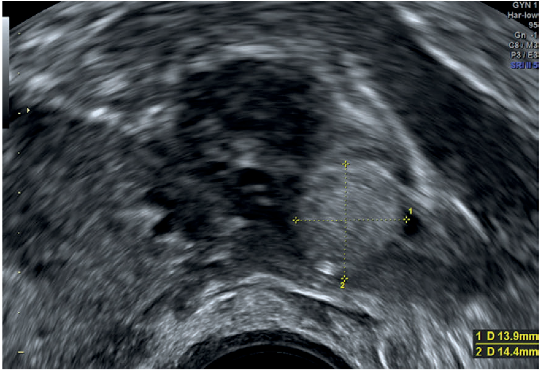
Ultrasound: Tubal ectopic pregnancy - Bailey & Love's Surgery
STEP 8: MANAGEMENT
Immediate Priorities (ABC)
- IV access + fluid resuscitation
- Vital signs monitoring
- Blood tests + crossmatch (prepare for surgery)
- Confirm pregnancy with β-hCG
Definitive Management Options
1. Surgical (most common)
- Laparoscopy (preferred) - shorter hospital stay, less blood loss, shorter recovery
- Laparotomy - if haemodynamically unstable/ruptured ectopic
- Salpingectomy (tube removal) vs. Salpingostomy (tube-conserving) - depends on contralateral tube status and future fertility desires
"Laparoscopy is superior to laparotomy for management of ectopic pregnancy in most cases."
- Berek & Novak's Gynecology
2. Medical (Methotrexate)
- Indications: haemodynamically STABLE, unruptured, small (<3.5 cm) ectopic, β-hCG <5000 mIU/mL, no fetal cardiac activity, patient compliant for follow-up
- Mechanism: Folate antagonist - inhibits rapidly dividing trophoblast cells
- Contraindications: haemodynamic instability, ruptured ectopic, liver/renal disease, breastfeeding, immunocompromise
3. Expectant Management
- Only for selected stable patients with low/falling β-hCG and no cardiac activity
Rh Status
- If the patient is Rh-negative, administer anti-D immunoglobulin (RhoGAM) to prevent isoimmunisation
STEP 9: DIAGNOSTIC ALGORITHM (Summary Flowchart Logic)
Positive β-hCG + Pain/Bleeding
        ↓
   Transvaginal Ultrasound
        ↓
IUP seen? → YES → Manage as threatened/incomplete abortion
        ↓ NO
Adnexal mass / free fluid? → YES → Ectopic pregnancy (likely)
        ↓ NO (indeterminate)
Serial β-hCG 48h later
        ↓
Rise ≥63% → Likely viable IUP, repeat USS
Fall or <53% rise → Ectopic or failed IUP → Manage accordingly
STEP 10: KEY LEARNING POINTS (PBL Take-Home)
- Any woman of reproductive age with pain + bleeding + amenorrhoea = rule out ectopic pregnancy first
- Ectopic pregnancy is the leading cause of maternal death in the 1st trimester - treat as emergency
- The classic triad (pain, amenorrhoea, bleeding) is present in only ~50% - high index of suspicion needed
- β-hCG + TVUS are the diagnostic cornerstones
- Dizziness and weakness in this patient = haemodynamic compromise = possible rupture
- Rh status must always be checked in early pregnancy bleeding
- Irregular cycles make LMP unreliable - always consider pregnancy even with vague history
Sources:
- Berek & Novak's Gynecology (Ectopic Pregnancy chapter, pp. 1757-1768)
- Roberts & Hedges' Clinical Procedures in Emergency Medicine (ectopic pregnancy, p. 4543-4589)
- Bailey & Love's Short Practice of Surgery 28th Edition (Chapter 87)
- Rosen's Emergency Medicine (Epidemiology, ectopic pregnancy)
- Textbook of Family Medicine 9e (Management of ectopic pregnancy)
This is a shared conversation. Sign in to Orris to start your own chat.