

f30f3627-bc4a-4634-a41b-0c9ebc52b7cc.jpeg
Answer for 2 nd question according to k narayanareddy textbook fmt mbbs 3rd year
Reading File
The question is a clinical scenario about a bike rider who hit his head on a road divider, had a brief loss of consciousness (lucid interval), then became unconscious the next day and died - a classic presentation of Epidural Hemorrhage. The question asks about:
- a) Most probable injury
- b) Mechanism and postmortem features
- c) Types of skull fractures
Let me search the K. Narayan Reddy FMT textbook now.
Searching sources...
Finding Sources
The K. Narayan Reddy textbook is listed as "The Essentials of Forensic Medicine and Toxicology 36th edition (latest 2026)" with ISBN 9789366165257. Let me search for epidural hemorrhage and skull fractures sections now.
Finding Sources
Finding Sources
Finding Sources
Finding Sources
Finding Sources
Reading File
Good - found the skull fractures section. Now let me read the Intracranial Hemorrhage section (especially for epidural/extradural hemorrhage) and continue the skull fracture types.
Reading File
Reading File
Excellent! Found the extradural hemorrhage section. Let me read more of it to get mechanism, postmortem features, and continue skull fracture types.
Reading File
Reading File
Reading File
Reading File
I now have comprehensive material from K. Narayan Reddy. Let me also fetch the extradural hemorrhage image for the answer.
Loading Image
Loading Image
I now have all the information needed from K. Narayan Reddy's textbook. Let me compile the complete answer.
Question 2 - Answer (as per K. Narayan Reddy - Essentials of FMT, 36th Edition)
Clinical Scenario Analysis
The rider struck his head on the road divider (left temporoparietal area), had a brief loss of consciousness (few seconds), then a lucid interval, then deteriorated and died the next day. The CT showed opacity at the left temporoparietal area.
(a) Most Probable Injury that Led to the Rider's Death
EXTRADURAL HEMORRHAGE (EDH) - also called Epidural Hematoma.
This is the classic "talk and die" scenario. The hallmark features in this case are:
- Blow to the left temporoparietal area (site of the middle meningeal artery)
- Brief unconsciousness at impact followed by a lucid interval
- CT showing opacity (hyperdense collection) at the left temporoparietal area
- Rapid deterioration and death the next day
The injury here is a coup injury - the blow at the temporoparietal area fractured the squamous temporal bone and ruptured the posterior branch of the middle meningeal artery, causing progressive arterial bleeding in the extradural space.
(b) Mechanism and Postmortem Features of Extradural Hemorrhage
Mechanism
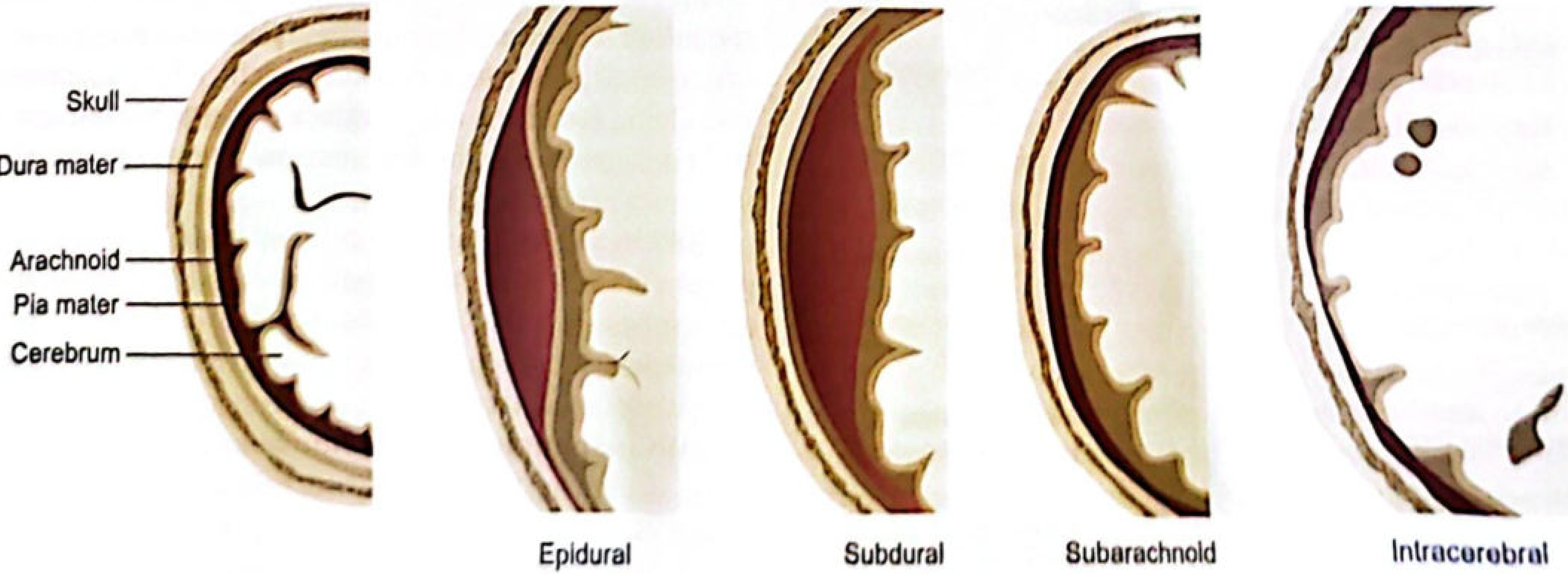
Anatomy: The dura is a strong, grey-bluish connective tissue membrane. Its outer layer is firmly attached to the skull; the inner layer merges with the arachnoid. It forms sinuses which drain venous blood from the brain, dura, and bone.
How it occurs:
- At the moment of impact, the skull moves relative to the dura beneath it
- The dura is stripped from the bone, producing an empty extradural space at the site of trauma
- A blood vessel is injured simultaneously
- A blow over the lateral convexity of the head fractures the squamous temporal bone and injures the middle meningeal artery (usually the posterior branch), especially as it passes upwards and backwards across the temporoparietal region
- The middle meningeal artery is a direct branch of the internal maxillary artery
- Arterial blood accumulates rapidly under high pressure in the extradural space, stripping the dura progressively away from the inner table
Types of vessels injured by site:
| Site of blow | Vessel injured |
|---|---|
| Lateral convexity (temporoparietal) | Middle meningeal artery (posterior branch) |
| Forehead | Anterior ethmoidal artery |
| Occiput / low behind ear | Transverse-sigmoid sinus |
| Vertex | Sagittal sinus |
Clinical course (Lucid Interval):
- Head injury causes temporary unconsciousness
- Patient recovers - this is the "LUCID INTERVAL" (typically 2-4 hours to a week; seen in 30-40% of cases)
- As blood accumulates, intracranial pressure rises
- Patient becomes confused, then develops contralateral limb weakness, ipsilateral pupil dilation (fixed and dilated)
- Progresses to bilateral pupil fixation, decerebrate rigidity, then death
- Death is due to respiratory failure from brainstem compression
Lucid interval is not seen if the brain injury is severe, because unconsciousness from brain injury overlaps with that from EDH pressure.
Postmortem Features (Autopsy Findings)
- External: Abrasions/laceration at the left temporoparietal area of scalp; no helmet
- Skull: Fissured fracture of the squamous temporal bone (90% cases are fissured type; rarely depressed)
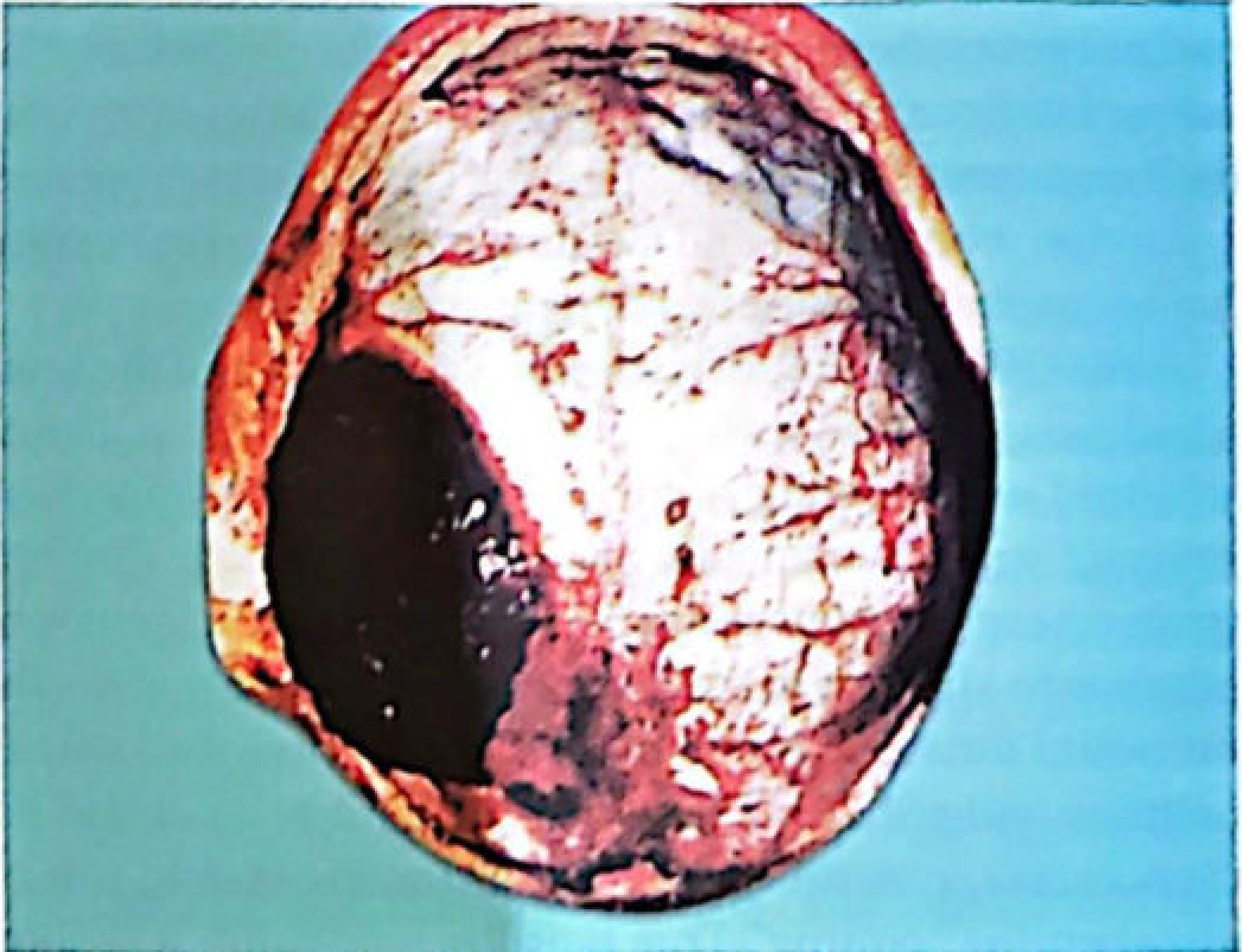
- Extradural space: A biconvex/lenticular blood clot in the extradural space, typically at the temporoparietal area
- Clot characteristics:
- Oval or circular shape
- Rubbery consistency
- Reddish-purple color
- About 10-20 cm in diameter, 2-6 cm thick
- Weighs 30-300 g (minimum ~100 mL associated with fatalities)
- Sharply defined with typical limitation due to dural attachments at suture lines
- Adherent to the dura mater
- Presses the dura inward, causing localized concavity of the external brain surface
- Brain: Cerebral edema; localized brain compression; secondary hemorrhages in the pons
- Herniation: Tentorial herniation - both from clot pressure and brain swelling beneath the hematoma
- Vessels: Gentle removal of clot shows the break in the middle meningeal artery and the fissured fracture of the nearby skull (sometimes confined to the inner table only)
- About 50% of cases have a second (associated) hemorrhage - subdural, subarachnoid, or intracerebral
Illustration from K. Narayan Reddy - Extradural Hemorrhage (Fig. 9.22):
Types of Intracranial Hemorrhage (Fig. 9.21):
(c) Types of Skull Fractures (with Notes)
K. Narayan Reddy describes two main mechanisms of skull fracture:
- Fractures due to LOCAL deformation - small, fast-moving object indents bone; inner table fractures first
- Fractures due to GENERAL deformation - skull compression laterally; distant parts fracture by bending
Types of Skull Fractures:
| # | Type | Key Features |
|---|---|---|
| 1 | Fissured (Linear) | Most common (~70%). Full-thickness crack. Outer table rebounds; brittle inner table fractures. Caused by broad-surface impacts (falls, ground contact). |
| 2 | Depressed | Fractured bone driven inward. "Fracture a la signature" - shape mirrors the weapon. Outer table into diploe; inner table comminuted. Caused by blunt objects with small surface area (hammer, axe, chopper). |
| 3 | Elevated | One end of fragment elevated above skull surface; other end depressed into cranial cavity. Caused by heavy sharp weapon that elevates fragment during retrieval. |
| 4 | Comminuted | Two or more intersecting fracture lines creating 3+ fragments. Caused by significant force over a broad area - crushing injuries, RTA, falls from height. |
| 5 | Gutter fracture | A groove or channel cut into the outer table by a tangential glancing blow. The bone is not completely perforated. |
| 6 | Pond (Depressed) fracture | Smooth, circular depression without fragmentation, like a ping-pong ball. More common in infants (elastic skulls). |
| 7 | Ring fracture | A fracture encircling the foramen magnum. Results from forces transmitted through the mandibular joints or sudden violent turning of the head on the spine. Associated with falls on the feet/buttocks or injuries to the mandible. |
| 8 | Perforating fracture | Caused by firearms or pointed sharp weapons (dagger, knife, axe). Clean-cut opening; size and shape corresponds to the weapon's cross-section. |
| 9 | Blowout fracture | Blunt trauma to the eye; forces transmitted via the globe to the bony orbit, fracturing the medial wall and floor of orbit. |
| 10 | Diastatic (Sutural) fracture | Separation of sutures; seen mainly in young persons. Usually at the sagittal suture. Associated with blunt weapon blows; common in traffic accidents. |
| 11 | Displaced fracture | Massive fragmentation/shattering of the skull; some pieces may be found outside the head. Due to massive trauma, close-range firearm injuries, or bomb blasts. |
Source: The Essentials of Forensic Medicine and Toxicology, K. Narayan Reddy, 36th Edition, Chapter 9 - Regional Injuries, pp. 245-248, 257.
This is a shared conversation. Sign in to Orris to start your own chat.