
Describe glial tumors in detail with role of IHC in their diagnosis from standard books like robins and Ackerman
glioblastoma histology GFAP immunohistochemistry pseudopalisading necrosis
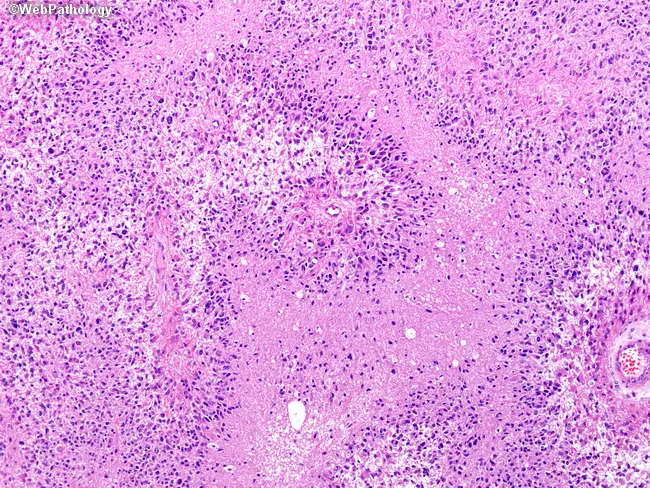
Histopathology image illustrating classic glioblastoma multiforme (GBM) architecture on an H&E stained brain tissue section. Microscopically, necrotic zones are evident and organized in serpentine, finger‑like channels that slice through the cellular matrix. Surrounding the necrosis, hypercellular, pleomorphic tumor cells crowd the periphery and frequently align in pseudopalisading arrangements, creating a distinctive columnar belt of tumor nuclei encircling the dead core. The intermingling of viable glial cells with necrotic debris produces a heterogeneous pattern characterized by dense cellularity, nuclear atypia, mitotic figures, and zones of microvascular proliferation that may be observed at higher magnification. The background stroma shows a mix of capillary networks and microhemorrhages, consistent with aggressive infiltration into adjacent brain parenchyma. This histology reflects the INFILTRATIVE nature of GBM and the hallmark combination of necrosis with pseudopalisading tumor cells. Clinically, these features correlate with rapid clinical progression, resistance to therapy, and a poor prognosis despite aggressive treatment. The image emphasizes the necessity of accurate histological grading (WHO grade IV) to guide multidisciplinary management, including maximal safe surgical resection, adjuvant radiotherapy, and chemotherapy. Recognition of serpentine and geographic necrosis, along with pseudopalisading cells, supports a GBM diagnosis and informs differential considerations such as anaplastic astrocytoma and other high‑grade gliomas. Variants.
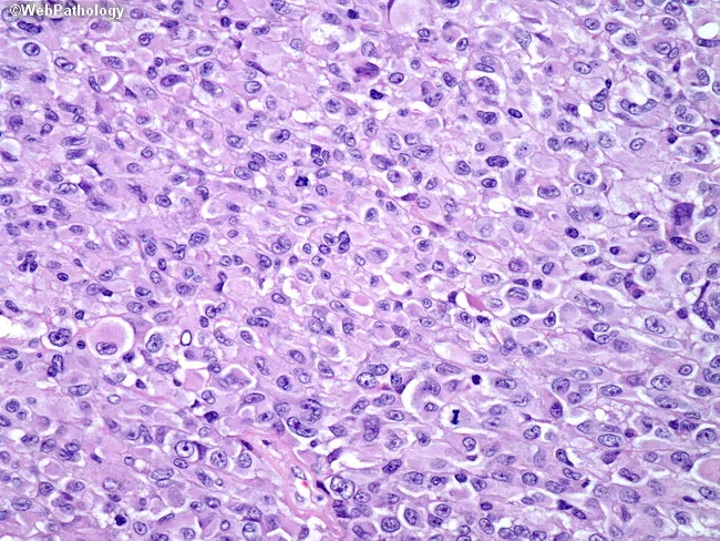
This is a brightfield histopathology image of a brain tumor tissue section, stained with hematoxylin and eosin. The specimen is from a 26-year-old female with left temporal lobe mass on MRI and presenting with headaches and sudden-onset hearing loss. The tumor demonstrates epithelioid glioblastoma morphology characterized by cells with abundant eosinophilic cytoplasm, vesicular nuclei with conspicuous nucleoli, and distinct cell borders. These tumor cells are arranged in cohesive, patternless sheets rather than classic glial fascicles. Necrosis and endothelial proliferation are present, accompanied by brisk mitotic activity, indicating high-grade malignancy. Immunophenotype shows strong GFAP positivity, weak CAM5.2 staining, and lack of melanoma-associated markers, supporting glial origin and arguing against metastatic melanoma or carcinoma. Approximately half of epithelioid GBMs harbor BRAF V600E mutations, a molecular feature with potential therapeutic implications; targeted therapy with BRAF inhibitors (e.g., vemurafenib) may be beneficial in mutation-positive cases. The described histology correlates with an aggressive phenotype and poor prognosis compared with conventional GBM, although targeted molecular therapy may modify outcomes. This image highlights diagnostic challenges where epithelioid features mimic metastatic carcinomas or melanoma, underscoring the need for immunohistochemistry and molecular profiling for accurate diagnosis and treatment planning. Clinical relevance includes guiding molecularly targeted therapy decisions for care.
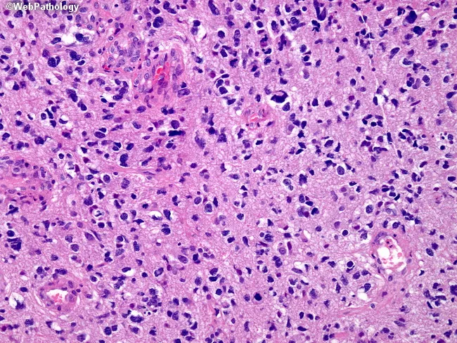
This image depicts a hematoxylin and eosin stained brain tissue section illustrating a glioblastoma multiforme (GBM), a WHO grade IV astrocytoma. Acquired by light microscopy, the slide shows high cellular density with marked nuclear pleomorphism and hyperchromasia, reflecting malignant astrocytic transformation. Numerous mitotic figures indicate active cell proliferation. The tumor infiltrates the surrounding neuropil, producing a markedly fibrillary background characteristic of astrocytic processes. In classic GBM, one may observe necrosis with pseudopalisading and microvascular proliferation, though the specific field here emphasizes the solid hypercellular cast rather than large necrotic zones. The presence of pleomorphic, irregular nuclei with prominent nucleoli and a disordered architectural arrangement confirms an infiltrative glioma of astrocytic lineage. Immunohistochemical confirmation is typically sought (GFAP positivity; IDH-wildtype in primary GBMs; MGMT promoter methylation status informs prognosis and therapy). Clinically, GBM carries a poor prognosis but requires definitive histological grading to guide treatment strategies, including maximal safe surgical resection followed by radiotherapy and temozolomide. This image is valuable for educational context in neuropathology, tumor biology, grading criteria, differential diagnosis with anaplastic astrocytoma or oligodendroglioma, and radiologic-pathologic correlation studies. This example underscores the importance of correlating histology with imaging features for accurate prognosis, therapy planning, and research into glioma biology.
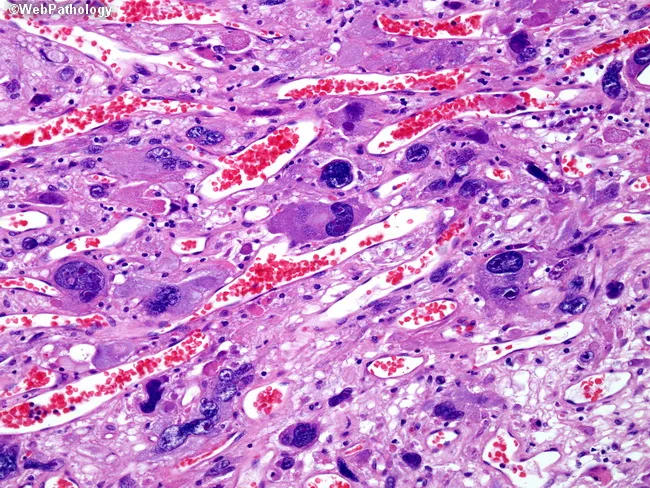
This histopathology image depicts a high-grade astrocytic neoplasm, specifically glioblastoma with giant cells (giant cell glioblastoma/monstrocellular glioblastoma). Prepared as a formalin-fixed paraffin-embedded brain tissue section, stained with Hematoxylin and Eosin (H&E) and examined under light microscopy at variable magnification (commonly 200–400x for cellular detail). The field shows numerous pleomorphic, giant astrocytic cells containing bizarre hyperchromatic nuclei, sometimes multinucleated, with abundant eosinophilic to pale cytoplasm. Tumor cells are embedded within a fibrillary glial background and interspersed with irregular, thickened blood vessels; microvascular proliferation is evident. Regions of necrosis with pseudopalisading necrosis are present, a hallmark of glioblastoma. Numerous mitotic figures may be observed, indicating active tumor proliferation. Intervening red blood cells in capillary channels are visible. The image demonstrates classic glioblastoma features: infiltrative growth, marked cellular pleomorphism, necrosis, microvascular proliferation, and astrocytic differentiation evidenced by GFAP-immunophenotypic concordance (implied). Clinically, this finding correlates with aggressive CNS malignancy in adults, presenting with seizures or focal deficits; treatment involves multidisciplinary management (maximal safe surgical resection, radiotherapy, temozolomide). Diagnostic significance includes distinguishing giant cell GBM variant from conventional GBM and other astrocytomas; prognosis is poor, though some studies suggest variable outcomes in co-occurring mutations. Consideration for targeted molecular profiling and immunohistochemistry is implied, and may guide prognosis and therapy decisions.
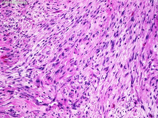
Imaging modality: Light microscopy of hematoxylin and eosin stained brain tumor tissue (formalin-fixed, paraffin-embedded; micrograph). The slide illustrates a gliosarcoma, a rare primary CNS neoplasm consisting of two malignant components: a glial, astrocytic element resembling glioblastoma and a mesenchymal, spindle-cell sarcomatous element. The histologic pattern is biphasic, with areas of highly cellular, pleomorphic astroglial cells showing brisk mitotic activity and microvascular proliferation, and separate fascicles of spindle-shaped cells with elongated nuclei, abundant eosinophilic cytoplasm, and occasional atypia. Notable features include necrosis with potential pseudopalisading arrangements in glial regions and intermingled mesenchymal regions that may exhibit collagen deposition. Immunophenotypic separation is typical, with GFAP positivity in glial areas and vimentin positivity in sarcomatous zones, reflecting divergent lineages within a single tumor. The overall architecture and cellular morphology support a high-grade, aggressive neoplasm; differential diagnoses include conventional glioblastoma, metastasis with sarcomatoid features, and other high-grade CNS tumors with mesenchymal differentiation. Clinically, gliosarcoma mirrors glioblastoma in epidemiology, presentation, and prognosis but may show a slight temporal lobe predilection. Diagnostic significance lies in recognizing biphasic histology, guiding surgical management and adjuvant therapy, and informing prognosis. This image emphasizes tumor heterogeneity and the need for combined histological and immunohistochemical confirmation to guide targeted therapy decisions.
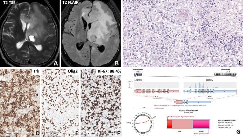
This composite image details the neuroradiological, histopathological, and molecular features of a glioblastoma (IDH-wildtype) harboring a BCR::NTRK2 fusion. (A-B) Axial T2-weighted turbo spin-echo and T2 FLAIR MRIs demonstrate a hyperintense mass involving the thalamus and bifrontal regions with extensive peritumoral edema. (C) H&E staining reveals a cellular tumor composed of oligodendroglioma-like round cells with clear cytoplasm, microvascular proliferation, and areas of pseudopalisading necrosis. (D-F) Immunohistochemical analysis shows diffuse cytoplasmic positivity for TRK, strong nuclear staining for Olig2, and a high Ki-67 proliferative index of 88.4%. (G) An Arriba plot illustrates the molecular findings, identifying an in-frame fusion between chromosome 22 (BCR gene) and chromosome 9 (NTRK2 gene), involving the retention of the protein tyrosine kinase domain. This case exemplifies the integration of advanced imaging, immunohistochemistry, and genomic sequencing for the diagnosis of high-grade NTRK-fused gliomas in neuro-oncology.
oligodendroglioma fried egg appearance IDH mutation 1p19q codeletion histology
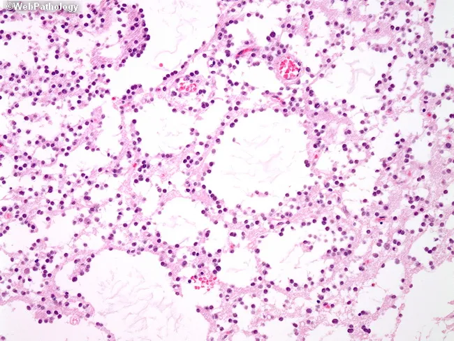
This histopathology image depicts a brain tissue biopsy stained with Hematoxylin and Eosin, viewed under light microscopy. The section shows diffuse infiltration by small, round to ovoid oligodendroglia-like cells with perinuclear halos and delicate capillary networks—classic fried-egg cytology with a chicken-wire vascular pattern. The tumor preferentially involves white matter and shows cortical invasion with perineuronal satellitosis, features that favor oligodendroglioma over other glial neoplasms in small samples. In this image, cellularity is moderate and nuclei are uniform with fine chromatin; the background is extracellular pink stroma. DNT (dysembryoplastic neuroepithelial tumor) is in the differential, particularly in young patients, but DNT often exhibits a distinctive columnar and meningioid component with specific architectural patterns not conspicuously evident here. The diagnostic significance lies in recognizing oligodendroglioma morphology and guiding ancillary testing, including molecular studies for IDH mutation and 1p/19q codeletion, which refine diagnosis and prognosis. Clinical implications include surgical resection planning, adjuvant radiotherapy, and alkylating chemotherapy considerations. Potential differential diagnoses include astrocytoma, ganglioglioma, and glioblastoma for lesions with higher-grade features. This image is valuable for pathology education, tumor classification discussions, and histology-based differential diagnosis in neuro-oncology. This contextualizes pathology findings for clinical decision-making and supports education in neuro-oncology curricula and research across disciplines.
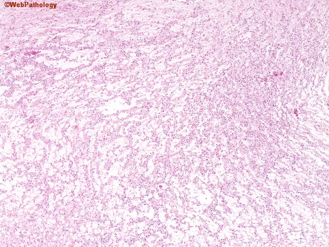
Wide-field histopathology of a neural tumor showing gelatinous nodules composed of ribbons of oligodendroglioma-like cells, often arranged around vasculature. Inter-nodular areas display mucin-rich pools in which scattered normal and dysmorphic ganglion cells are seen, described as floating neurons among the neoplastic glial population. The oligodendroglia-like cells exhibit uniform, round nuclei with perinuclear clearing (fried-egg appearance) set in a myxoid to mucinous background. Blood vessels may be surrounded by tumor cells, underscoring a perivascular growth pattern. The overall architecture is nodular and gelatinous, with alternating densely cellular glial nodules and less cellular mucinous zones. These features suggest a glioneuronal tumor with oligodendroglial differentiation and neuronal components; differential diagnoses include oligodendroglioma with neuronal differentiation or a mixed glial-neuronal tumor such as oligodendroglioma/ganglioglioma spectrum, or oligoastrocytoma depending on molecular context. The presence of mucin pools and floating ganglion cells is characteristic of ganglioglioma-like maturation. Molecular correlates (not shown) such as IDH mutation and 1p/19q co-deletion would support oligodendroglioma, whereas absence could favor ganglioglioma or other dysembryoplastic lesions. Clinically relevant for prognosis and treatment planning, as 1p/19q status informs chemo-sensitivity and survival. This histology would be essential for neuropathology conference discussion and guiding surgical and adjuvant therapy decisions.
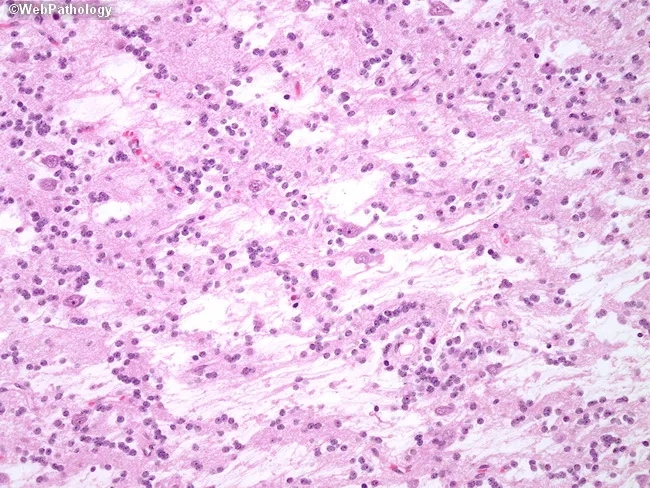
Comprehensive Description: This histopathology slide depicts a glial tumor of the brain with oligodendroglioma-like morphology. The intranodular or inter-nodular areas are composed of small, round to oval cells with relatively dispersed chromatin and prominent perinuclear halos giving a fried-egg appearance. The tumor cells are arranged in sheets and subtle neuropil-like stroma; capillary networks may be present but conspicuous mitotic activity and necrosis are not observed. In a subset of cases, foci resembling pilocytic astrocytoma or ganglioglioma with astrocytic processes or neuronal components can be identified, though these features are not universally present. Endothelial hyperplasia and necrosis are typically absent, supporting a low to intermediate grade phenotype. The pattern suggests a glial neoplasm with oligodendroglial differentiation; the presence of calcifications is not clearly evident in this field but would have diagnostic significance if seen. Clinically, these findings warrant correlation with neuroimaging to assess tumor location and frontal or temporal lobe involvement, calcifications, and cortical invasion. Immunohistochemistry and molecular studies (e.g., IDH mutation status, 1p/19q co-deletion) would provide definitive classification and prognostic information, distinguishing oligodendroglioma from pilocytic astrocytoma or ganglioglioma. This image is relevant for educational teaching, differential diagnosis, and research into glial-neuronal tumors.
pilocytic astrocytoma Rosenthal fibers biphasic pattern ependymoma perivascular pseudorosettes
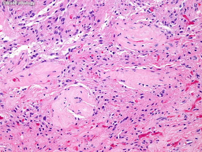
This histopathology image shows pilocytic astrocytoma tissue characterized by a biphasic architecture with alternating dense, fibrillary areas and looser, microcystic regions. The tumor cells are astrocytic with long, hairlike processes and piloid cytoplasm, arranged in bipolar, threadlike formations. Rosenthal fibers—eosinophilic, elongated inclusions within processes—are evident, contributing to the diagnostic pattern. Vascular hyalinization is conspicuous, with thickened blood vessel walls persisting amidst the tumor. Occasional infarct-like necrosis may be present in a minority of cases (5–10%), though the image primarily demonstrates viable tumor. The background shows a mixed inflammatory infiltrate and microglial activation in some fields. The staining is hematoxylin and eosin, highlighting nuclei in dark blue/purple against pink eosinophilic cytoplasm and connective tissue; mitotic figures are rare, consistent with a low-grade neoplasm. Clinically, pilocytic astrocytomas are associated with MAPK pathway activation, frequently via BRAF alterations or FGFR1/PTPN11 mutations, underscoring the genetic etiology described in contemporary genomics studies. The histologic silhouette supports a low-grade glioma diagnosis with favorable prognosis following gross-total resection; differential considerations include pilomyxoid astrocytoma and oligodendroglioma in atypical presentations. This image exemplifies key diagnostic features used in neuropathology practice and research, as well as molecular correlation with targeted therapies.
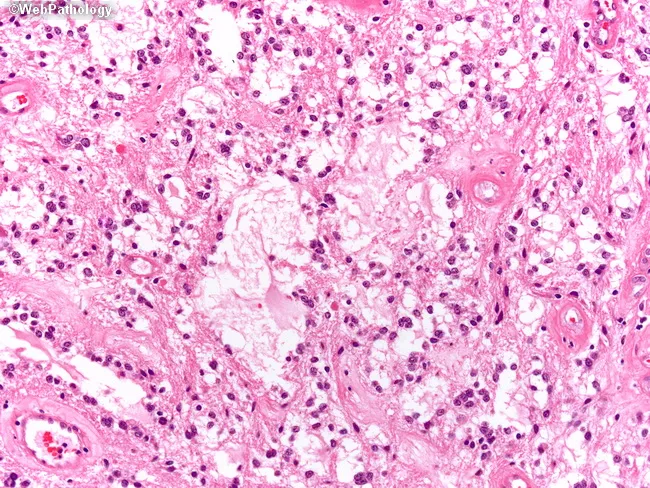
This is a high-magnification histopathology image (Hematoxylin and Eosin stain) of pilocytic astrocytoma, a World Health Organization Grade 1 glioma. The tissue shows biphasic architecture with microcystic, loose backgrounds interposed with compact, piloid areas composed of spindle-shaped astrocytic cells bearing long, hair-like processes. The cellular regions resemble fibrillary astrocytoma but are distinguished by their abundant, slender cytoplasmic processes and cohesive, monomorphic nuclei with minimal atypia. Rosenthal fibers—eosinophilic, corkscrew-like inclusions—may be evident within the fibrillary matrix. The microcystic zones contain small vacuolated spaces; vessels are typically capillary and inconspicuous. Overall, there is low mitotic activity, absent necrosis, and mild nuclear pleomorphism, all supportive of a benign, slow-growing tumor. Clinically, pilocytic astrocytomas arise predominantly in children, most commonly in the posterior fossa, but can involve cerebral hemispheres or spinal cord; neurofibromatosis type 1 predisposes to optic pathway gliomas. The lesion’s image-based histology correlates with favorable prognosis after gross total resection and a low recurrence rate. Differential diagnoses include fibrillary astrocytoma, oligodendroglioma, ganglioglioma, and DNET; however, the biphasic microcystic/piloid pattern, Rosenthal fibers, and low-grade cytology strongly support pilocytic astrocytoma. This slide aids diagnostic confirmation and informs surgical planning and prognostic assessment. Knowledge of this pattern guides tailored therapy and long-term surveillance, improving outcomes.
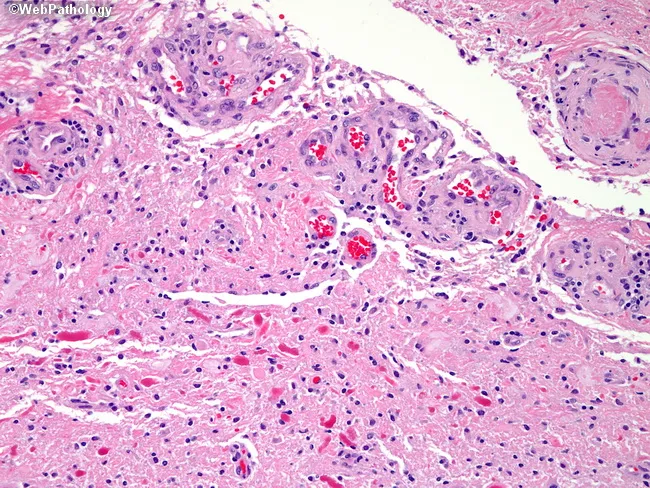
Brightfield hematoxylin and eosin stained brain tumor tissue (FFPE) illustrating pilocytic astrocytoma morphology. The tumor shows cystic architecture with loose, interspersed piloid astrocytes, featuring hair-like processes and elongated bipolar cells. Adjacent solid areas merge with the cyst wall, creating a biphasic pattern typical of pilocytic astrocytoma. A prominent feature is glomeruloid microvascular proliferation: tortuous, lumen-rich endothelial tufts with multiple lumina arranged around cystic spaces. While rare multilayered endothelial hyperplasia can occur in pilocytic astrocytoma, it does not impart the ominous prognostic significance seen in diffuse high-grade gliomas. Rosenthal fibers are present in the lower portion of the image as brightly eosinophilic, thick, corkscrew-like inclusions within astrocytic processes. The background matrix shows microcystic change and a mild inflammatory infiltrate; mitotic activity is rare and nuclear atypia minimal, consistent with a low-grade glioma. Overall, the histology supports a diagnosis of pilocytic astrocytoma in a cyst-containing cerebral lesion. The documented features—piloid cells, Rosenthal fibers, and glomeruloid vessels—are critical for differential diagnosis, helping distinguish from high-grade gliomas where endothelial hyperplasia carries grave prognosis. Correlation with age, imaging, and clinical course is essential to confirm the diagnosis and guide management.
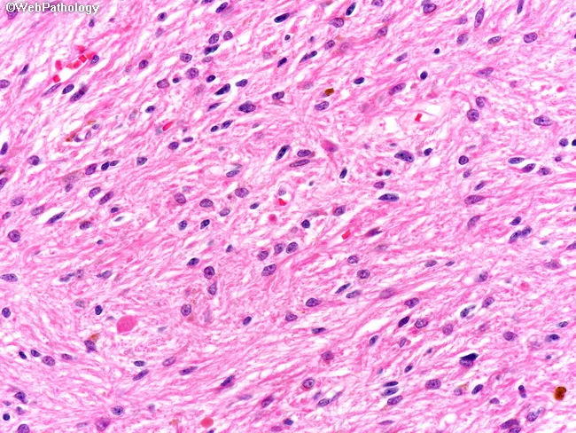
Histology (brightfield) of brain parenchyma stained with Hematoxylin and Eosin (H&E) at approximately 400x magnification reveals classic pilocytic astrocytoma features. The cellular component comprises piloid astrocytes with elongated, slender processes that extend in hair-like cylindrical strands, creating a delicate, feathery background within a variably fibrillary stroma. The biphasic architecture often shows loosely textured, microcystic areas interspersed with more compact, densely packed zones, producing a biphasic appearance. Nuclei are typically bland with modest eccentricity, fine chromatin, and low mitotic activity, supporting a low-grade glioma diagnosis in a pediatric or young adult brain specimen. Rosenthal fibers, thick eosinophilic corkscrew inclusions within processes, may be present but are not universally seen in every field. Background astrocytic processes contribute to a pink, fibrillar matrix, and occasional eosinophilic granular bodies can occur. The observed morphology is highly characteristic of pilocytic astrocytoma and correlates with indolent clinical behavior following maximal safe surgical resection. Clinically, these features distinguish pilocytic astrocytoma from higher-grade astrocytomas and other glial neoplasms. This image is relevant for educational discussion of glial tumors, neuropathology teaching, differential diagnosis, and surgical planning, providing keyword-rich descriptors including piloid cells, hair-like processes, Rosenthal fibers, biphasic pattern, and low-grade glioma. Heightened searchability is achieved through alternative terminology and related terms.
ependymoma perivascular pseudorosettes true rosettes GFAP EMA histology
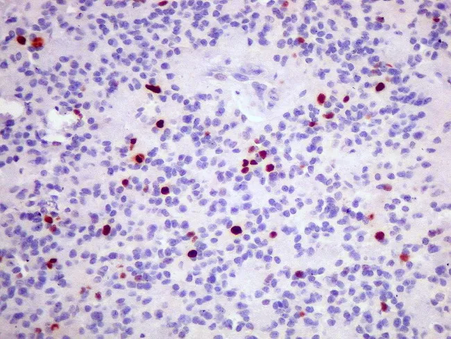
Light microscopy of a formalin-fixed paraffin-embedded brain tumor section stained with Hematoxylin and Eosin (H&E), complemented by Ki-67 immunohistochemistry (MIB-1). The histology demonstrates an ependymal neoplasm with relatively uniform, small round to oval nuclei, scant cytoplasm, and minimal pleomorphism. Architectural features may include perivascular pseudorosettes; true rosettes can be variably evident depending on the field. The tumor shows low to moderate cellularity and rare mitotic figures, consistent with a World Health Organization (WHO) Grade II designation. Ki-67 labeling is low overall, with nuclear positivity up to approximately 5% in some foci and largely negative elsewhere, indicating a relatively indolent proliferative rate. The background is not overtly hyperchromatic, and there is no overt necrosis. Correlation with radiologic imaging and clinical parameters supports a low-grade central nervous system tumor prognosis, though intratumoral heterogeneity may harbor focal proliferative hotspots. This image is valuable for pathology education, neural tumor grading, and multidisciplinary case discussions, including neurosurgery, neuropathology, and neuro-oncology teams. Keywords: ependymoma, CNS tumor, brain tumor, WHO Grade II, Ki-67, MIB-1, proliferation index, perivascular pseudorosette, histology, H&E, duplicate staining, FFPE, brain pathology, neuropathology education. This descriptive caption supports targeted search terms for education, diagnosis, prognosis assessment, and research on CNS ependymal tumors and outcomes.
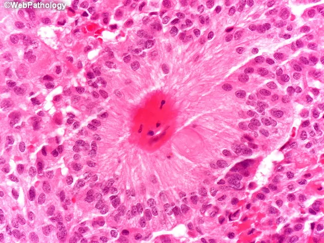
This histopathology image uses hematoxylin and eosin staining to show a central nervous system tumor with classic ependymal differentiation. The tissue exhibits small to medium-sized round to oval nuclei with finely granular chromatin set in a fibrillary, eosinophilic background. A hallmark feature is the arrangement of tumor cells around blood vessels producing perivascular pseudorosettes, with a central zone around the vascular lumen that is relatively nucleus-free. True ependymal rosettes, containing a central lumen lined by tumor cells, are present in a minority of cases (approximately 10%). Overall cellularity is variable, with prominent perivascular organization and occasional ependymal canals. The lesion supports an ependymal lineage; immunohistochemistry would typically show glial fibrillary acidic protein (GFAP) positivity, and EMA often reveals characteristic dot-like perinuclear staining (not evident on H&E). Clinically, classic ependymomas are usually WHO grade II, while the myxopapillary variant is grade I and the anaplastic variant is grade III. The presence of rosettes and pseudorosettes is a defining feature for diagnosis and aids in differentiating from astrocytomas or primitive neuroectodermal tumors. Clinically relevant context includes intraventricular or spinal canal localization, hydrocephalus risk, and prognosis after surgical resection with radiotherapy decisions influenced by location and histologic grade.
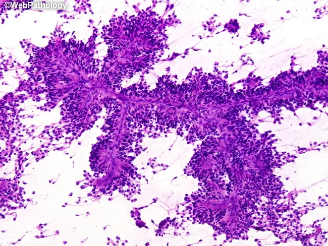
Imaging modality: Cytology smear derived from a central nervous system tumor. Preparation: Hematoxylin and Eosin (H&E) stained smear prepared from a CNS lesion, most consistent with ependymal lineage. The smear shows high cellularity of small, round to oval tumor cells with scant cytoplasm and finely stippled chromatin. The most distinctive feature is perivascular pseudorosette formation: tumor cells arranged in radial cuffs around slender blood vessel cores, creating spoke‑like patterns with intervening delicate fibrillary processes. These cytologic rosettes correspond to the histologic perivascular pseudorosettes seen in ependymoma. The background is fibrillary and slightly basophilic, reflecting glial-type stroma. Nuclei are generally uniform with mild pleomorphism; mitotic activity is limited in this smear. Overall architecture mirrors the histopathology, including ependymal differentiation and possible true rosettes in some fields. Diagnostic significance: The presence of perivascular pseudorosettes strongly supports ependymal differentiation and favors ependymoma, particularly in pediatric or young adult patients. Differential considerations include medulloblastoma with Homer-Wright rosettes and other glial neoplasms; rosette pattern around vessels helps distinguish from true vascular channels. Clinical correlation with imaging findings (ventricular or intraventricular lesion) and patient age enhances diagnostic accuracy and guides management. This description supports educational teaching, cytology-pathology correlation, and differential diagnosis in neuropathology for clinical practice.
Overview and Classification
- No morphologically evident premalignant or in situ lesions
- Even low-grade lesions can infiltrate vast brain regions and be unresectable
- Malignant gliomas rarely metastasize outside the CNS
- Location independently influences prognosis
I. DIFFUSE GLIOMAS (Adult-Type)
| Feature | Astrocytoma, IDH-mutant (WHO 2-4) | Oligodendroglioma, IDH-mutant and 1p/19q-codeleted (WHO 2-3) | Glioblastoma, IDH-wildtype (WHO 4) |
|---|---|---|---|
| IDH status | Mutant | Mutant | Wildtype |
| Other genetics | TP53-mut, ATRX-mut, CDKN2A-HD (grade 4) | 1p/19q-codeleted, TERT-mut | +7/-10, pTERT-mut, EGFR-amp |
| Typical morphology | Nuclear atypia; mitoses (grade 3); MVP/necrosis (grade 4) | Round nuclei, clear halos; mitoses, MVP, necrosis (grade 3) | Nuclear atypia, mitoses, MVP, necrosis |
- Robbins & Cotran, Table 28.5, p. 1194
II. ASTROCYTOMA, IDH-Mutant (WHO Grades 2-4)
Epidemiology and Location
- Predominant in the cerebral hemispheres of young to middle-aged adults (median age 38 years)
- Present with seizures, headaches, focal neurologic deficits
- Much better prognosis than IDH-wildtype glioblastoma
Pathogenesis
- IDH1 or IDH2 mutation - the earliest and defining tumorigenic event; produces the oncometabolite 2-hydroxyglutarate
- TP53 mutation - inactivating mutation contributing to genomic instability
- ATRX inactivation - leads to alternative lengthening of telomeres (mechanism of senescence evasion)
Morphology
- Gross (Grade 2-3): Poorly defined, gray, infiltrative tumors that expand and distort the brain without forming a discrete mass; grade 4 lacks the large necrosis zones of glioblastoma
- Gross (Grade 4, IDH-mutant): Diffuse infiltration without necrosis/hemorrhage (contrast with IDH-WT GBM)
- Grade 2: Mildly to moderately hypercellular; enlarged, elongated or irregular hyperchromatic nuclei; fibrillar background; perineuronal satellitosis (tumor cells surrounding neurons); transition to normal tissue is indistinct
- Grade 3: Denser cellularity, greater nuclear pleomorphism, mitotic figures present
- Grade 4: Greater cellular crowding, cytological atypia, increased proliferative activity, microvascular proliferation (MVP) and/or necrosis
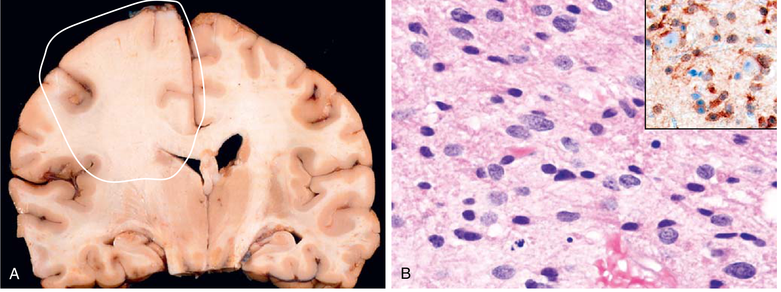
IHC in Diagnosis
| Marker | Result | Significance |
|---|---|---|
| GFAP | Positive (fibrillar cytoplasmic) | Confirms astrocytic lineage; fibrillar background highlights cell processes |
| IDH1 R132H | Positive (up to 90% of cases) | The R132H antibody detects the most common IDH1 mutation; highlights tumor cells against background brain - "perineuronal satellitosis" is beautifully demonstrated |
| IDH1/IDH2 sequencing | Performed when R132H IHC negative | Detects less common IDH mutations not covered by R132H antibody |
| ATRX | Loss of nuclear expression | Reflects ATRX gene inactivation; tumor cells lose ATRX staining while normal endothelial/lymphoid cells retain it (internal positive control) |
| p53 | Overexpression (nuclear) | Reflects TP53 mutation leading to protein accumulation |
| Ki-67 (MIB-1) | Elevated (especially grade 3-4) | Proliferation index; helps grade assessment |
-
Grade 2: >10 years
-
Grade 3: 5-10 years
-
Grade 4: ~3 years
-
Robbins & Kumar Basic Pathology, p. 857-860
-
Robbins & Cotran Pathologic Basis of Disease, p. 1193-1196
III. GLIOBLASTOMA, IDH-Wildtype (WHO Grade 4)
Epidemiology
- Predominantly affects adults in their 6th to 8th decades
- Common locations: temporal, parietal, frontal lobes; basal ganglia and thalamus
- Butterfly glioma: rapid infiltration of corpus callosum → bilateral symmetric lesion
Pathogenesis
| Alteration | Effect |
|---|---|
| +7/-10 (gain chr 7, loss chr 10) | Most common copy number alteration |
| TERT promoter mutation | Evasion of senescence via telomerase activation |
| EGFR gene amplification | Activation of growth factor signaling |
| CDKN2A biallelic deletion | Loss of p16 → escape from growth controls |
| TP53 mutation | Resistance to apoptosis |
| MGMT promoter methylation | Loss of DNA repair enzyme; predicts response to temozolomide |
| PTEN loss | PI3K pathway activation |
Morphology
- Gross: Highly variable - firm white areas, soft yellow necrotic zones, cystic degeneration, hemorrhage; ring-enhancing on MRI
- Histology:
- High cellularity
- Poorly differentiated pleomorphic cells with nuclear atypia
- Brisk mitotic activity
- Pseudopalisading necrosis - tumor nuclei palisade (line up) around serpiginous/geographic bands of necrosis (pathognomonic feature)
- Microvascular proliferation (MVP) - glomeruloid tufts of proliferating endothelial cells
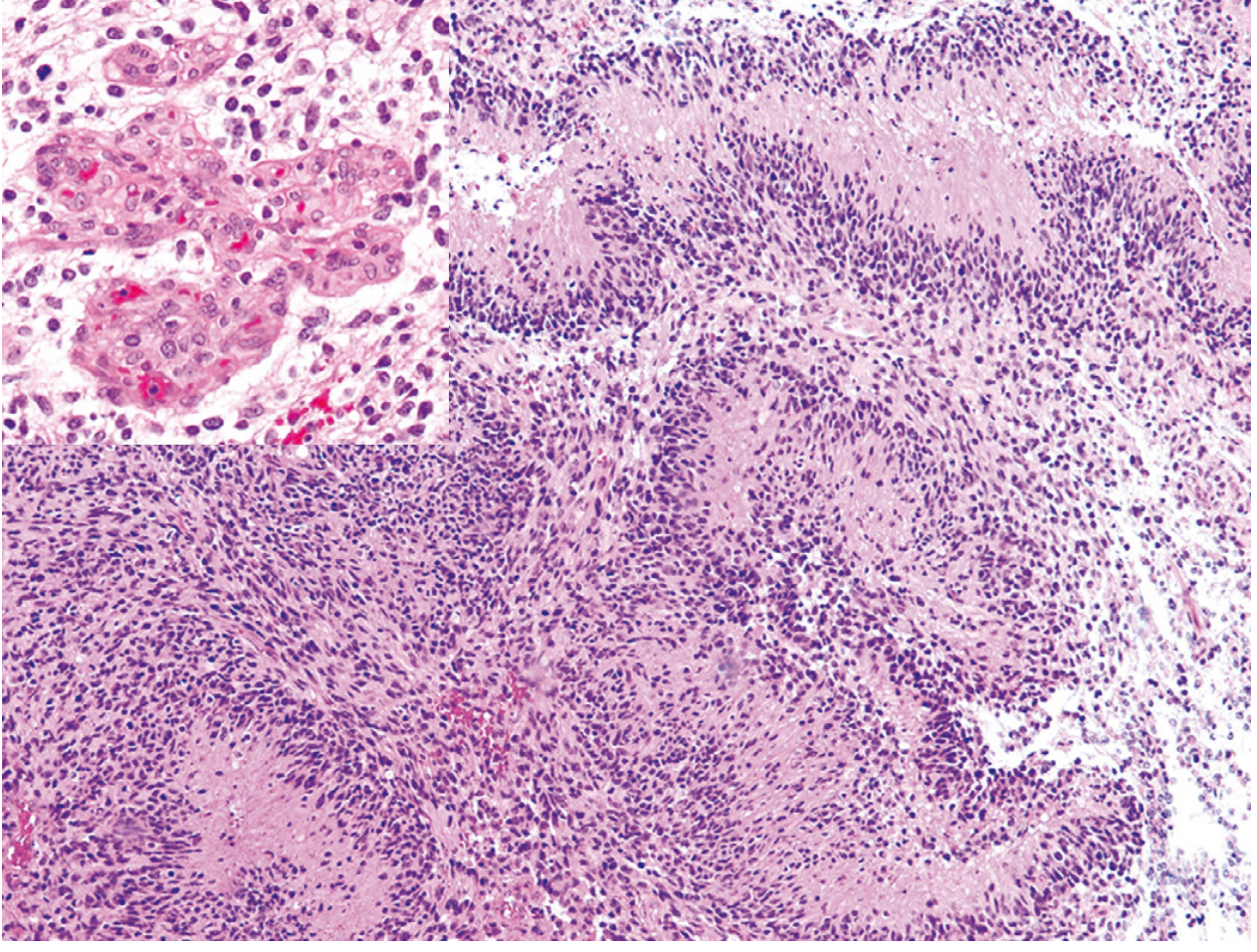
IHC in Diagnosis of GBM
| Marker | Result | Significance |
|---|---|---|
| GFAP | Positive | Confirms glial (astrocytic) origin; may be focally reduced in poorly differentiated areas |
| IDH1 R132H | Negative | Critical - IDH-wildtype status is a defining feature; negative IDH IHC (confirmed by sequencing) + GBM histology = primary GBM |
| EGFR | Overexpression/amplification | Seen in ~50%; associated with aggressive behavior |
| PTEN | Loss | Correlates with poor prognosis |
| Ki-67 (MIB-1) | High (often >20%) | Reflects aggressive proliferation |
| MGMT (methylation) | IHC unreliable; PCR-based testing preferred | Methylated MGMT = better response to temozolomide |
| p53 | Variable | Less consistent than in IDH-mutant astrocytoma |
IV. OLIGODENDROGLIOMA, IDH-Mutant and 1p/19q-Codeleted (WHO Grades 2-3)
Overview
Pathogenesis
- IDH1 or IDH2 mutation (same as astrocytoma)
- Whole-arm codeletion of chromosomes 1p and 19q (defining molecular hallmark; these must BOTH be present)
- TERT promoter mutation (vast majority)
- EGFR amplification is NOT seen (contrast with GBM)
- Loss of chromosome 9p (CDKN2A deletion) occurs in higher-grade tumors but is less common than in IDH-mutant astrocytoma
Morphology
- Gross: Well-circumscribed tumors; may show calcifications, mucoid degeneration, small cysts; cortical invasion common
- Histology:
- Round, uniform nuclei with cleared cytoplasm forming perinuclear halos ("fried-egg" appearance) - a processing artifact from formalin fixation that is nonetheless highly characteristic
- Thin-walled capillaries in an interlacing ("chicken-wire") vascular pattern
- Calcification is common (microcalcifications)
- Grade 3 (anaplastic): Higher mitotic activity, MVP, and/or necrosis
IHC in Diagnosis
| Marker | Result | Significance |
|---|---|---|
| IDH1 R132H | Positive (tumor cells) | Same as IDH-mutant astrocytoma; highlights tumor cells including perineuronal satellitosis |
| GFAP | Variable, usually weaker | Less strongly positive than astrocytomas |
| Olig2 | Positive (nuclear) | Strong Olig2 positivity supports oligodendrocytic lineage |
| ATRX | Retained (positive) | Key distinguishing point: ATRX is RETAINED in oligodendroglioma but LOST in IDH-mutant astrocytoma - vital in differential diagnosis |
| p53 | Usually negative/low | Helps distinguish from IDH-mutant astrocytoma |
| Ki-67 | Low in grade 2, elevated in grade 3 | Grading |
| 1p/19q codeletion | By FISH or PCR | Definitive molecular test required for diagnosis; IHC alone insufficient |
- IDH-mutant + ATRX loss + p53 overexpression = Astrocytoma
- IDH-mutant + ATRX retained + p53 low + 1p/19q codeletion = Oligodendroglioma
- Grade 2: 15-20 years
- Grade 3: 5-10 years (Robbins & Kumar Basic Pathology, p. 859-861)
V. PILOCYTIC ASTROCYTOMA (WHO Grade 1)
Overview
Pathogenesis
- KIAA1549-BRAF gene fusion/duplication - the most common alteration, activates the MAPK signaling pathway
- Other MAPK pathway alterations (FGFR1, PTPN11 mutations)
- No IDH mutations - fundamental distinction from adult diffuse gliomas
Morphology
- Gross: Often cystic with a contrast-enhancing mural nodule in the cyst wall; well demarcated
- Histology (biphasic pattern):
- Loose "microcystic" areas with stellate cells
- Compact densely fibrillar areas with bipolar cells having long, thin "hairlike" processes
- Rosenthal fibers - eosinophilic, corkscrew-shaped/elongated inclusions within astrocytic processes (characteristic but not exclusive)
- Eosinophilic granular bodies (EGB) - mulberry-like inclusions
- Microvascular proliferation may be present but does NOT imply poor prognosis (unlike diffuse gliomas)
- Necrosis and mitoses are rare
IHC in Diagnosis
| Marker | Result | Significance |
|---|---|---|
| GFAP | Strongly positive | Bipolar cell processes intensely positive; confirms astrocytic lineage |
| IDH1 R132H | Negative | Key distinction from adult infiltrative astrocytomas |
| BRAF V600E | Positive in some non-cerebellar cases (~10%) | Therapeutic implications (BRAF inhibitors) |
| BRAF KIAA1549 fusion | Detected by FISH/PCR | Most common in cerebellar pilocytic astrocytomas |
| Ki-67 | Low (<5%) | Reflects benign biology; consistent with Grade 1 |
| Olig2 | Variable | Less useful than in infiltrative gliomas |
- Robbins & Cotran, p. 1196-1197
VI. EPENDYMOMA (WHO Grades 1-3)
Overview
- Supratentorial: ZFTA::RELA fusion (poor prognosis) or YAP1 fusion (favorable)
- Posterior fossa: Group A (PFA - poor prognosis, H3K27me3 loss) vs Group B (PFB - better prognosis)
- Spinal: NF2 mutation
Morphology
- Gross (4th ventricle): Solid or papillary masses extending from ventricular floor; may extend through foramina into cisterns ("plastic ependymoma")
- Histology:
- Regular round to oval nuclei with abundant granular chromatin
- Variably dense fibrillary background
- Perivascular pseudorosettes - tumor cells radially arranged around blood vessels with an anuclear fibrillar zone between cells and vessel wall (most constant and characteristic feature)
- True ependymal rosettes - tumor cells arranged around a central lumen/canal (less common, ~10% of cases, but highly specific)
- Anaplastic ependymoma (Grade 3): Increased cellularity, mitoses, necrosis, MVP
IHC in Diagnosis
| Marker | Result | Significance |
|---|---|---|
| GFAP | Positive | Glial origin; especially strong in perivascular pseudorosette processes |
| EMA (Epithelial Membrane Antigen) | Dot-like perinuclear or ring-like staining | Highly characteristic of ependymoma; reflects microlumen formation; useful in poorly differentiated cases |
| Olig2 | Variable/weak | Less useful |
| IDH1 R132H | Negative | Ependymomas do not carry IDH mutations |
| Ki-67 | Variable; elevated in anaplastic type | Grading |
| H3K27me3 | Loss in posterior fossa group A | Identifies PFA ependymoma (poor prognosis) |
| L1CAM | Positive | Seen in ZFTA::RELA fusion type; associated with aggressive behavior |
| YAP1 | Positive (nuclear) | Identifies YAP1 fusion type (favorable prognosis) |
- Robbins & Kumar Basic Pathology, p. 861-862; Robbins & Cotran, p. 1197-1198
VII. COMPREHENSIVE IHC PANEL SUMMARY FOR GLIAL TUMORS
| IHC Marker | Astrocytoma IDH-mut | GBM IDH-wt | Oligodendroglioma | Pilocytic Astrocytoma | Ependymoma |
|---|---|---|---|---|---|
| GFAP | + (fibrillar) | + (variable) | + (weak/variable) | ++ (strong, fibrillar) | + (esp. pseudorosettes) |
| IDH1 R132H | ++ (positive) | - (negative) | ++ (positive) | - | - |
| ATRX | Loss | Retained (usually) | Retained | N/A | N/A |
| p53 | Overexpressed | Variable | Negative/low | Negative | Negative |
| Olig2 | + | + | ++ (strong) | +/- | +/- |
| Ki-67 | Low (G2) → High (G4) | High (>20%) | Low (G2) → High (G3) | Low (<5%) | Variable |
| EMA | Negative | Negative | Negative | Negative | Dot/ring pattern |
| 1p/19q codeletion (FISH) | Absent | Absent | Present (defining) | Absent | Absent |
| EGFR | Not amplified | Amplified (~50%) | Not amplified | N/A | N/A |
| BRAF V600E / fusion | Negative | Rare | Negative | KIAA1549-BRAF fusion | Negative |
| H3K27me3 | Retained | Retained | Retained | Retained | Loss in PFA |
VIII. KEY DIAGNOSTIC ALGORITHMS USING IHC
Approach to a Diffuse Infiltrating Glioma:
-
Perform IDH1 R132H IHC first
- If positive: tumor is IDH-mutant → proceed to ATRX and p53
- ATRX loss + p53 overexpression → Astrocytoma, IDH-mutant
- ATRX retained + p53 low → Oligodendroglioma (confirm with 1p/19q FISH)
- If negative: perform IDH1/IDH2 sequencing
- If IDH wildtype + GBM histology + any of {+7/-10, TERT-mut, EGFR-amp} → Glioblastoma, IDH-wildtype
- If positive: tumor is IDH-mutant → proceed to ATRX and p53
-
GFAP establishes glial lineage throughout
-
Ki-67 assists grading
Approach to a Pediatric Circumscribed Cerebellar Tumor:
- GFAP+, IDH-, Ki-67 low, Rosenthal fibers → Pilocytic Astrocytoma
- Confirm BRAF KIAA1549 fusion by FISH
Approach to a Ventricular/Spinal Mass:
- GFAP+, EMA dot pattern, pseudorosettes, IDH- → Ependymoma
- H3K27me3 loss → PFA group (posterior fossa)
- L1CAM positive → ZFTA::RELA fusion type
IX. PEDIATRIC-TYPE DIFFUSE GLIOMAS (Brief)
- Pediatric low-grade diffuse gliomas: Mostly indolent; harbor MAPK pathway alterations
- Pediatric high-grade diffuse gliomas: Include Diffuse Midline Glioma, H3K27-altered (previously DIPG) - defined by H3K27M mutation detectable by IHC; Diffuse Hemispheric Glioma, H3G34-mutant
- IHC for H3K27M (anti-H3K27M antibody): Strongly positive in diffuse midline gliomas; this single marker can identify the tumor in a biopsy and is associated with WHO Grade 4 by definition
X. PRACTICAL IHC PEARLS (Examination-Relevant)
- GFAP is the cornerstone marker of glial differentiation - both astrocytic and ependymal tumors express it
- IDH1 R132H IHC is the most important single marker that separates IDH-mutant gliomas (better prognosis) from IDH-wildtype GBM (poor prognosis)
- ATRX loss is the IHC-detectable surrogate for ATRX gene mutation - present in IDH-mutant astrocytoma but NOT oligodendroglioma; normal internal controls (endothelial cells, lymphocytes) retain ATRX staining
- EMA dot/ring pattern is unique to ependymomas among CNS glial tumors
- Pseudopalisading necrosis on H&E is the histologic hallmark of GBM; IHC is less critical for the diagnosis compared to the pathognomonic morphology
- Ki-67/MIB-1 helps grade - low in Grade 1-2, high in Grade 3-4
- p53 IHC shows diffuse strong nuclear positivity in IDH-mutant astrocytoma (due to protein accumulation from TP53 mutation); scattered weak positivity is non-specific
- Olig2 is expressed in all gliomas but is strongest in oligodendrogliomas; it is a general glioma marker not a lineage-specific discriminator