
Reasons for tonsils
function and purpose of tonsils immune system
tonsils anatomy location throat lymphoid tissue
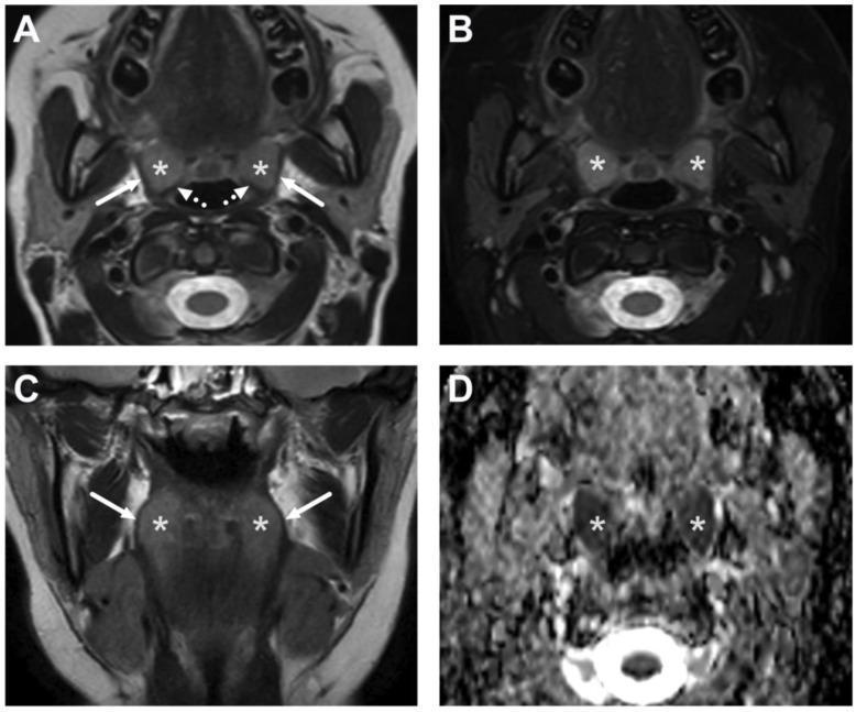
This diagnostic imaging panel demonstrates the normal MRI anatomy of the oropharynx in a pediatric patient across four sequences: (A) axial T2-weighted, (B) axial fat-suppressed T2-weighted, (C) coronal T2-weighted, and (D) an Apparent Diffusion Coefficient (ADC) map. The primary focus is the palatine tonsils (asterisks), which exhibit intermediate to high signal intensity on T2-weighted images due to lymphoid tissue content. Anatomical relationships are clearly demarcated: the tonsils are bounded laterally by the superior constrictor muscles (solid white arrows) and posteriorly by the palatopharyngeus muscles (dotted white arrows). In sequence B, fat suppression enhances the contrast between the tonsillar tissue and surrounding soft tissues. Image D shows the ADC map where the palatine tonsils demonstrate low signal intensity, indicating innately restricted diffusion characteristic of highly cellular lymphoid tissue. This comparison is clinically significant for differentiating normal physiologic prominent tonsils in children from pathological processes like tonsillitis or peritonsillar abscesses.
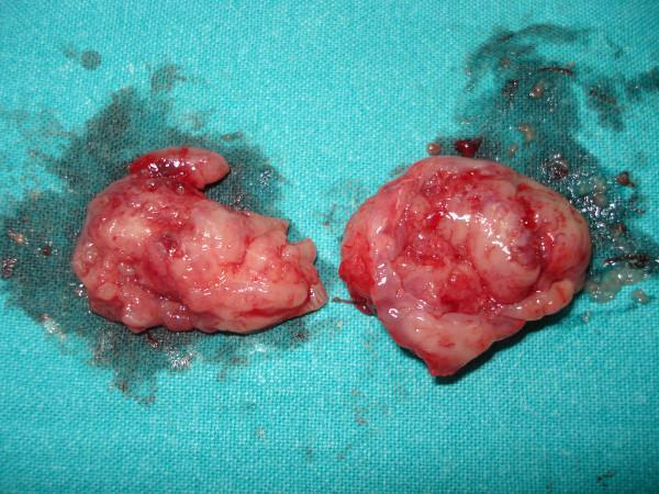
This clinical photograph shows a macroscopically resected specimen of two palatine tonsils following a tonsillectomy, placed on a blue surgical drape. The specimens exhibit an irregular, lobulated, and fleshy surface characteristic of lymphoid tissue. The color is mottled, ranging from pale pink to deep red, indicating varying levels of vascularity and focal areas of hemorrhage or congestion. The surface appears wet and glistening, likely due to serosanguinous fluid and surgical residue. Surrounding the tissues on the drape are visible stains and small fragments of clotted blood. This image serves as a pathological specimen representation of chronic tonsillitis, illustrating the anatomical appearance of inflamed tonsillar tissue post-surgical removal in a clinical setting.
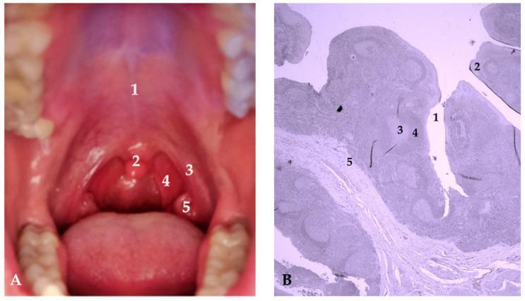
Educational panel illustrating the anatomy and histology of the palatine tonsils. Figure A is a clinical photograph of the oral cavity (oropharynx) showing key landmarks: 1—hard palate; 2—uvula; 3—anterior faucial pillar (palatoglossal arch); 4—posterior faucial pillar (palatopharyngeal arch); and 5—palatine tonsil, located within the tonsillar fossa between the pillars. Figure B is a low-power (200x) light micrograph of a histological section of tonsillar tissue stained with hematoxylin and eosin (H&E). Labeled features include: 1—tonsillar crypt, an invagination of the stratified squamous epithelium; 2—lymphoepithelium; 3—germinal center of a lymphatic follicle, indicating active B-cell proliferation; 4—mantle zone, the peripheral denser layer of the follicle; and 5—interfollicular area containing diffuse lymphoid tissue. The combination of macro-anatomical and micro-anatomical views highlights the tonsils' role as immunocompetent lymphoepithelial organs at the intersection of the upper respiratory and digestive tracts. This material is suitable for medical education regarding ENT anatomy, immunology, and histology.
What Are Tonsils?
- Palatine tonsils - the two visible lumps at the back of the throat
- Pharyngeal tonsil (adenoids) - at the roof of the pharynx
- Lingual tonsil - at the base of the tongue
- Tubal tonsils - near the opening of the auditory (Eustachian) tube
Reasons / Functions of Tonsils
1. First Line of Immune Defense
2. Antigen Sampling and Immune Activation
3. B Lymphocyte Proliferation and Antibody Production
4. T Cell Activation
5. Immune "Education" in Childhood
6. Part of the Mucosal Immune System (MALT)
7. Lymphatic Drainage
Why Are They Sometimes Removed?
Any relation to STD?
STD sexually transmitted infection tonsils throat pharyngeal gonorrhea syphilis HPV
Removal of tomsils lead to supreesing your immune system?
tonsillectomy immune function long term effects
does tonsil removal suppress immune system tonsillectomy effects immunity
Does Removing Tonsils Suppress Your Immune System?
What the Textbooks Say
"There is no evidence to suggest that tonsillectomy results in impaired immunity, presumably as a result of the extensive 'back-up' in the immune system."
- Scott-Brown's Otorhinolaryngology Head & Neck Surgery
"To date there is no proven negative immunologic impact of adenoidectomy and tonsillectomy."
- K.J. Lee's Essential Otolaryngology
Why the Immune System Copes Fine
- Adenoids and lingual tonsils - the other components of Waldeyer's ring remain and continue their immune surveillance role.
- Cervical lymph nodes - take over local antigen sampling and immune response for the throat region.
- Gut-associated lymphoid tissue (GALT) and lymph nodes throughout the body carry the bulk of systemic immunity anyway - tonsils were never a dominant player in total immune function.
- Bone marrow and thymus - continue producing and maturing B and T cells regardless of tonsil presence.
What the Research Shows
"The removal of tonsils and adenoids does not negatively affect cellular and humoral immunity."
Is There Any Nuance?
- Local throat immunity may be slightly reduced immediately after surgery, as the tonsils were the local first-line sentinel for the oropharynx. This is usually temporary.
- In young children (under 3-4 years), tonsils are at their most immunologically active, so some ENT guidelines recommend caution about early removal unless strictly necessary - not because of immune suppression per se, but because of their heightened role at that developmental stage.
- A minority of older studies (~11%) found some measurable reduction in certain antibody levels post-tonsillectomy, but these changes were transient and not clinically significant.