
Atherosclerosis notes pathology by robbins mbbs 10 marks with flow chart and diagrams
Reading File
Finding Sources
Finding Sources
I have both Robbins books. Let me read the full atherosclerosis sections from both, plus search for risk factors, morphology, and complications.
Reading File
Reading File
Finding Sources
Excellent - I have the key sections. Now let me read the pathogenesis, morphology, and complications sections in detail from both Robbins books simultaneously.
Reading File
Reading File
Now let me read the morphology and complications sections from both books, and also fetch the key diagrams.
Reading File
Reading File
Loading Image
Loading Image
Excellent! I have the key diagrams and content. Now let me read the morphology section and stable vs vulnerable plaque image:
Reading File
Loading Image
I now have all the key content, diagrams, and images needed. Let me also read the morphology section from Robbins Pathologic Basis of Disease for the gross and microscopic features.
Reading File
I now have comprehensive content from both Robbins textbooks plus all the key diagrams. Let me compile the full 10-mark notes.
ATHEROSCLEROSIS - Pathology Notes (Robbins) - 10 Marks
DEFINITION
Atherosclerosis (Greek: athero = gruel/porridge; sclerosis = hardening) is a chronic inflammatory disease of large and medium-sized arteries characterized by intimal lesions called atheromas (atherosclerotic plaques) - raised lesions with a soft, grumous lipid core covered by a fibrous cap that protrude into vessel lumens and can rupture.
It is the most common cause of morbidity and mortality in the Western world, responsible for ~50% of all deaths.
EPIDEMIOLOGY
- Sites most affected (in decreasing order): Abdominal aorta > coronary arteries > popliteal arteries > descending thoracic aorta > internal carotid arteries > Circle of Willis vessels
- Causes: Ischemic heart disease (MI), stroke, aortic aneurysm, peripheral vascular disease
- Death rate in Eastern Europe is 3-5x higher than the USA; Africa, India, Southeast Asia rates now exceed the USA
RISK FACTORS
FLOW CHART: Risk Factors
RISK FACTORS FOR ATHEROSCLEROSIS
│
├── NON-MODIFIABLE (Constitutional)
│   ├── Genetics / Family history (most important independent RF)
│   ├── Age (incidence of MI increases 5x between ages 40-60)
│   ├── Male sex (premenopausal females relatively protected by estrogen)
│   └── Clonal hematopoiesis (CHIP - pro-inflammatory monocytes)
│
└── MODIFIABLE
    ├── MAJOR
    │   ├── Hyperlipidemia (↑LDL, ↓HDL, ↑Lp[a]) ← most important
    │   ├── Hypertension
    │   ├── Cigarette smoking
    │   └── Diabetes mellitus
    │
    └── ADDITIONAL / EMERGING
        ├── Inflammation (↑CRP, IL-6)
        ├── Hyperhomocysteinemia
        ├── ↑Plasminogen activator inhibitor-1
        ├── Lipoprotein(a) [Lp(a)]
        ├── Obesity
        └── Physical inactivity
Key point - Hyperlipidemia:
- ↑LDL cholesterol = most important modifiable RF
- ↓HDL = independent risk factor (HDL promotes cholesterol efflux/reverse transport)
- LDL-lowering with statins reduces risk of MI by ~30%
- Oxidized LDL is far more atherogenic than native LDL
PATHOGENESIS
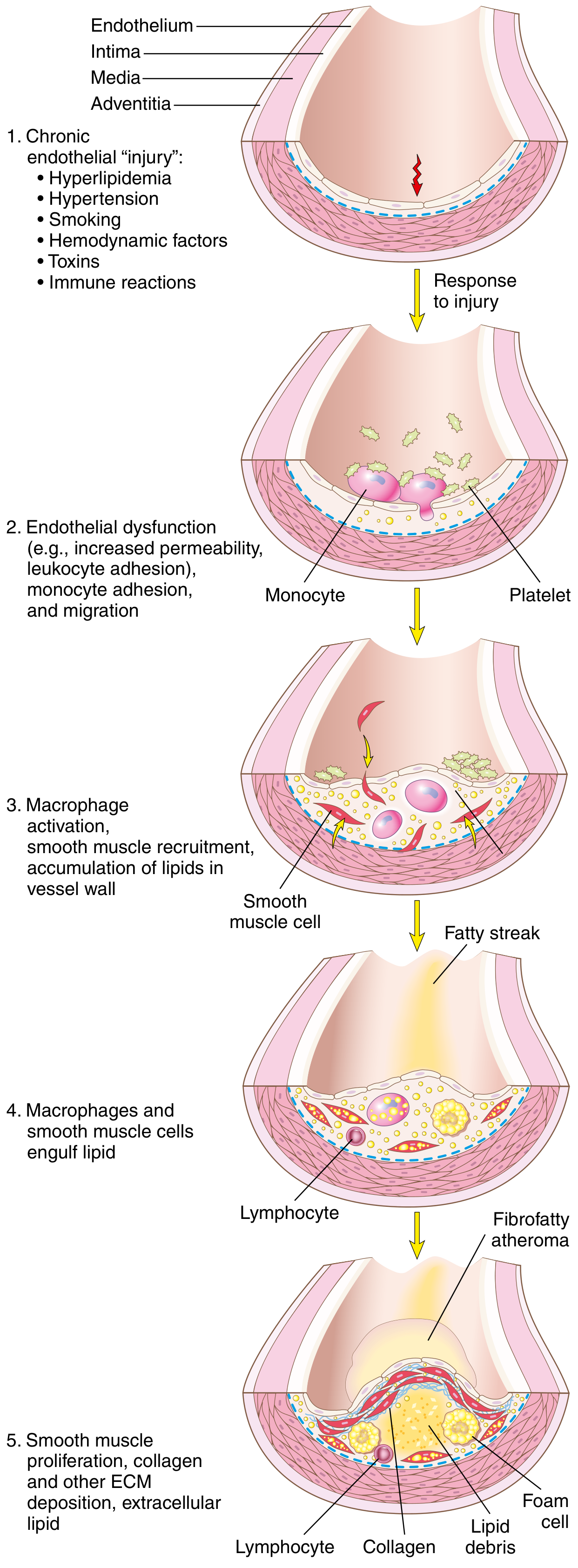
The currently accepted model is the "Response to Injury" Hypothesis (Ross, 1976, updated). Atherosclerosis is viewed as a chronic inflammatory response of the arterial wall to endothelial injury.
FLOW CHART: Pathogenesis of Atherosclerosis
ENDOTHELIAL INJURY / DYSFUNCTION
(Hyperlipidemia, HTN, Smoking, Hemodynamic turbulence, Toxins)
        │
        ▼
ENDOTHELIAL DYSFUNCTION
• ↑Permeability to lipoproteins
• ↑Leukocyte adhesion molecule expression (VCAM-1, ICAM-1)
• Pro-thrombotic state
• ↓Nitric oxide (vasodilatory, anti-inflammatory)
        │
        ├──────────────────────────────────────────┐
        ▼                                          ▼
LDL ACCUMULATION IN INTIMA             MONOCYTE ADHESION & MIGRATION
• LDL oxidized by free radicals        into intima
• Oxidized LDL is cytotoxic to ECs     ↓
• Triggers further inflammation        MACROPHAGES (via M-CSF)
        │                              + FOAM CELLS
        └────────────────┬─────────────┘
                         ▼
              FATTY STREAK FORMATION
        (earliest lesion - yellow intimal discoloration)
        Foam cells = macrophages + SMCs engorged with lipid
                         │
                         ▼
              PLATELET ADHESION
        (exposed subendothelial collagen)
                         │
                         ▼
        RELEASE OF GROWTH FACTORS & CYTOKINES
        (PDGF from platelets, macrophages, ECs)
        (FGF, TGF-α, TNF, IL-1, MCP-1)
                         │
                         ▼
        SMC RECRUITMENT (from media → intima)
        SMC PROLIFERATION + ECM SYNTHESIS
        (collagen, proteoglycans → FIBROUS CAP)
                         │
                         ▼
        ADVANCED ATHEROSCLEROTIC PLAQUE
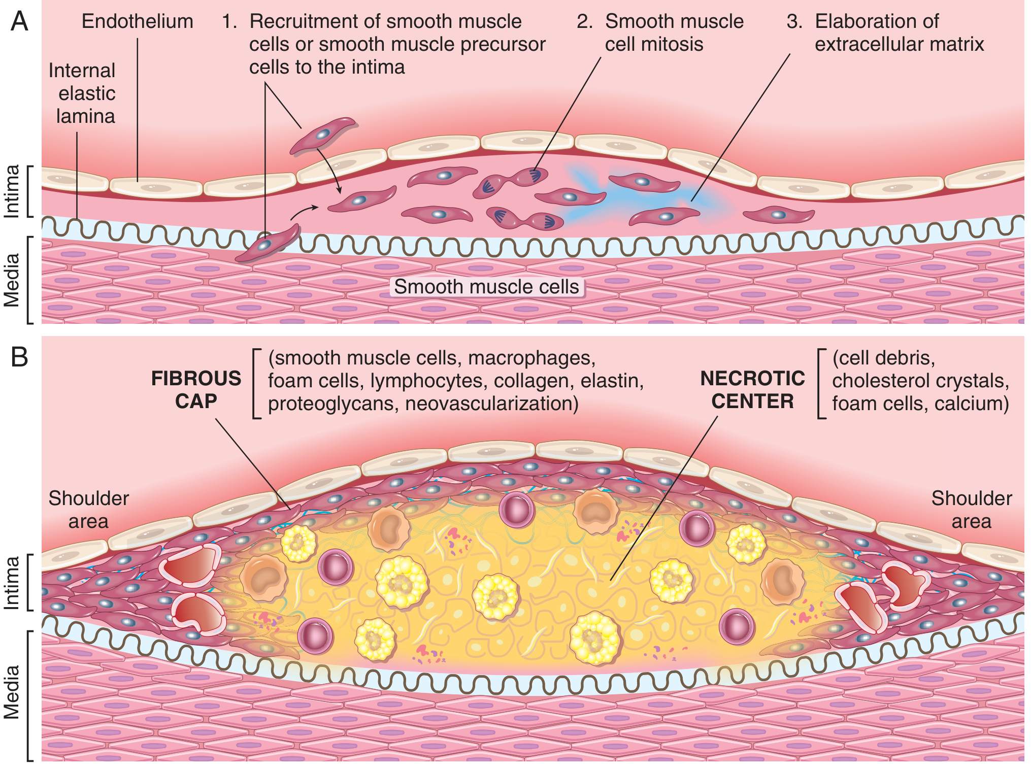
        • Fibrous cap (collagen, SMCs, macrophages)
        • Necrotic/lipid core (foam cell debris,
          cholesterol crystals, calcium)
        • Shoulder region (most inflammatory)
Key cellular players:
| Cell | Role |
|---|---|
| Endothelial cells | Injury triggers the whole cascade; become dysfunctional |
| Monocytes/Macrophages | Engulf oxidized LDL → foam cells; release cytokines (TNF, IL-1, MMP) |
| Smooth muscle cells | Migrate from media → intima; proliferate; synthesize collagen (fibrous cap) |
| T lymphocytes | Produce IFN-γ → inhibit SMC collagen synthesis → plaque destabilization |
| Platelets | Adhere to injured endothelium; release PDGF, TGF-β |
MORPHOLOGY
Gross Appearance
1. Fatty Streak (earliest lesion)
- Flat or slightly raised yellow intimal streak or spot
- Composed of foam cells (lipid-laden macrophages + SMCs)
- Reversible - can appear in aorta of infants/children (harmless at this stage)
- Precursor to atherosclerotic plaques
2. Atherosclerotic (Fibrous/Atheromatous) Plaque
- Yellow-white raised intimal lesion, 0.3-1.5 cm diameter
- May coalesce → larger plaques
- Eccentric distribution (not circumferential)
- Patchy, irregular distribution throughout the vessel
Components of a Mature Plaque (Microscopic)
LUMEN
    │
    ▼
┌─────────────────────────────────┐
│        FIBROUS CAP              │
│  • SMCs, macrophages, foam      │
│    cells, T lymphocytes         │
│  • Collagen, elastin,           │
│    proteoglycans                │
│  • Neovascularization           │
│  SHOULDER (most vulnerable      │
│   to rupture - most inflamed)   │
├─────────────────────────────────┤
│       NECROTIC/LIPID CORE       │
│  • Cell debris                  │
│  • Cholesterol crystals         │
│  • Foam cells                   │
│  • Calcium deposits             │
│  • Intraplaque hemorrhage       │
│    (rupture of vasa vasorum)    │
└─────────────────────────────────┘
    │
MEDIA (compressed, may atrophy)
    │
ADVENTITIA
Textbook Robbins Diagram - Atheromatous Plaque Structure:
PATHOGENESIS DIAGRAM (Robbins Classic)
STABLE VS. VULNERABLE PLAQUE
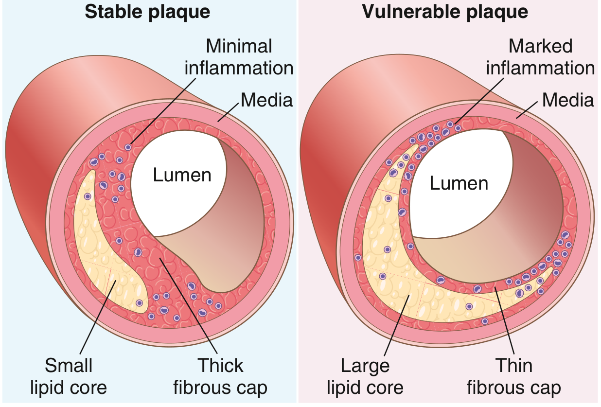
| Feature | Stable Plaque | Vulnerable Plaque |
|---|---|---|
| Fibrous cap | Thick | Thin |
| Lipid core | Small | Large |
| Inflammation | Minimal | Marked (foamy macrophages, T cells) |
| SMC content | High | Low |
| Collagen | Abundant | Reduced (MMPs degrade it) |
| Risk | Angina (stable) | ACS, MI, sudden death |
CONSEQUENCES / COMPLICATIONS
FLOW CHART: Complications of Atherosclerosis
ATHEROSCLEROTIC PLAQUE
        │
        ├──── GRADUAL STENOSIS (>70% occlusion)
        │     → Chronic ischemia
        │     → Stable angina
        │     → Intermittent claudication
        │     → Mesenteric ischemia
        │
        ├──── ACUTE PLAQUE CHANGE (rupture/erosion)
        │         │
        │    THREE TYPES:
        │    1. Rupture/Fissuring → exposed thrombogenic core
        │                         → THROMBOSIS (occlusive)
        │                         → MYOCARDIAL INFARCTION
        │                         → ISCHEMIC STROKE
        │                         → SUDDEN CARDIAC DEATH
        │    2. Erosion/Ulceration → exposed subendothelial
        │                           basement membrane
        │                           → partial thrombosis
        │    3. Intraplaque Hemorrhage → rupture of vasa vasorum
        │                               → rapid plaque expansion
        │
        ├──── ATHEROEMBOLISM
        │     • Plaque debris → microemboli downstream
        │     • Can cause renal failure, bowel ischemia
        │
        └──── ANEURYSM FORMATION
              • Plaque ischemia of media
              • SMC loss + ECM degradation in media
              • Weakens wall → dilatation
              → Abdominal Aortic Aneurysm (AAA) most common
              → Risk of rupture → hemorrhage → death
LESION SEQUENCE (AHA Classification)
TYPE I - Initial lesion
(isolated macrophage foam cells)
        ↓
TYPE II - Fatty streak
(multiple foam cell layers; visible yellow streaks)
[REVERSIBLE STAGE]
        ↓
TYPE III - Intermediate lesion
(foam cells + small extracellular lipid pools)
        ↓
TYPE IV - Atheroma
(large lipid core = "gruel"; endothelium intact)
        ↓
TYPE V - Fibroatheroma
(lipid core + fibrous cap = classic plaque)
[MOST CLINICALLY SIGNIFICANT]
        ↓
TYPE VI - Complicated lesion
(surface defect, hematoma, hemorrhage, thrombosis)
[CAUSES ACUTE EVENTS - MI, STROKE]
KEY MOLECULES SUMMARY TABLE
| Molecule | Source | Effect |
|---|---|---|
| Oxidized LDL | Oxidation of LDL in intima | Cytotoxic to ECs; stimulates foam cell formation |
| MCP-1 (CCL2) | Endothelial cells, SMCs | Recruits monocytes into intima |
| VCAM-1 | Dysfunctional endothelium | Adhesion of monocytes and T cells |
| PDGF | Platelets, macrophages, ECs | Stimulates SMC migration + proliferation |
| M-CSF | ECs, macrophages | Macrophage differentiation and activation |
| IFN-γ | T lymphocytes | Inhibits SMC collagen synthesis → cap weakening |
| MMPs (metalloproteinases) | Macrophages, foam cells | Degrade collagen → plaque instability |
| IL-1, TNF | Macrophages | Pro-inflammatory; upregulate adhesion molecules |
| HDL | Liver | Reverse cholesterol transport; protective |
| hsCRP | Liver (acute phase) | Marker of inflammation; independent risk predictor |
IMPORTANT EXAM POINTS (10-Mark Answer Tips)
- Definition: Chronic inflammatory disease of large/medium arteries - always mention "response to injury hypothesis"
- Fatty streak = earliest lesion - foam cells - reversible
- Plaque components: Fibrous cap (collagen, SMCs) + Necrotic core (lipid, calcium, debris)
- Vulnerable plaque = thin cap + large lipid core + marked inflammation = rupture → ACS
- Sites: Abdominal aorta > coronary > popliteal arteries
- Complications: Stenosis, thrombosis (MI, stroke), aneurysm, atheroembolism
- Role of oxidized LDL as central pathogenic molecule
- Foam cells = macrophages + SMCs engorged with oxidized LDL via scavenger receptors
REFERENCES
- Robbins & Kumar Basic Pathology, 11th ed. - Chapter 8 (Vascular Diseases), pp. 316-324
- Robbins, Cotran & Kumar Pathologic Basis of Disease, 10th ed. - Chapter 11 (Blood Vessels), pp. 466-478
Harsh mohan notes
Reading File
Finding Sources
Finding Sources
Harsh Mohan's Textbook of Pathology is not available in the library - only the two Robbins titles are in the pathology collection. However, since you asked for Harsh Mohan notes on atherosclerosis (continuing from the previous topic), I can give you comprehensive notes in Harsh Mohan's style and format, drawing from both Robbins titles in the library (which cover the same content Harsh Mohan covers, often in a similar structured format popular with Indian MBBS students).
ATHEROSCLEROSIS
(As per Harsh Mohan - Textbook of Pathology, 8th Edition Style)
Note: Harsh Mohan's Textbook of Pathology is not in our digital library. The notes below are compiled from both Robbins Pathology books in our collection and structured in Harsh Mohan's characteristic format - numbered points, tabular summaries, morphology boxes, and Indian exam-oriented style. The content is identical across both texts.
DEFINITION
Atherosclerosis is a disease of large and medium-sized muscular arteries characterized by endothelial dysfunction, vascular inflammation, and the build-up of lipids, cholesterol, cellular debris, calcium, and fibrin in the intima of large arteries, producing atheromatous plaques (also called atheromas).
- Name derived from Greek: athere = porridge/gruel (soft lipid core) + sclerosis = hardening (fibrous component)
INCIDENCE AND SITES AFFECTED
Sites (in order of frequency):
| Rank | Site | Main Complication |
|---|---|---|
| 1st | Abdominal aorta | Aneurysm |
| 2nd | Coronary arteries | Myocardial infarction |
| 3rd | Popliteal arteries | Gangrene of leg |
| 4th | Descending thoracic aorta | Aneurysm |
| 5th | Internal carotid arteries | Stroke |
| 6th | Circle of Willis | Stroke |
Plaques tend to form at:
- Ostia of exiting vessels
- Branch points
- Posterior wall of abdominal aorta
- These are sites of turbulent blood flow (hemodynamic stress)
ETIOPATHOGENESIS
Theory: "Response to Injury" Hypothesis (Ross, 1976 - revised)
The most widely accepted theory. Atherosclerosis is a chronic inflammatory response of the arterial wall initiated by injury to the endothelium.
FLOW CHART: Pathogenesis
STEP 1: ENDOTHELIAL INJURY / DYSFUNCTION
Causes:
• Hyperlipidemia (↑LDL, oxidized LDL)
• Hypertension (shear stress)
• Cigarette smoking (free radicals, CO)
• Hemodynamic factors (turbulence at bifurcations)
• Homocysteine, toxins, viruses (CMV, HSV)
• Immune mechanisms
        ↓
STEP 2: ENDOTHELIAL DYSFUNCTION
• ↑ Permeability to lipoproteins (LDL enters intima)
• ↑ Expression of adhesion molecules:
  VCAM-1, ICAM-1, E-selectin
• ↓ Nitric oxide (vasodilatory + anti-atherogenic)
• ↑ Pro-inflammatory cytokines
        ↓
STEP 3: LIPOPROTEIN ACCUMULATION
• LDL enters intima → oxidized by free radicals
• Oxidized LDL (ox-LDL):
  - Cytotoxic to ECs and SMCs
  - Chemoattractant for monocytes
  - Stimulates MCP-1 release
  - Ingested by macrophages via SCAVENGER RECEPTORS
    (not downregulated like LDL receptors → uncontrolled uptake)
        ↓
STEP 4: MONOCYTE RECRUITMENT & FOAM CELL FORMATION
• Monocytes adhere to activated endothelium (via VCAM-1)
• Monocytes migrate into intima
• Differentiate into MACROPHAGES (under M-CSF influence)
• Macrophages engulf ox-LDL → become FOAM CELLS
• SMCs also engulf lipid → become foam cells
        ↓
STEP 5: FATTY STREAK FORMATION
(collections of foam cells = earliest visible lesion)
        ↓
STEP 6: PLATELET ADHESION & ACTIVATION
• Dysfunctional/denuded endothelium
• Platelets adhere → release PDGF, TGF-β, serotonin
        ↓
STEP 7: SMC MIGRATION AND PROLIFERATION
• PDGF, FGF from platelets/macrophages/ECs stimulate SMCs
• SMCs migrate from MEDIA → INTIMA
• SMCs proliferate in intima
• SMCs synthesize COLLAGEN, elastin, proteoglycans
  → forms FIBROUS CAP
        ↓
STEP 8: PLAQUE GROWTH & PROGRESSION
• Lipid accumulation (extracellular + intracellular)
• T lymphocyte infiltration
• IFN-γ from T cells inhibits SMC collagen synthesis
• MMPs from macrophages degrade collagen
• Calcification of necrotic debris
        ↓
ADVANCED ATHEROSCLEROTIC PLAQUE
(Fibrous cap + Necrotic/lipid core)
MORPHOLOGY
A. GROSS MORPHOLOGY
1. Fatty Streak
- Earliest grossly visible lesion
- Slightly raised, yellow, 1-2 mm flat spots or streaks on intima
- Composed entirely of lipid-laden foam cells
- Occur in infancy and childhood - seen in aorta of children as young as 1 year
- Reversible - may not progress to plaque
- Do not cause clinical symptoms
2. Atherosclerotic (Atheromatous) Plaque - Fibrofatty Plaque
- Yellow-white, raised, irregular plaques
- Size: 0.3 cm to several cm in diameter
- Protrude into the vessel lumen
- Distribution: patchy and eccentric (not circumferential)
- Advanced plaques may show:
- Calcification (chalky white, gritty deposits)
- Ulceration of surface
- Thrombosis on top
- Intraplaque hemorrhage
B. MICROSCOPIC MORPHOLOGY
Components of an Atheromatous Plaque:
┌────────────────────────────────────────────────┐
│               FIBROUS CAP                       │
│  • Smooth muscle cells (modified, secretory)    │
│  • Macrophages and foam cells                   │
│  • T lymphocytes                                │
│  • Collagen (type I and III)                    │
│  • Elastic fibers, proteoglycans                │
│  • Neovascularization (vasa vasorum)            │
├────────────────────────────────────────────────┤
│            SHOULDER REGION                      │
│  (junction of cap and normal intima)            │
│  • Most cellular and inflammatory region        │
│  • Most prone to RUPTURE                        │
├────────────────────────────────────────────────┤
│         NECROTIC / LIPID CORE                   │
│  • Extracellular lipid (cholesterol esters)     │
│  • Cholesterol clefts (needle-shaped crystals)  │
│  • Foam cells (dead macrophages + SMCs)         │
│  • Cellular debris / necrotic material          │
│  • Calcium deposits                             │
│  • Hemorrhage (from ruptured vasa vasorum)      │
└────────────────────────────────────────────────┘
Histological Features:
- Cholesterol clefts - elongated needle-shaped spaces left after dissolution of cholesterol during processing - pathognomonic
- Foam cells - macrophages with abundant foamy cytoplasm (lipid-laden)
- Lymphocytic infiltration - mostly T cells
- SMC proliferation in intima (spindle-shaped cells)
- Calcification - dystrophic calcification in necrotic core
- Neovascularization from adventitial vasa vasorum
Textbook Diagrams:
CLASSIFICATION OF ATHEROSCLEROTIC LESIONS (AHA, Stary 1995)
| Type | Name | Morphology | Clinical |
|---|---|---|---|
| I | Initial | Isolated macrophage foam cells | Silent |
| II | Fatty streak | Multiple foam cell layers | Silent, reversible |
| III | Intermediate | Foam cells + small extracellular lipid pools | Silent |
| IV | Atheroma | Large confluent lipid core | Silent/unstable |
| V | Fibroatheroma | Lipid core + thick fibrous cap | Stable angina |
| VI | Complicated | Rupture, hemorrhage, thrombosis | ACS, MI, Stroke |
RISK FACTORS
Constitutional (Non-modifiable)
- Genetic factors - family history (most important independent RF); familial hypercholesterolemia
- Age - incidence increases with age; MI incidence increases 5-fold between age 40-60
- Male sex - premenopausal women relatively protected (estrogen effect); risk equalizes after menopause
- CHIP (Clonal Hematopoiesis of Indeterminate Potential) - pro-inflammatory monocyte clones
Modifiable (Major)
- Hyperlipidemia - most important modifiable RF
- ↑LDL (atherogenic), ↓HDL (protective)
- Lipoprotein(a) [Lp(a)] - independent RF
- Hypertension - increases shear stress on endothelium
- Cigarette smoking - free radicals oxidize LDL; CO injures ECs; vasoconstriction
- Diabetes mellitus - accelerated atherosclerosis (2-3x risk); glycation of LDL
Additional Risk Factors
- Hyperhomocysteinemia (injures ECs)
- Elevated CRP / markers of inflammation
- Elevated PAI-1 (↓fibrinolysis)
- Obesity, physical inactivity
- Type A personality / stress
COMPLICATIONS OF ATHEROSCLEROSIS
Flow Chart: Complications
ATHEROSCLEROTIC PLAQUE
        │
        ├─── 1. STENOSIS (Gradual lumen narrowing)
        │         >75% stenosis → Critical stenosis
        │         → ISCHEMIA of supplied organ
        │         Coronary: Stable Angina, Chronic IHD
        │         Peripheral: Intermittent claudication
        │         Renal: Renovascular hypertension
        │         Mesenteric: Bowel ischemia
        │
        ├─── 2. ACUTE PLAQUE CHANGE (Rupture/Erosion)
        │    Three forms:
        │    (a) Plaque RUPTURE/FISSURE
        │        Thrombogenic core exposed → Thrombus formation
        │        → Complete vessel occlusion
        │        → ACUTE MI / STROKE / SUDDEN CARDIAC DEATH
        │    (b) Plaque EROSION/ULCERATION
        │        Exposed basement membrane → Partial thrombosis
        │    (c) INTRAPLAQUE HEMORRHAGE
        │        Rupture of vasa vasorum within plaque
        │        → Rapid plaque expansion
        │        → Near-total occlusion
        │
        ├─── 3. ATHEROEMBOLISM
        │    Plaque rupture → Cholesterol crystals/debris
        │    → Microemboli in distal vessels
        │    → Blue toe syndrome, renal failure
        │
        └─── 4. ANEURYSM FORMATION
             Ischemia of media (from plaque compression)
             → Medial SMC loss
             → Wall weakening
             → ANEURYSM (especially Abdominal Aortic Aneurysm)
             → Risk of rupture → fatal hemorrhage
STABLE vs. VULNERABLE PLAQUE
| Feature | Stable Plaque | Vulnerable (Unstable) Plaque |
|---|---|---|
| Fibrous cap | Thick | Thin (<65 μm) |
| Lipid core | Small (<40% of plaque volume) | Large (>40%) |
| Inflammation | Minimal | Marked (macrophages, T cells) |
| SMC content | High | Low |
| Collagen | Abundant | Reduced (MMPs degrade it) |
| Calcification | Variable | Often present |
| IFN-γ effect | - | Inhibits SMC collagen synthesis |
| MMP activity | Low | High |
| Clinical outcome | Stable angina | ACS, MI, sudden death |
Mechanism of plaque destabilization:
- Macrophages release matrix metalloproteinases (MMP-1, 8, 13) → degrade collagen
- T cells release IFN-γ → inhibit SMC collagen synthesis
- Combined effect → fibrous cap thinning → rupture
PATHOGENESIS DIAGRAM
DIFFERENCES: FATTY STREAK vs. ATHEROMATOUS PLAQUE
| Feature | Fatty Streak | Atheromatous Plaque |
|---|---|---|
| Age | Infancy/childhood | Middle age onwards |
| Appearance | Flat yellow spots/streaks | Raised yellow-white plaque |
| Composition | Foam cells only | Fibrous cap + lipid core |
| Reversibility | Reversible | Irreversible (advanced) |
| Symptoms | None | Can cause symptoms |
| Progression | May or may not progress | Progressive |
SUMMARY BOX (Quick Revision)
ATHEROSCLEROSIS - KEY FACTS
━━━━━━━━━━━━━━━━━━━━━━━━━━━━━━━━━━━━━━━━━
Definition    : Chronic intimal inflammatory disease
               of large/medium arteries
Theory        : Response to Injury Hypothesis
Earliest lesion: Fatty streak (foam cells)
Classic lesion : Atheromatous plaque
               (fibrous cap + necrotic core)
Key cell      : Foam cell (macrophage + SMC
               engorged with ox-LDL)
Pathognomonic : Cholesterol clefts (histology)
Most imp. RF  : Hyperlipidemia (↑LDL)
Most imp. site: Abdominal aorta > Coronary arteries
Key danger    : Vulnerable plaque rupture → ACS
Fatal comps   : MI, Stroke, Aortic rupture
━━━━━━━━━━━━━━━━━━━━━━━━━━━━━━━━━━━━━━━━━
Sources used:
- Robbins & Kumar Basic Pathology, 11th ed. - Chapter 8, pp. 316-326 (Vascular Diseases)
- Robbins, Cotran & Kumar Pathologic Basis of Disease, 10th ed. - Chapter 11, pp. 463-480 (Blood Vessels)
Harsh Mohan's Textbook of Pathology (8th edition) is not in the digital library, but the content above covers all topics that Harsh Mohan covers on atherosclerosis, presented in his style (numbered points, morphology boxes, classification tables, diagrams). The facts are identical - Harsh Mohan draws heavily from Robbins for the atherosclerosis chapter.
This is a shared conversation. Sign in to Orris to start your own chat.