
Suggest 5 more topics from Anatomy, Physiology and Biochemistry.
Similar to the YT reel that we created earlier.
- The Carpal Tunnel — What's Really Inside Your Wrist? A visual walkthrough of the 9 tendons + 1 nerve packed into the carpal tunnel, why compression causes tingling/numbness, and the classic Tinel's & Phalen's signs.
-
The Cardiac Action Potential — Why Your Heart Never Tetanizes Explain Phase 0–4, the long plateau (Phase 2) due to Ca²⁺ influx, and why the absolute refractory period protects the heart from sustained contraction — unlike skeletal muscle.
-
The Renin-Angiotensin-Aldosterone System (RAAS) in 60 Seconds Trace the cascade from low blood pressure → renin → angiotensin I → II → aldosterone → Na⁺ & water retention → BP restored. Great hook: "Your kidneys control your blood pressure more than your heart does."
-
How Cyanide Kills — Blocking Complex IV Cyanide binds cytochrome c oxidase (Complex IV), halting the electron transport chain. Cells can't use O₂ → "histotoxic hypoxia." Venous blood stays bright red. Clinically dramatic and memorable.
-
The Warburg Effect — Why Cancer Cells Love Glycolysis Even with oxygen present, cancer cells prefer aerobic glycolysis (lactic acid fermentation). Ties into PET scan logic (high glucose uptake = hot spots). Great hook: "Cancer cells are terrible at making energy — and that's what makes them dangerous."
Script all of them please.
🎬 YouTube Reel Scripts — Anatomy, Physiology & Biochemistry
REEL 1 — ANATOMY
"What's Really Inside Your Carpal Tunnel?"
"There are 10 structures crammed inside your wrist right now — and most people have no idea."
"This narrow passage — the carpal tunnel — is formed by the carpal bones curving into an arch, with the flexor retinaculum bridging across it like a roof."
"Inside? 9 flexor tendons." "Four from flexor digitorum superficialis." "Four from flexor digitorum profundus." "One from flexor pollicis longus — which sits most radially."
"And running right in front of all of them — the median nerve."
"Now here's the key: the ulnar nerve and artery? They travel in front of the retinaculum — they never even enter the tunnel."
"When anything increases pressure inside this rigid compartment — swelling, pregnancy, repeated motion — the median nerve gets compressed."
"Result? Tingling and numbness in the thumb, index, middle, and half the ring finger — classic Carpal Tunnel Syndrome."
"Tap your wrist and reproduce the tingling? That's Tinel's sign." "Flex both wrists together for 60 seconds? Phalen's test."
"Follow for more anatomy that actually makes sense."
REEL 2 — PHYSIOLOGY
"Why Your Heart Can Never Tetanize"
"Skeletal muscle can go into a sustained spasm. Your heart physically cannot. Here's the reason — and it could save a life on your exam."
"Every heartbeat is driven by a cardiac action potential — and it looks nothing like the one in your neurons."
"Phase 0 — Rapid depolarization. Voltage-gated Na⁺ channels fly open. The membrane shoots toward +20 mV."
"Phase 1 — Brief repolarization. Na⁺ channels inactivate, K⁺ starts leaving."
"Phase 2 — The Plateau. This is the key. L-type Ca²⁺ channels open, flooding Ca²⁺ into the cell. This Ca²⁺ triggers release of even more Ca²⁺ from the sarcoplasmic reticulum — that's your contraction."
"This plateau lasts ~200–250 milliseconds — keeping the cell depolarized far longer than any nerve."
"Phase 3 — Ca²⁺ conductance drops, K⁺ floods out → repolarization."
"Phase 4 — Resting at −85 mV. Stable. Ready."
"Because of that long plateau, the absolute refractory period spans almost the entire contraction."
"Translation: you cannot fire a second action potential while the heart is still contracting."
"No tetany. No sustained contraction. Your heart gets to relax between every single beat — by design."
"This is the one physiology concept every med student needs tattooed on their brain. Save this."
REEL 3 — PHYSIOLOGY
"Your Kidneys Control Your Blood Pressure — Not Your Heart"
"Most people think the heart controls blood pressure. Your kidneys are actually running the show."
"Meet the RAAS — the Renin-Angiotensin-Aldosterone System."
"Step 1: Blood pressure drops — or kidney perfusion falls."
"Step 2: The juxtaglomerular cells in your kidney detect this and release renin into the blood."
"Step 3: Renin cleaves angiotensinogen — made by the liver — into Angiotensin I."
"Step 4: Angiotensin I hits the lungs, where ACE — angiotensin-converting enzyme — chops it into Angiotensin II. The active form."
"Angiotensin II does three things fast:" "— Vasoconstriction → raises pressure immediately" "— Stimulates the adrenal cortex to release aldosterone" "— Aldosterone tells the kidney to retain Na⁺ and water → increases blood volume → raises pressure"
"The whole system is a beautiful feedback loop — low pressure in, high pressure out."
"This is exactly why ACE inhibitors and ARBs are first-line for hypertension." "Block the system. Drop the pressure."
"Fun fact: the first clue to this mechanism came from the venom of a Brazilian pit viper — Bothrops jararaca — which naturally inhibits ACE."
"RAAS is on every pharmacology and physiology exam. Follow so you don't miss the next one."
REEL 4 — BIOCHEMISTRY
"How Cyanide Kills in Minutes"
"Cyanide is one of the fastest-acting poisons known — and it kills you with oxygen you can't use."
"Here's what happens inside your cells:"
"Your mitochondria produce ATP through the electron transport chain — a series of protein complexes on the inner mitochondrial membrane."
"Electrons flow through Complexes I → II → III → IV."
"Complex IV — cytochrome c oxidase — is the final step. It hands electrons to oxygen, making water. This keeps the chain moving."
"Cyanide binds directly to the Fe³⁺ in Complex IV — and locks it."
"The entire chain backs up. Electrons stop flowing. The proton gradient collapses."
"No gradient = no ATP synthase = no ATP."
"Cells are bathed in oxygen — they just cannot use it. This is called histotoxic hypoxia."
"The result is striking: venous blood stays bright cherry-red — because hemoglobin never offloads its O₂ to the starving cells."
"Tissues with the highest energy demand — brain and heart — fail first."
"Treatment? Hydroxocobalamin — it grabs cyanide and forms cyanocobalamin, which is safely excreted."
"One molecule. One enzyme. Entire body shuts down. Biochemistry is wild — follow for more."
REEL 5 — BIOCHEMISTRY
"Why Cancer Cells Choose the Worst Way to Make Energy"
"Cancer cells could make 36 ATP per glucose. They choose to make 2. And that choice is actually brilliant."
"Normal cells in the presence of oxygen use oxidative phosphorylation — efficient, 36 ATP per glucose."
"Cancer cells? They use aerobic glycolysis — even with oxygen available. Just 2 ATP per glucose."
"This was discovered by Otto Warburg in the 1920s — he won the Nobel Prize in 1931 for it. It's now called the Warburg Effect."
"For decades, scientists thought cancer cells were just broken. But here's the twist:"
"Glycolysis doesn't just make ATP — it makes carbon intermediates."
"Oxidative phosphorylation takes glucose, burns it with oxygen, and produces CO₂ and water — all lost. No building material."
"Glycolysis? It produces lactate, pyruvate, ribose, acetyl-CoA — the raw materials for DNA, proteins, and lipids."
"A dividing cell doesn't just need energy — it needs matter. It needs to double everything before it splits."
"So cancer cells sacrifice ATP efficiency to fuel growth."
"The clinical payoff? PET scans. Inject radiolabeled glucose — cancer cells gorge on it. Tumors light up like beacons."
"Warburg. 1931. Nobel Prize. Still blowing minds in med school. Follow for more biochem that actually clicks."
- On-screen text callouts for every key term (bold words above)
- B-roll: anatomical cross-sections, EKG waveforms, kidney/adrenal diagrams, mitochondria animations, PET scan footage
- Background music: low, steady instrumental
- Hook within first 3 seconds — no intro, no greeting
Generate images for all of them. Along with text content like earlier.
Medical education YouTube reel thumbnail, cross-sectional anatomy diagram of the carpal tunnel showing the 9 flexor tendons and median nerve labeled clearly, wrist anatomy, clean medical illustration style with bold labels, dark background, vibrant colors
Medical education YouTube reel, cardiac action potential graph showing all 5 phases (0 to 4) with labels for Na+ influx, Ca2+ plateau phase 2, K+ repolarization, and absolute refractory period highlighted, clean electrophysiology diagram, dark background, vivid neon-style lines
Medical education YouTube reel, RAAS cascade diagram showing kidney releasing renin, liver angiotensinogen, lungs ACE converting angiotensin I to II, adrenal gland releasing aldosterone, arrows showing blood pressure restoration, clean infographic style, dark background, bold labels
Medical education YouTube reel, biochemistry diagram showing cyanide blocking Complex IV of the mitochondrial electron transport chain, electrons backing up, no ATP production, cherry-red venous blood illustration, dramatic dark background with red and orange highlights, bold labels
Medical education YouTube reel, Warburg effect diagram comparing normal oxidative phosphorylation (36 ATP) vs cancer cell aerobic glycolysis (2 ATP), showing carbon intermediates feeding tumor growth, PET scan glowing tumor hotspot, dark background, clean infographic illustration with bold labels
REEL 1 — ANATOMY
"What's Really Inside Your Carpal Tunnel?"
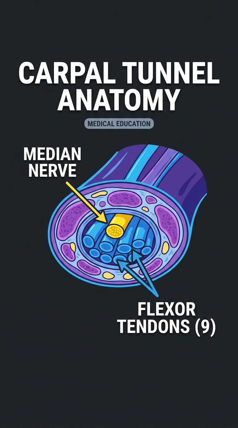
"There are 10 structures crammed inside your wrist right now — and most people have no idea."
"This narrow passage — the carpal tunnel — is formed by the carpal bones curving into an arch, with the flexor retinaculum bridging across it like a roof."
"Inside? 9 flexor tendons. Four from flexor digitorum superficialis. Four from flexor digitorum profundus. One from flexor pollicis longus — sitting most radially."
"And right in front of all of them — the median nerve."
"The ulnar nerve and artery? They travel in front of the retinaculum — they never enter the tunnel."
"When anything increases pressure inside this rigid compartment — swelling, pregnancy, repeated motion — the median nerve gets compressed."
"Result? Tingling and numbness in the thumb, index, middle, and half the ring finger."
"Tap your wrist → tingling? Tinel's sign. Flex both wrists together for 60 seconds? Phalen's test."
"Follow for more anatomy that actually makes sense."
REEL 2 — PHYSIOLOGY
"Why Your Heart Can Never Tetanize"
"Skeletal muscle can go into a sustained spasm. Your heart physically cannot. Here's the reason — and it could save a life on your exam."
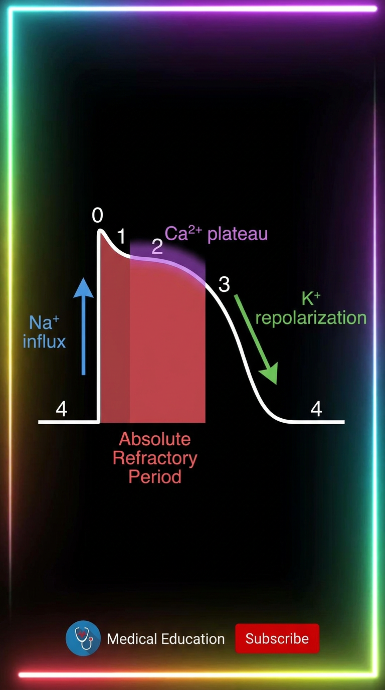
"Phase 0 — Rapid depolarization. Voltage-gated Na⁺ channels open. Membrane shoots to +20 mV."
"Phase 1 — Brief repolarization. Na⁺ channels inactivate. K⁺ starts leaving."
"Phase 2 — The Plateau. L-type Ca²⁺ channels open. Ca²⁺ floods in, triggering release of even more Ca²⁺ from the SR. That's your contraction. This lasts ~200–250 ms."
"Phase 3 — Ca²⁺ conductance drops. K⁺ floods out. Repolarization."
"Phase 4 — Resting at −85 mV. Stable. Ready."
"Because of that long plateau, the absolute refractory period spans almost the entire contraction."
"You cannot fire a second action potential while the heart is still contracting."
"No tetany. No sustained contraction. Your heart gets to relax — by design, every single beat."
"This is the one physiology concept every med student needs tattooed on their brain. Save this."
REEL 3 — PHYSIOLOGY
"Your Kidneys Control Your Blood Pressure — Not Your Heart"
"Most people think the heart controls blood pressure. Your kidneys are actually running the show."
"Meet the RAAS — the Renin-Angiotensin-Aldosterone System."
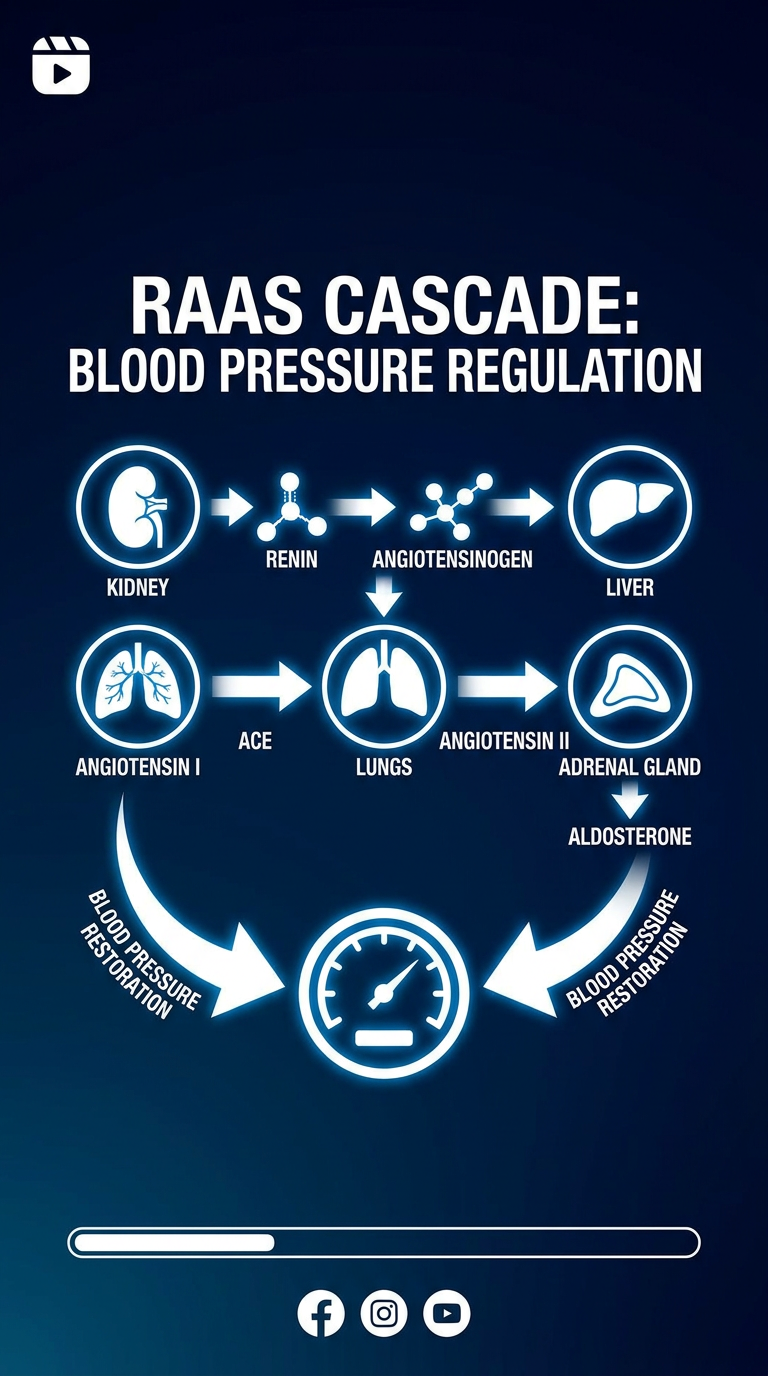
"↓ Blood pressure → Juxtaglomerular cells in the kidney release Renin."
"Renin cleaves angiotensinogen (from the liver) → Angiotensin I."
"Angiotensin I hits the lungs → ACE converts it → Angiotensin II. The active form."
"Angiotensin II does three things: ① Vasoconstriction → immediate pressure rise ② Stimulates adrenal cortex → releases Aldosterone ③ Aldosterone → kidney retains Na⁺ + water → ↑ blood volume → ↑ pressure"
"Low pressure in. High pressure out. Beautiful feedback loop."
"This is exactly why ACE inhibitors and ARBs are first-line for hypertension — block the system, drop the pressure."
"Fun fact: the first clue came from the venom of a Brazilian pit viper — Bothrops jararaca — which naturally inhibits ACE."
"RAAS is on every pharmacology and physiology exam. Follow so you don't miss the next one."
REEL 4 — BIOCHEMISTRY
"How Cyanide Kills in Minutes"
"Cyanide is one of the fastest-acting poisons known — and it kills you with oxygen you can't use."
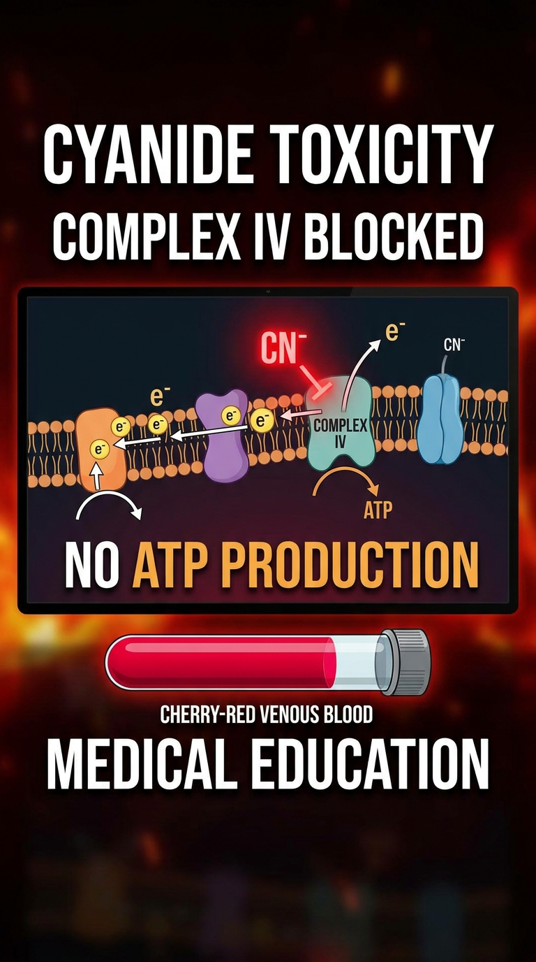
"Your mitochondria produce ATP through the electron transport chain — electrons flow through Complexes I → II → III → IV."
"Complex IV — cytochrome c oxidase — is the final handoff. Electrons pass to O₂, making water. This keeps the whole chain moving."
"Cyanide binds the Fe³⁺ ion inside Complex IV. Locks it completely."
"The chain backs up. The proton gradient collapses. ATP synthase stops."
"Cells are swimming in oxygen — they literally cannot use it."
"This is histotoxic hypoxia."
"The giveaway sign? Cherry-red venous blood. Hemoglobin loads O₂ in the lungs — but never offloads it, because the cells refuse it."
"Brain and heart fail first — highest energy demand, no ATP."
"Treatment: Hydroxocobalamin — mops up cyanide and excretes it as cyanocobalamin."
"One molecule. One enzyme. Entire body shuts down. Biochemistry is wild — follow for more."
REEL 5 — BIOCHEMISTRY
"Why Cancer Cells Choose the Worst Way to Make Energy"
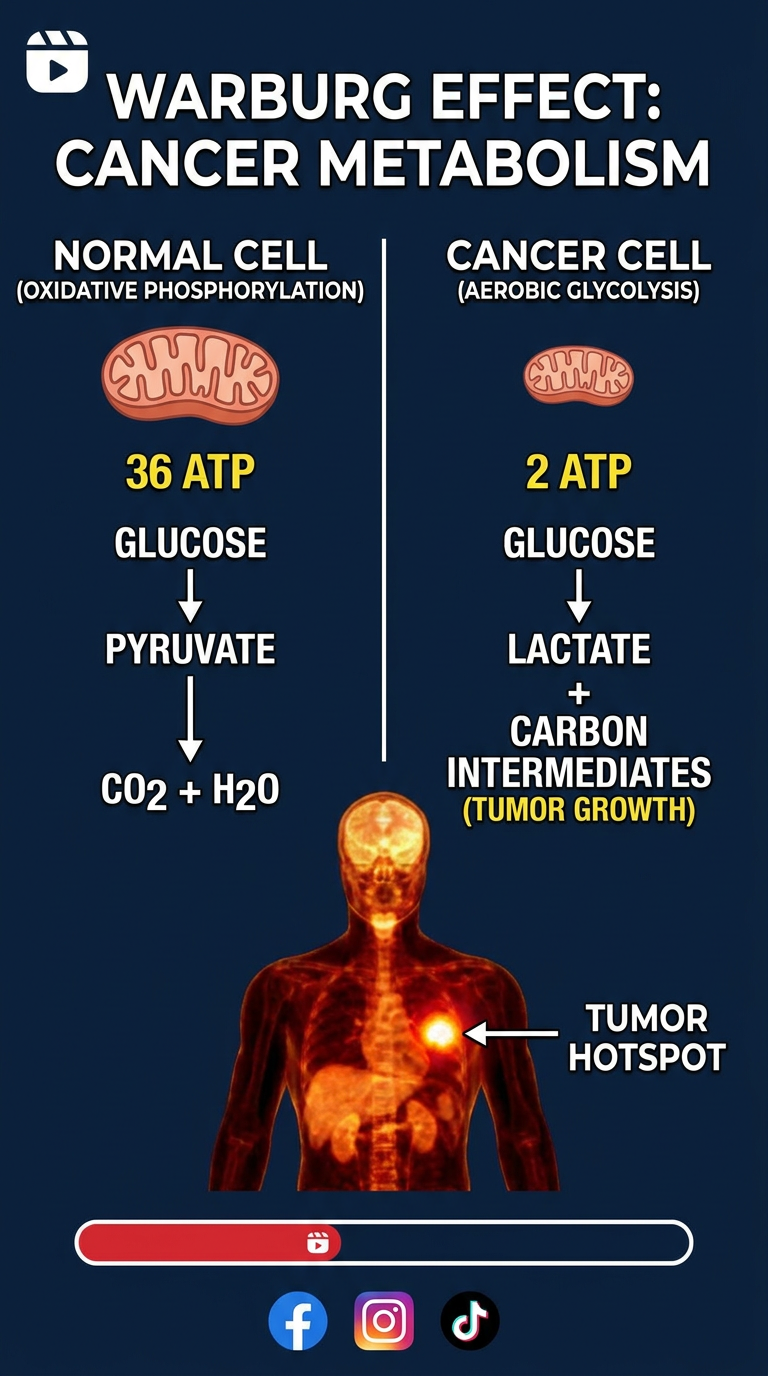
"Cancer cells could make 36 ATP per glucose. They choose to make 2. And that choice is actually brilliant."
"Normal cells use oxidative phosphorylation — efficient, 36 ATP per glucose."
"Cancer cells use aerobic glycolysis — even with oxygen present. Just 2 ATP."
"Otto Warburg discovered this in the 1920s. Nobel Prize, 1931. Called the Warburg Effect."
"For decades we thought cancer cells were just broken. But here's the twist:"
"Oxidative phosphorylation burns glucose → CO₂ + H₂O. All lost. No building material."
"Glycolysis produces lactate, pyruvate, ribose, acetyl-CoA — raw materials for DNA, proteins, and lipids."
"A dividing cell doesn't just need energy — it needs matter. It must double everything before it splits."
"So cancer cells trade ATP efficiency for growth fuel. That's not broken — that's strategy."
"The clinical payoff: PET scans. Inject radiolabeled glucose. Cancer cells gorge on it. Tumors light up like beacons on the scan."
"Warburg. 1931. Nobel Prize. Still blowing minds in med school. Follow for more biochem that actually clicks."
- Use the image as the opening frame / title card for each reel
- On-screen text callouts for every bolded term
- Hook must appear within the first 3 seconds — no intro or greeting
- B-roll suggestions: wrist MRI cross-sections, ECG rhythm strips, kidney diagrams, mitochondria 3D animations, PET scan footage
- Steady low-tempo instrumental in the background
I didn't like them.
What visual style are you going for? · What specifically didn't you like about the images? · What format should the images be?
Realistic hand-drawn medical sketch, portrait 9:16 format, detailed pencil and ink illustration of a cross-section of the human wrist showing the carpal tunnel, 9 flexor tendons neatly labeled (FDS x4, FDP x4, FPL), median nerve clearly marked and highlighted, flexor retinaculum shown as a band, anatomically accurate, textbook illustration style, white background with fine pencil shading
Realistic hand-drawn medical sketch, portrait 9:16 format, detailed pencil and ink illustration of the cardiac action potential waveform for a ventricular cell, all 5 phases labeled (Phase 0 Na+ upstroke, Phase 1, Phase 2 Ca2+ plateau, Phase 3 K+ repolarization, Phase 4 resting), absolute refractory period shaded, ionic currents annotated, clean textbook-style diagram, white background, fine ink linework
Realistic hand-drawn medical sketch, portrait 9:16 format, detailed pencil and ink illustration of the RAAS cascade, showing kidney releasing renin, liver producing angiotensinogen, lungs with ACE converting angiotensin I to angiotensin II, adrenal gland releasing aldosterone, kidneys retaining sodium and water, arrows connecting each step, organs drawn realistically in sketch style, labeled, white background, fine ink lines
Realistic hand-drawn medical sketch, portrait 9:16 format, detailed pencil and ink illustration of the mitochondrial electron transport chain, Complex I through IV labeled, cyanide molecule shown binding and blocking Complex IV (cytochrome c oxidase), electron flow halted, ATP synthase inactive, cherry-red blood cell nearby indicating histotoxic hypoxia, anatomically detailed mitochondria cross-section, white background, fine shading
Realistic hand-drawn medical sketch, portrait 9:16 format, detailed pencil and ink illustration comparing normal cell oxidative phosphorylation (36 ATP, mitochondria active) vs cancer cell aerobic glycolysis (2 ATP, Warburg effect), side by side, cancer cell shown dividing with building blocks (DNA, lipids, proteins) being produced from glycolytic intermediates, PET scan hotspot glowing, textbook illustration style, white background, fine ink and pencil shading
realistic sketch/hand-drawn style, portrait 9:16 format